Components of the Blood
By the end of this section, you will be able to:
- List the basic components of the blood
- Compare red and white blood cells
- Describe blood plasma and serum
Hemoglobin is responsible for distributing oxygen, and to a lesser extent, carbon dioxide, throughout the circulatory systems of humans, vertebrates, and many invertebrates. The blood is more than the proteins, though. Blood is actually a term used to describe the liquid that moves through the vessels and includes plasma (the liquid portion, which contains water, proteins, salts, lipids, and glucose) and the cells (red and white cells) and cell fragments called platelets. Blood plasma is actually the dominant component of blood and contains the water, proteins, electrolytes, lipids, and glucose. The cells are responsible for carrying the gases (red cells) and the immune response (white). The platelets are responsible for blood clotting. Interstitial fluid that surrounds cells is separate from the blood, but in hemolymph, they are combined. In humans, cellular components make up approximately 45 percent of the blood and the liquid plasma 55 percent. Blood is 20 percent of a person’s extracellular fluid and eight percent of weight.
The Role of Blood in the Body
Blood, like the human blood illustrated below, is important for regulation of the body’s systems and homeostasis. Blood helps maintain homeostasis by stabilizing pH, temperature, osmotic pressure, and by eliminating excess heat. Blood supports growth by distributing nutrients and hormones, and by removing waste. Blood plays a protective role by transporting clotting factors and platelets to prevent blood loss and transporting the disease-fighting agents or white blood cells to sites of infection.
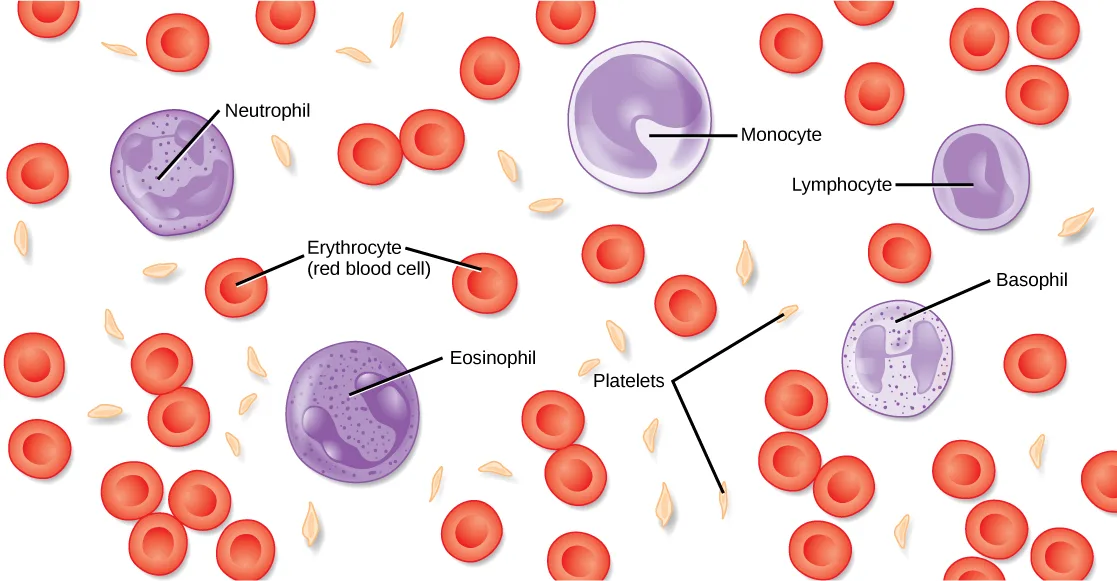
Extended description
Reading left to right, top to bottom: one leader line labels Neutrophil, a purple cell with a granular, multi-lobed nucleus; one labels Monocyte, the largest purple cell, with a broad U-shaped nucleus; one labels Lymphocyte, a smaller purple cell with an oblong nucleus; one labels Erythrocyte (red blood cell), one of the many disc-shaped red cells; one labels Basophil, at the right, a paler purple cell with an irregular, multi-lobed nucleus; one labels Eosinophil, a purple cell with a granular, U-shaped nucleus; one label, Platelets, forks into two lines pointing to two of the small, spindle-shaped fragments scattered among the red cells. Unlabeled red blood cells and platelets fill the rest of the scene.
Red Blood Cells
Red blood cells, or erythrocytes (erythro- = “red”; -cyte = “cell”), are specialized cells that circulate through the body delivering oxygen to cells; they are formed from stem cells in the bone marrow. In mammals, red blood cells are small biconcave cells that at maturity do not contain a nucleus or mitochondria and are only 7–8 µm in size. In birds and non-avian reptiles, a nucleus is still maintained in red blood cells.
The red coloring of blood comes from the iron-containing protein hemoglobin, illustrated below (a). The principle job of this protein is to carry oxygen, but it also transports carbon dioxide as well. Hemoglobin is packed into red blood cells at a rate of about 250 million molecules of hemoglobin per cell. Each hemoglobin molecule binds four oxygen molecules so that each red blood cell carries one billion molecules of oxygen. There are approximately 25 trillion red blood cells in the five liters of blood in the human body, which could carry up to 25 sextillion () molecules of oxygen in the body at any time. In mammals, the lack of organelles in erythrocytes leaves more room for the hemoglobin molecules, and the lack of mitochondria also prevents use of the oxygen for metabolic respiration. Only mammals have anucleated red blood cells, and some mammals (camels, for instance) have unusually shaped oval red blood cells, though still without nuclei. (Source note: the source says camels “even have nucleated red blood cells”; camelid erythrocytes are anucleate ellipsoids (Schalm’s Veterinary Hematology, 6th ed., “Hematology of Camelids”).) The advantage of nucleated red blood cells is that these cells can undergo mitosis. Anucleated red blood cells metabolize anaerobically (without oxygen), making use of a primitive metabolic pathway to produce ATP and increase the efficiency of oxygen transport.
Not all organisms use hemoglobin as the method of oxygen transport. Invertebrates that utilize hemolymph rather than blood use different pigments to bind to the oxygen. These pigments use copper or iron to bind to the oxygen. Invertebrates have a variety of other respiratory pigments. Hemocyanin, a blue-green, copper-containing protein, illustrated below (b), is found in mollusks, crustaceans, and some of the arthropods. Chlorocruorin, a green-colored, iron-containing pigment is found in four families of polychaete tubeworms. Hemerythrin, a red, iron-containing protein is found in some polychaete worms and annelids and is illustrated below (c). Despite the name, hemerythrin does not contain a heme group and its oxygen-carrying capacity is poor compared to hemoglobin.
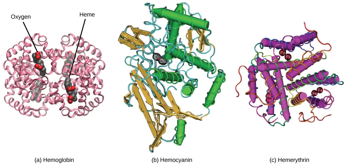
Extended description
In panel (a), Hemoglobin appears as a four-lobed, pink ribbon structure; one leader line labeled Oxygen and one labeled Heme each point to one of the two visible ball-and-stick clusters embedded in the ribbons, one in the left half of the molecule and one in the right. In panel (b), Hemocyanin is drawn as intertwined green cylindrical helices, gold ribbon sheets, and teal coils, with two gray spheres marked Cu near the model’s center. In panel (c), Hemerythrin is drawn as purple cylindrical helices threaded with a thin, multicolored coil, with six spheres marked Fe, drawn as three pairs, distributed through the structure.
The small size and large surface area of red blood cells allows for rapid diffusion of oxygen and carbon dioxide across the plasma membrane. In the lungs, carbon dioxide is released and oxygen is taken in by the blood. In the tissues, oxygen is released from the blood and carbon dioxide is bound for transport back to the lungs. Studies have found that hemoglobin also binds nitrous oxide (NO). NO is a vasodilator that relaxes the blood vessels and capillaries and may help with gas exchange and the passage of red blood cells through narrow vessels. Nitroglycerin, a heart medication for angina and heart attacks, is converted to NO to help relax the blood vessels and increase oxygen flow through the body.
A characteristic of red blood cells is their glycolipid and glycoprotein coating; these are lipids and proteins that have carbohydrate molecules attached. In humans, the surface glycoproteins and glycolipids on red blood cells vary between individuals, producing the different blood types, such as A, B, and O. Red blood cells have an average life span of 120 days, at which time they are broken down and recycled in the liver and spleen by phagocytic macrophages, a type of white blood cell.
White Blood Cells
White blood cells, also called leukocytes (leuko = white), make up approximately one percent by volume of the cells in blood. The role of white blood cells is very different than that of red blood cells: they are primarily involved in the immune response to identify and target pathogens, such as invading bacteria, viruses, and other foreign organisms. White blood cells are formed continually; some only live for hours or days, but some live for years.
The morphology of white blood cells differs significantly from red blood cells. They have nuclei and do not contain hemoglobin. The different types of white blood cells are identified by their microscopic appearance after histologic staining, and each has a different specialized function. The two main groups, both illustrated below, are the granulocytes, which include the neutrophils, eosinophils, and basophils, and the agranulocytes, which include the monocytes and lymphocytes.
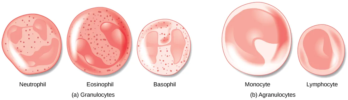
Granulocytes contain granules in their cytoplasm; the agranulocytes are so named because of the lack of granules in their cytoplasm. Some leukocytes become macrophages that either stay at the same site or move through the bloodstream and gather at sites of infection or inflammation where they are attracted by chemical signals from foreign particles and damaged cells. Lymphocytes are the primary cells of the immune system and include B cells, T cells, and natural killer cells. B cells destroy bacteria and inactivate their toxins. They also produce antibodies. T cells attack viruses, fungi, some bacteria, transplanted cells, and cancer cells. T cells attack viruses by releasing toxins that kill the viruses. Natural killer cells attack a variety of infectious microbes and certain tumor cells.
One reason that HIV poses significant management challenges is because the virus directly targets T cells by gaining entry through a receptor. Once inside the cell, HIV then multiplies using the T cell’s own genetic machinery. After the HIV virus replicates, it is transmitted directly from the infected T cell to macrophages. The presence of HIV can remain unrecognized for an extensive period of time before full disease symptoms develop.
Platelets and Coagulation Factors
Blood must clot to heal wounds and prevent excess blood loss. Small cell fragments called platelets (thrombocytes) are attracted to the wound site where they adhere by extending many projections and releasing their contents. These contents activate other platelets and also interact with other coagulation factors, which convert fibrinogen, a water-soluble protein present in blood serum into fibrin (a non-water soluble protein), causing the blood to clot. Many of the clotting factors require vitamin K to work, and vitamin K deficiency can lead to problems with blood clotting. Many platelets converge and stick together at the wound site forming a platelet plug (also called a fibrin clot), as illustrated below (b). The plug or clot lasts for a number of days and stops the loss of blood. Platelets are formed from the disintegration of larger cells called megakaryocytes, like that shown below (a). For each megakaryocyte, 2000–3000 platelets are formed with 150,000 to 400,000 platelets present in each cubic millimeter of blood. Each platelet is disc shaped and 2–4 µm in diameter. They contain many small vesicles but do not contain a nucleus.
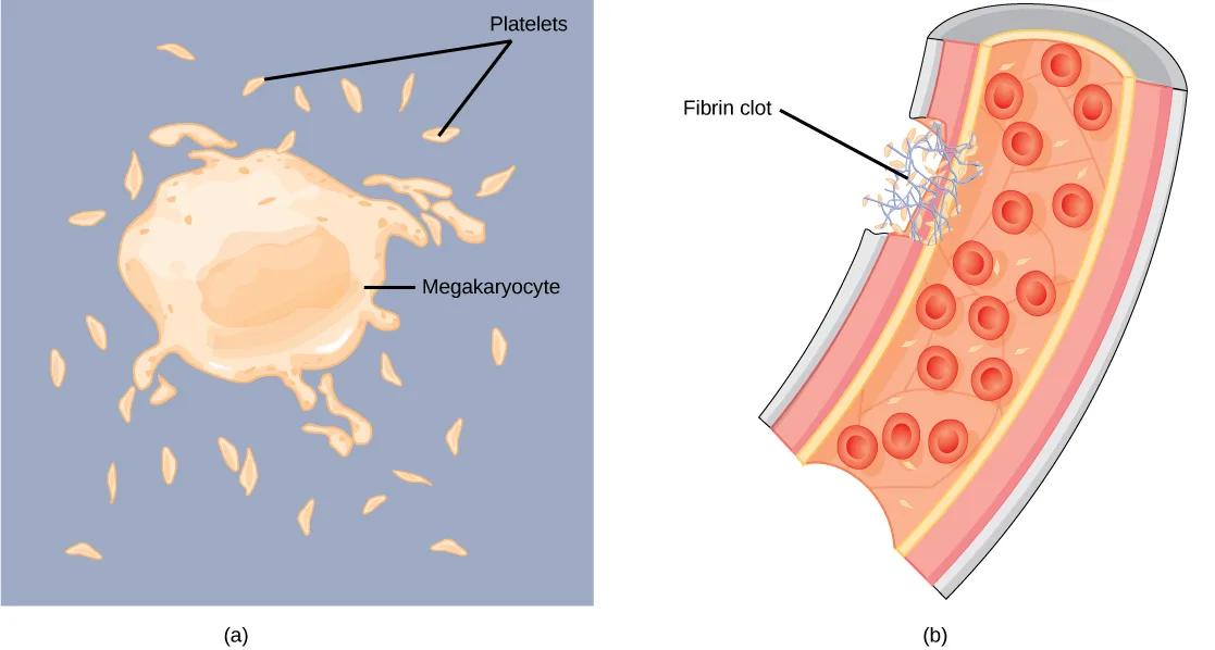
Extended description
In panel (a), one leader line labels Megakaryocyte, the single large, irregularly lobed cell at center; a second leader line labeled Platelets forks into two branches, each pointing to one of the many small, spindle-shaped fragments scattered around it. In panel (b), one leader line labels Fibrin clot, a tangled, thread-like mass at a wedge-shaped breach in the vessel wall; the vessel’s interior is otherwise filled with numerous unlabeled red blood cells.
Plasma and Serum
The liquid component of blood is called plasma, and it is separated by spinning or centrifuging the blood at high rotations (3000 rpm or higher). The blood cells and platelets are separated by centrifugal forces to the bottom of a specimen tube. The upper liquid layer, the plasma, consists of 90 percent water along with various substances required for maintaining the body’s pH, osmotic load, and for protecting the body. The plasma also contains the coagulation factors and antibodies.
The plasma component of blood without the coagulation factors is called the serum. Serum is similar to interstitial fluid in which the correct composition of key ions acting as electrolytes is essential for normal functioning of muscles and nerves. Other components in the serum include proteins that assist with maintaining pH and osmotic balance while giving viscosity to the blood. The serum also contains antibodies, specialized proteins that are important for defense against viruses and bacteria. Lipids, including cholesterol, are also transported in the serum, along with various other substances including nutrients, hormones, metabolic waste, plus external substances, such as, drugs, viruses, and bacteria.
Human serum albumin is the most abundant protein in human blood plasma and is synthesized in the liver. Albumin, which constitutes about half of the blood serum protein, transports hormones and fatty acids, buffers pH, and maintains osmotic pressures. Immunoglobulin is a protein antibody produced in the mucosal lining and plays an important role in antibody mediated immunity.
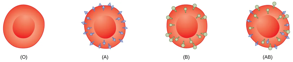
Evolution Connection. Blood Types Related to Proteins on the Surface of the Red Blood Cells. Red blood cells are coated in antigens made of glycolipids and glycoproteins. The composition of these molecules is determined by genetics, which have evolved over time. In humans, the different surface antigens are grouped into 24 different blood groups with more than 100 different antigens on each red blood cell. The two most well known blood groups are the ABO, shown below, and Rh systems. The surface antigens in the ABO blood group are glycolipids, called antigen A and antigen B. People with blood type A have antigen A, those with blood type B have antigen B, those with blood type AB have both antigens, and people with blood type O have neither antigen. Antibodies called agglutinogens are found in the blood plasma and react with the A or B antigens, if the two are mixed. When type A and type B blood are combined, agglutination (clumping) of the blood occurs because of antibodies in the plasma that bind with the opposing antigen; this causes clots that coagulate in the kidney causing kidney failure. Type O blood has neither A or B antigens, and therefore, type O blood can be given to all blood types. Type O negative blood is the universal donor. Type AB positive blood is the universal acceptor because it has both A and B antigen. The ABO blood groups were discovered in 1900 and 1901 by Karl Landsteiner at the University of Vienna.
The Rh blood group was first discovered in Rhesus monkeys. Most people have the Rh antigen (Rh+) and do not have anti-Rh antibodies in their blood. The few people who do not have the Rh antigen and are Rh– can develop anti-Rh antibodies if exposed to Rh+ blood. This can happen after a blood transfusion or after an Rh– person has an Rh+ baby. The first exposure does not usually cause a reaction; however, at the second exposure, enough antibodies have built up in the blood to produce a reaction that causes agglutination and breakdown of red blood cells. An injection can prevent this reaction.
Link to Learning
Play a blood typing game on the Nobel Prize website to solidify your understanding of blood types.
Summary
Specific components of the blood include red blood cells, white blood cells, platelets, and the plasma, which contains coagulation factors and serum. Blood is important for regulation of the body’s pH, temperature, osmotic pressure, the circulation of nutrients and removal of waste, the distribution of hormones from endocrine glands, and the elimination of excess heat; it also contains components for blood clotting. Red blood cells are specialized cells that contain hemoglobin and circulate through the body delivering oxygen to cells. White blood cells are involved in the immune response to identify and target invading bacteria, viruses, and other foreign organisms; they also recycle waste components, such as old red blood cells. Platelets and blood clotting factors cause the change of the soluble protein fibrinogen to the insoluble protein fibrin at a wound site forming a plug. Plasma consists of 90 percent water along with various substances, such as coagulation factors and antibodies. The serum is the plasma component of the blood without the coagulation factors.
Key terms
- plasma — liquid component of blood that is left after the cells are removed.
- platelet — (also, thrombocyte) small cellular fragment that collects at wounds, cross-reacts with clotting factors, and forms a plug to prevent blood loss.
- red blood cell — small (7–8 µm) biconcave cell without mitochondria (and in mammals without nuclei) that is packed with hemoglobin, giving the cell its red color; transports oxygen through the body.
- serum — plasma without the coagulation factors.
- white blood cell — large (12–20 µm) cell with nuclei (Source note: the source glossary says 30 µm; human leukocytes measure about 12–20 µm (Junqueira’s Basic Histology, 15th ed., chapter 12).) of which there are many types with different roles including the protection of the body from viruses and bacteria, and cleaning up dead cells and other waste.
Practice
List the basic components of the blood
Platelet plug formation occurs at which point?
Trace platelet activity in order: where do they start, where are they dispersed, and where do they finally converge and stick? The plug forms at the last of these steps.The liquid component of blood that is left after the cells are removed is called ________.
This is the 90-percent-water portion left in the tube after centrifuging separates out the cells and platelets.A small cellular fragment that collects at wounds and forms a plug to prevent blood loss is called a ________.
Megakaryocytes break apart into thousands of these tiny fragments, which converge at a wound to help it clot.List some of the functions of blood in the body.
Show model answer
Did your answer mention:
Compare red and white blood cells
White blood cells:
Check the three statements about white blood cells separately — their two structural groups, their defensive role, and their alternate name — before deciding whether all three hold up.The red blood cells of birds differ from mammalian red blood cells because:
The Red Blood Cells section describes what mammalian red blood cells lack at maturity, then notes what birds and non-avian reptiles keep instead.A small biconcave cell packed with hemoglobin that transports oxygen through the body is called a(n) ________.
In mammals, this cell type lacks a nucleus and mitochondria at maturity, leaving more room for the oxygen-carrying pigment.A large cell with a nucleus that protects the body from viruses and bacteria and cleans up dead cells and waste is called a(n) ________.
Unlike the oxygen-carrying cells, this type has a nucleus and multiplies continually to fight invaders.How does the lymphatic system work with blood flow?
Show model answer
Did your answer mention:
Describe blood plasma and serum
In humans, the plasma comprises what percentage of the blood?
The opening paragraph gives one percentage for the cellular components and the complementary percentage for the liquid portion — which one matches plasma?Plasma without the coagulation factors is called ________.
Draw blood into a tube with no anticoagulant, let it clot, and what separates out on top still carries antibodies but none of the clotting proteins.Describe the cause of different blood type groups.
Show model answer
Did your answer mention:
This section is adapted from Biology 2e, Section 40.2: Components of the Blood by Mary Ann Clark, Jung Choi, Matthew Douglas, and OpenStax, © OpenStax, licensed under CC BY-NC-SA 4.0. Access the original for free at openstax.org. Changes: two source misspellings corrected (“Immunoglobin” to “immunoglobulin”, “agglutinougens” to “agglutinogens”, both reported as source defects); figures re-encoded as WebP with the source alt text edited where noted; all five figures re-kinded from the manifest’s file-extension “photo” guess to “diagram” (Figure_40_02_01, Figure_40_02_02abc, Figure_40_02_03ab, Figure_40_02_04ab, and Figure_40_02_05 are each a hand-drawn or computer-rendered illustration, not a captured photograph); a longdesc added to the three figures whose printed leader-line labels are not carried by their one-line caption — the blood-cell-types illustration, the hemoglobin/hemocyanin/hemerythrin molecular models, and the megakaryocyte/platelet-clot illustration — transcribing each drawing’s own labels and leader-line counts, in reading order; ion charges (Fe²⁺, Fe³⁺) set in Unicode; the numeric exponent (25 × 10 to the 21st power) set in KaTeX per the house notation rule; in-text pointers to figures (“Figure 40.5” through “Figure 40.9”) replaced with “shown below,” “illustrated below,” or “shown below (a)/(b)/(c),” since Hugo does not number figures; the evolution note rendered as an Evolution Connection callout and the interactive note as a Link to Learning callout, keeping the module’s own openstax.org/l/blood_typing redirect URL; the end-of-section Review Questions and Critical Thinking Questions adapted into the closing interactive Practice block (multiple choice and self-check respectively) — all four Review Questions and all three Critical Thinking Questions are used; rubric checkpoints added to each self-check, decomposing its model answer (the source solution) into check-off clauses with no new claims; five key-term recall items added from the glossary (plasma, platelet, red blood cell, white blood cell, serum), covering every glossary term in the section. This module carries no comparison table, so no sortbins exercise applies. Two claims are corrected with visible Source notes: camel red blood cells are oval but anucleate, not nucleated, and white blood cells are 12–20 µm rather than 30 µm (errata 446–447).