Mammalian Heart and Blood Vessels
By the end of this section, you will be able to:
- Describe the structure of the heart and explain how cardiac muscle is different from other muscles
- Describe the cardiac cycle
- Explain the structure of arteries, veins, and capillaries, and how blood flows through the body
The heart is a complex muscle that pumps blood through the three divisions of the circulatory system: the coronary (vessels that serve the heart), pulmonary (heart and lungs), and systemic (systems of the body), shown below. Coronary circulation intrinsic to the heart takes blood directly from the main artery (aorta) coming from the heart. For pulmonary and systemic circulation, the heart has to pump blood to the lungs or the rest of the body, respectively. In vertebrates, the lungs are relatively close to the heart in the thoracic cavity. The shorter distance to pump means that the muscle wall on the right side of the heart is not as thick as the left side which must have enough pressure to pump blood all the way to your big toe.
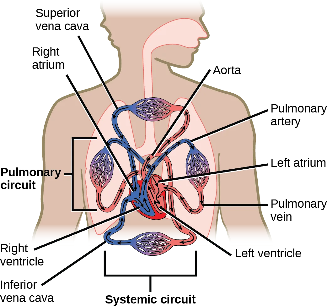
Extended description
At upper left, leader lines label Superior vena cava and Right atrium, pointing to the vessel and chamber entering the top of the heart. At upper right, four leader lines label Aorta, Pulmonary artery, Left atrium, and Pulmonary vein, pointing to the vessels arcing over the heart’s upper chambers. At lower left, leader lines label Right ventricle and Inferior vena cava; at lower right, one leader line labels Left ventricle. A bracket at the left, spanning the vessels between the heart and the drawn lungs, is labeled Pulmonary circuit. A second bracket at the bottom, spanning the vessel loop beneath the heart, is labeled Systemic circuit. Arrowheads trace one loop: from the venae cavae into the right atrium and ventricle, up through the pulmonary artery to the lungs, back through the pulmonary vein to the left atrium and ventricle, and out through the aorta.
Which of the following statements about the circulatory system is false?
For each vessel, ask two things: does it carry blood toward the lungs or away from them, and does blood pick up oxygen or release it while it is inside the lungs? The module’s own sentences about the pulmonary vein and the pulmonary arteries, later in this section, settle both questions.Structure of the Heart
The heart muscle is asymmetrical as a result of the distance blood must travel in the pulmonary and systemic circuits. Since the right side of the heart sends blood to the pulmonary circuit it is smaller than the left side which must send blood out to the whole body in the systemic circuit, shown below. In humans, the heart is about the size of a clenched fist; it is divided into four chambers: two atria and two ventricles. There is one atrium and one ventricle on the right side and one atrium and one ventricle on the left side. The atria are the chambers that receive blood, and the ventricles are the chambers that pump blood. The right atrium receives deoxygenated blood from the superior vena cava, which drains blood from the jugular vein that comes from the brain and from the veins that come from the arms, as well as from the inferior vena cava which drains blood from the veins that come from the lower organs and the legs. In addition, the right atrium receives blood from the coronary sinus which drains deoxygenated blood from the heart itself. This deoxygenated blood then passes to the right ventricle through the atrioventricular valve or the tricuspid valve, a flap of connective tissue that opens in only one direction to prevent the backflow of blood. The valve separating the chambers on the left side of the heart valve is called the bicuspid or mitral valve. After it is filled, the right ventricle pumps the blood through the semilunar valve (or pulmonic valve or pulmonary) to the pulmonary arteries and on to the lungs for re-oxygenation. After blood passes through the pulmonary arteries, the right semilunar valves close preventing the blood from flowing backwards into the right ventricle. The left atrium then receives the oxygen-rich blood from the lungs via the pulmonary veins. This blood passes through the bicuspid valve or mitral valve (the atrioventricular valve on the left side of the heart) to the left ventricle where the blood is pumped out through the aorta, the major artery of the body, taking oxygenated blood to the organs and muscles of the body. Once blood is pumped out of the left ventricle and into the aorta, the aortic semilunar valve (or aortic valve) closes preventing blood from flowing backward into the left ventricle. This pattern of pumping is referred to as double circulation and is found in all mammals.
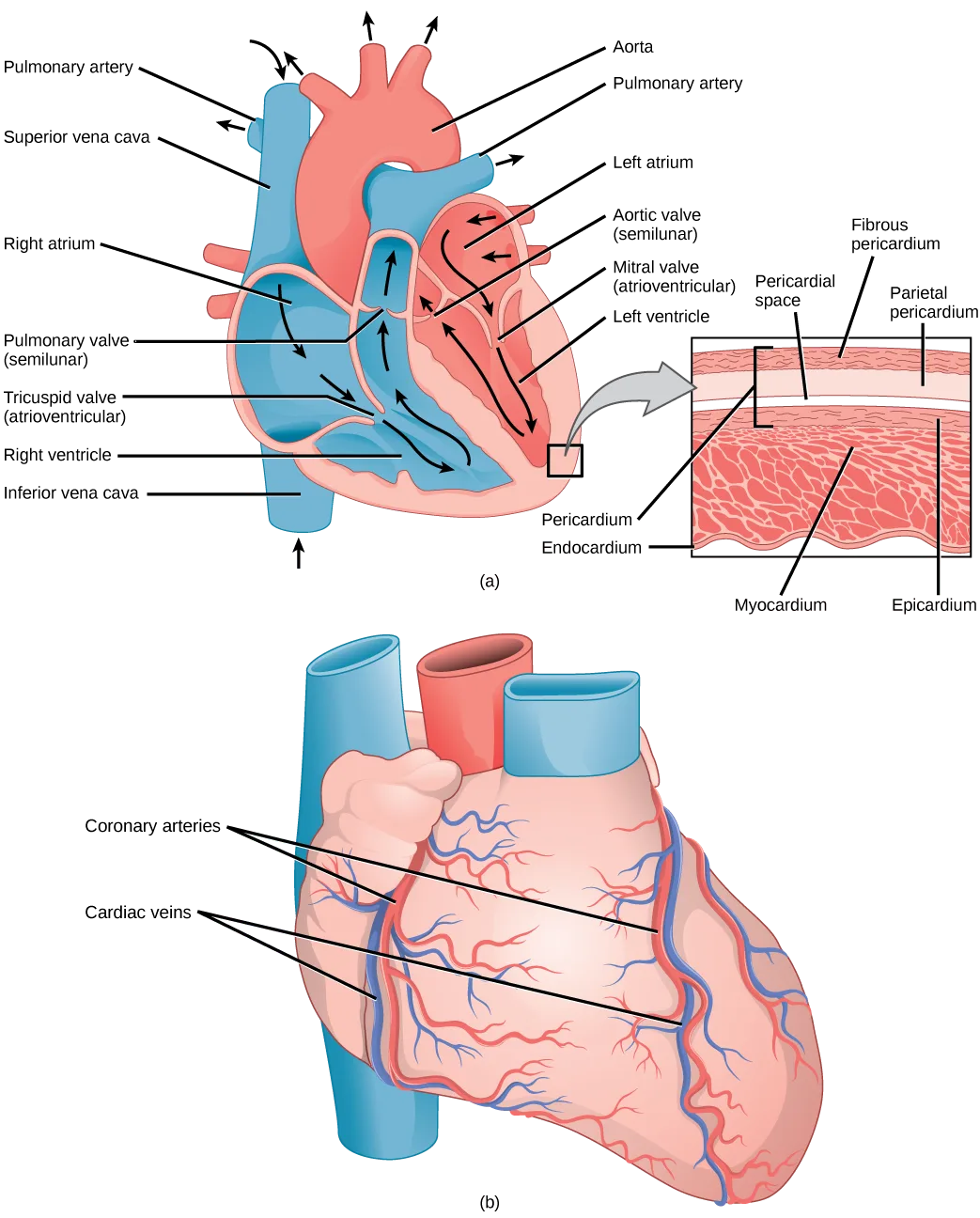
Extended description
Panel (a)’s left side carries seven leader lines, top to bottom: Pulmonary artery, Superior vena cava, Right atrium, Pulmonary valve (semilunar), Tricuspid valve (atrioventricular), Right ventricle, and Inferior vena cava. Its right side carries six leader lines, top to bottom: Aorta, Pulmonary artery, Left atrium, Aortic valve (semilunar), Mitral valve (atrioventricular), and Left ventricle. A gray arrow leads from a small boxed region of the heart wall to an inset labeling, top to bottom, Fibrous pericardium, Parietal pericardium, and Pericardial space, then Pericardium and Endocardium on the inner edge of the muscle band and Myocardium and Epicardium across and beyond it. Panel (b), below, shows the heart’s outer surface with the label Coronary arteries pointing to two vessels and Cardiac veins pointing to three, both running down from the same great vessels at the top.
Which of the following statements about the heart is false?
Trace where the bicuspid valve sits in the sequence blood follows through the left side of the heart — between which two chambers does the labeled diagram place it, and in which direction does blood move through a valve on its way toward the body?The heart is composed of three layers; the epicardium, the myocardium, and the endocardium, illustrated above. The inner wall of the heart has a lining called the endocardium. The myocardium consists of the heart muscle cells that make up the middle layer and the bulk of the heart wall. The outer layer of cells is called the epicardium, of which the second layer is a membranous layered structure called the pericardium that surrounds and protects the heart; it allows enough room for vigorous pumping but also keeps the heart in place to reduce friction between the heart and other structures.
The heart has its own blood vessels that supply the heart muscle with blood. The coronary arteries branch from the aorta and surround the outer surface of the heart like a crown. They diverge into capillaries where the heart muscle is supplied with oxygen before converging again into the coronary veins to take the deoxygenated blood back to the right atrium where the blood will be re-oxygenated through the pulmonary circuit. The heart muscle will die without a steady supply of blood. Atherosclerosis is the blockage of an artery by the buildup of fatty plaques. Because of the size (narrow) of the coronary arteries and their function in serving the heart itself, atherosclerosis can be deadly in these arteries. The slowdown of blood flow and subsequent oxygen deprivation that results from atherosclerosis causes severe pain, known as angina, and complete blockage of the arteries will cause myocardial infarction: the death of cardiac muscle tissue, commonly known as a heart attack. Marie M. Daly was the first to associate cholesterol, high blood pressure, and the causes of atheroclerosis, which led to a deeper understanding of ways to prevent heart attack and treat heart disease.
The Cardiac Cycle
The main purpose of the heart is to pump blood through the body; it does so in a repeating sequence called the cardiac cycle. The cardiac cycle is the coordination of the filling and emptying of the heart of blood by electrical signals that cause the heart muscles to contract and relax. The human heart beats over 100,000 times per day. In each cardiac cycle, the heart contracts (systole), pushing out the blood and pumping it through the body; this is followed by a relaxation phase (diastole), where the heart fills with blood, as illustrated below. The atria contract at the same time, forcing blood through the atrioventricular valves into the ventricles. Closing of the atrioventricular valves produces a monosyllabic “lup” sound. Following a brief delay, the ventricles contract at the same time forcing blood through the semilunar valves into the aorta and the artery transporting blood to the lungs (via the pulmonary artery). Closing of the semilunar valves produces a monosyllabic “dup” sound.
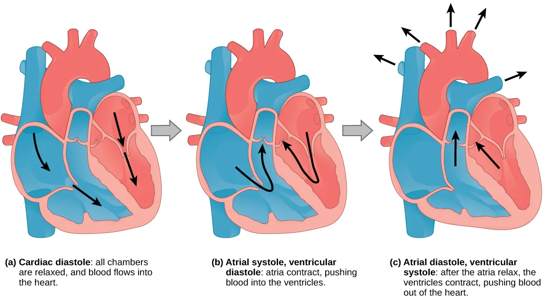
Extended description
Panel (a), Cardiac diastole, shows three black arrows pointing down into the relaxed chambers — two in the blue-toned right heart and one in the pink-toned left heart — as blood flows in from the great vessels above. Panel (b), Atrial systole and ventricular diastole, shows two black arrows curving upward, one from each atrium into its ventricle — the blue-toned right atrium into the right ventricle, the pink-toned left atrium into the left ventricle — as the atria contract. Panel (c), Atrial diastole and ventricular systole, shows two black arrows pointing straight up from the ventricles toward the valves, one on each side, plus five more black arrows fanning outward from the tops of the aorta and pulmonary artery, as the ventricles contract and blood leaves the heart.
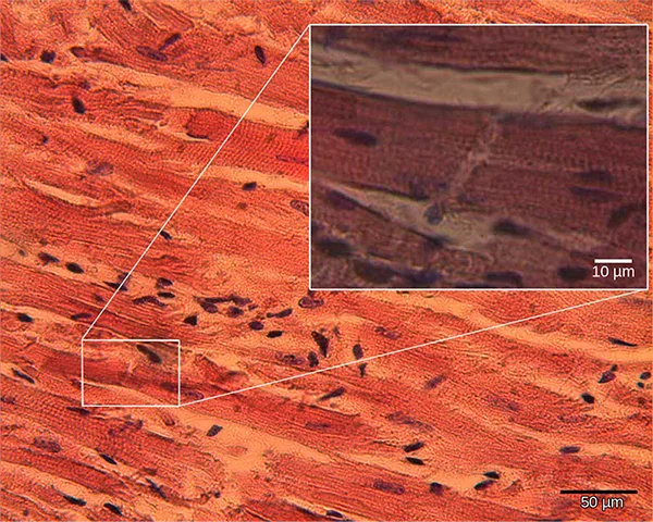
The pumping of the heart is a function of the cardiac muscle cells, or cardiomyocytes, that make up the heart muscle. Cardiomyocytes, shown below, are distinctive muscle cells that are striated like skeletal muscle but pump rhythmically and involuntarily like smooth muscle; they are connected by intercalated disks exclusive to cardiac muscle. They are self-stimulated for a period of time and isolated cardiomyocytes will beat if given the correct balance of nutrients and electrolytes.
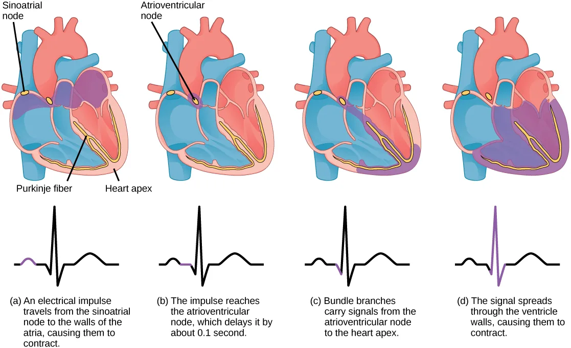
The autonomous beating of cardiac muscle cells is regulated by the heart’s internal pacemaker that uses electrical signals to time the beating of the heart. The electrical signals and mechanical actions, illustrated below, are intimately intertwined. The internal pacemaker starts at the sinoatrial (SA) node, which is located near the wall of the right atrium. Electrical charges spontaneously pulse from the SA node causing the two atria to contract in unison. The pulse reaches a second node, called the atrioventricular (AV) node, between the right atrium and right ventricle where it pauses for approximately 0.1 second before spreading to the walls of the ventricles. From the AV node, the electrical impulse enters the bundle of His, then to the left and right bundle branches extending through the interventricular septum. Finally, the Purkinje fibers conduct the impulse from the apex of the heart up the ventricular myocardium, and then the ventricles contract. This pause allows the atria to empty completely into the ventricles before the ventricles pump out the blood. The electrical impulses in the heart produce electrical currents that flow through the body and can be measured on the skin using electrodes. This information can be observed as an electrocardiogram (ECG)—a recording of the electrical impulses of the cardiac muscle.
Extended description
One leader line above panel (a) labels Sinoatrial node, pointing to a small oval at the top of that heart; one leader line above panel (b) labels Atrioventricular node, pointing to a second oval lower in that heart. Below panel (a), two more leader lines label Purkinje fiber, tracing a curved line down the septum and around the ventricle floor, and Heart apex, the bottom tip of the heart. Each of the four panels shows the same heart shape with purple shading spreading further across it than the last. Below each heart is its own ECG trace: a black line with one short purple segment marking that panel’s event. In (a) the purple segment is the small hump before the sharp spike, as the impulse spreads through the atria. In (b) it is the flat segment right after that hump, as the impulse is delayed at the atrioventricular node. In (c) it is a brief rise just before the spike, as bundle branches carry the signal toward the heart apex. In (d) the sharp spike itself is purple, as the signal spreads through the ventricle walls.
Link to Learning
Watch an animation of the heart’s electrical pacemaker in action.
Arteries, Veins, and Capillaries
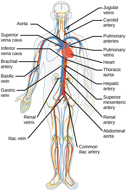
The blood from the heart is carried through the body by a complex network of blood vessels (shown below). Arteries take blood away from the heart. The main artery is the aorta that branches into major arteries that take blood to different limbs and organs. These major arteries include the carotid artery that takes blood to the brain, the brachial arteries that take blood to the arms, and the thoracic artery that takes blood to the thorax and then into the hepatic, renal, and gastric arteries for the liver, kidney, and stomach, respectively. The iliac artery takes blood to the lower limbs. The major arteries diverge into minor arteries, and then smaller vessels called arterioles, to reach more deeply into the muscles and organs of the body.
Extended description
On the figure’s left side, eight leader lines label, top to bottom: Aorta, Superior vena cava, Inferior vena cava, Brachial artery, Basilic vein, Gastric vein, Renal veins, and Iliac vein. On its right side, eleven leader lines label, top to bottom: Jugular veins, Carotid artery, Pulmonary arteries, Pulmonary veins, Heart, Thoracic aorta, Hepatic artery, Superior mesenteric artery, Renal artery, Abdominal aorta, and Common iliac artery. Red vessels branch from the aorta down through the neck, arms, trunk, and legs; blue vessels return along the same routes to the venae cavae.
Arterioles diverge into capillary beds. Capillary beds contain a large number (10 to 100) of capillaries that branch among the cells and tissues of the body. Capillaries are narrow-diameter tubes that can fit red blood cells through in single file and are the sites for the exchange of nutrients, waste, and oxygen with tissues at the cellular level. Fluid also crosses into the interstitial space from the capillaries. The capillaries converge again into venules that connect to minor veins that finally connect to major veins that take blood high in carbon dioxide back to the heart. Veins are blood vessels that bring blood back to the heart. The major veins drain blood from the same organs and limbs that the major arteries supply. Fluid is also brought back to the heart via the lymphatic system.
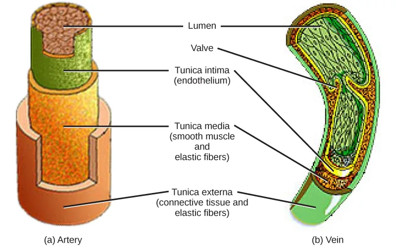
The structure of the different types of blood vessels reflects their function or layers. There are three distinct layers, or tunics, that form the walls of blood vessels (shown below). The first tunic is a smooth, inner lining of endothelial cells that are in contact with the red blood cells. The endothelial tunic is continuous with the endocardium of the heart. In capillaries, this single layer of cells is the location of diffusion of oxygen and carbon dioxide between the endothelial cells and red blood cells, as well as the exchange site via endocytosis and exocytosis. The movement of materials at the site of capillaries is regulated by vasoconstriction, narrowing of the blood vessels, and vasodilation, widening of the blood vessels; this is important in the overall regulation of blood pressure.
Extended description
Leader lines from Lumen point to the open central channel of both the artery, at left, and the vein, at right. Leader lines from Tunica intima (endothelium) point to the innermost layer of each vessel; from Tunica media (smooth muscle and elastic fibers), to the thick middle layer of the artery and the thinner corresponding layer of the vein; from Tunica externa (connective tissue and elastic fibers), to the outermost layer of each. A leader line from Valve points only to the vein, where two flap-like folds of the tunica intima project into the lumen. The artery’s three layers are visibly thicker than the vein’s same three layers.
Veins and arteries both have two further tunics that surround the endothelium: the middle tunic is composed of smooth muscle and the outermost layer is connective tissue (collagen and elastic fibers). The elastic connective tissue stretches and supports the blood vessels, and the smooth muscle layer helps regulate blood flow by altering vascular resistance through vasoconstriction and vasodilation. The arteries have thicker smooth muscle and connective tissue than the veins to accommodate the higher pressure and speed of freshly pumped blood. The veins are thinner walled as the pressure and rate of flow are much lower. In addition, veins are structurally different than arteries in that veins have valves to prevent the backflow of blood. Because veins have to work against gravity to get blood back to the heart, contraction of skeletal muscle assists with the flow of blood back to the heart.
Summary
The heart muscle pumps blood through three divisions of the circulatory system: coronary, pulmonary, and systemic. There is one atrium and one ventricle on the right side and one atrium and one ventricle on the left side. The pumping of the heart is a function of cardiomyocytes, distinctive muscle cells that are striated like skeletal muscle but pump rhythmically and involuntarily like smooth muscle. The internal pacemaker starts at the sinoatrial node, which is located near the wall of the right atrium. Electrical charges pulse from the SA node causing the two atria to contract in unison; then the pulse reaches the atrioventricular node between the right atrium and right ventricle. A pause in the electric signal allows the atria to empty completely into the ventricles before the ventricles pump out the blood. The blood from the heart is carried through the body by a complex network of blood vessels; arteries take blood away from the heart, and veins bring blood back to the heart.
Key terms
- angina — pain caused by partial blockage of the coronary arteries by the buildup of plaque and lack of oxygen to the heart muscle
- aorta — major artery of the body that takes blood away from the heart
- arteriole — small vessel that connects an artery to a capillary bed
- artery — blood vessel that takes blood away from the heart
- atherosclerosis — buildup of fatty plaques in the coronary arteries in the heart
- atrioventricular valve — one-way membranous flap of connective tissue between the atrium and the ventricle in the right side of the heart; also known as tricuspid valve
- bicuspid valve — (also, mitral valve; left atrioventricular valve) one-way membranous flap between the atrium and the ventricle in the left side of the heart
- capillary — smallest blood vessel that allows the passage of individual blood cells and the site of diffusion of oxygen and nutrient exchange
- capillary bed — large number of capillaries that converge to take blood to a particular organ or tissue
- cardiac cycle — filling and emptying the heart of blood by electrical signals that cause the heart muscles to contract and relax
- cardiomyocyte — specialized heart muscle cell that is striated but contracts involuntarily like smooth muscle
- coronary artery — vessel that supplies the heart tissue with blood
- coronary vein — vessel that takes blood away from the heart tissue back to the chambers in the heart
- diastole — relaxation phase of the cardiac cycle when the heart is relaxed and the ventricles are filling with blood
- electrocardiogram (ECG) — recording of the electrical impulses of the cardiac muscle
- endocardium — innermost layer of tissue in the heart
- epicardium — outermost tissue layer of the heart
- inferior vena cava — drains blood from the veins that come from the lower organs and the legs
- myocardial infarction — (also, heart attack) complete blockage of the coronary arteries and death of the cardiac muscle tissue
- myocardium — heart muscle cells that make up the middle layer and the bulk of the heart wall
- pericardium — membrane layer protecting the heart; also part of the epicardium
- semilunar valve — membranous flap of connective tissue between the aorta and a ventricle of the heart (the aortic or pulmonary semilunar valves)
- sinoatrial (SA) node — the heart’s internal pacemaker; located near the wall of the right atrium
- superior vena cava — drains blood from the jugular vein that comes from the brain and from the veins that come from the arms
- systole — contraction phase of cardiac cycle when the ventricles are pumping blood into the arteries
- tricuspid valve — one-way membranous flap of connective tissue between the atrium and the ventricle in the right side of the heart; also known as atrioventricular valve
- vasoconstriction — narrowing of a blood vessel
- vasodilation — widening of a blood vessel
- vein — blood vessel that brings blood back to the heart
- vena cava — major vein of the body returning blood from the upper and lower parts of the body; see the superior vena cava and inferior vena cava
- venule — blood vessel that connects a capillary bed to a vein
Practice
Describe the structure of the heart and explain how cardiac muscle is different from other muscles
Cardiomyocytes are similar to skeletal muscle because:
Look at what a microscope shows about the two tissues’ banding pattern, not at how each one behaves in the body — involuntary rhythmic beating is something cardiac muscle shares with a different tissue type instead.The one-way membranous flap of connective tissue between the atrium and the ventricle on the right side of the heart is called the ________.
Think about which valve sits between an atrium and a ventricle, not between a ventricle and a great vessel — it goes by two names in the text, and either one is accepted here.The heart muscle cells that make up the middle layer and the bulk of the heart wall are called the ________.
This layer sits between the heart’s inner lining and its outer tissue layer, and it is thicker than either of them.The buildup of fatty plaques in the coronary arteries of the heart is called ________.
The name combines a root meaning ‘hardening’ with a root for a fatty, gruel-like deposit.Describe the cardiac cycle
The heart’s internal pacemaker beats by:
Two options name the same two nodes in opposite order — check which node the pacemaker signal reaches first.During the systolic phase of the cardiac cycle, the heart is ________.
This phase’s name shares a root with a word meaning ’to contract’ — match that root to exactly one of the four options.Describe the cardiac cycle.
Show model answer
Did your answer mention:
The relaxation phase of the cardiac cycle, when the heart is relaxed and the ventricles fill with blood, is called ________.
This phase’s Greek root means ’to draw apart’ — the opposite of the cycle’s contraction phase.Explain the structure of arteries, veins, and capillaries, and how blood flows through the body
How do arteries differ from veins?
Arteries handle blood fresh from a forceful pump; that pressure difference is what the vessel wall itself has to be built for.What happens in capillaries?
Show model answer
Did your answer mention:
The small vessel that connects an artery to a capillary bed is called a(n) ________.
This vessel sits on the artery side of a capillary bed, one step smaller than a minor artery.The blood vessel that connects a capillary bed to a vein is called a(n) ________.
This vessel sits on the vein side of a capillary bed, one step larger than a capillary.This section is adapted from Biology 2e, Section 40.3: Mammalian Heart and Blood Vessels by Mary Ann Clark, Jung Choi, Matthew Douglas, and OpenStax, © OpenStax, licensed under CC BY-NC-SA 4.0. Access the original for free at openstax.org. Changes: figures re-encoded as WebP with the source alt text edited where noted; four figures re-kinded from the manifest’s file-extension “photo” guess to “diagram” (Figure_40_03_03, Figure_40_03_05, Figure_40_03_06, and Figure_40_03_07 are each a hand-drawn or computer-rendered illustration, not a captured photograph), and Figure_B40_03_04 re-kinded the other way, from the manifest’s “diagram” guess to “photo” (it is an actual light micrograph of cardiac tissue, and its alt was rewritten to name the inset magnification the source alt omitted); a longdesc added to every diagram whose printed labels or arrows are not carried by its one-line caption — the systemic/pulmonary circuit illustration, the labeled heart cutaway with pericardial-layer inset, the three-panel cardiac-cycle illustration, the four-panel pacemaker/ECG illustration, the major-vessels body illustration, and the artery/vein cross-section illustration — transcribing each drawing’s own labels, leader-line counts, and (for the cardiac-cycle panels only, which carry no hidden exercise key) arrow counts and positions, in reading order; the two body Visual Connections’ longdescs (the systemic/pulmonary circuit figure and the labeled heart cutaway) transcribe only the printed labels and never state which of that item’s four statements is false; in-text pointers to figures (“Figure 40.10” through “Figure 40.16”) replaced with “shown below,” “shown above,” or “illustrated below,” since Hugo does not number figures; both notes wrapping a Visual Connection rendered as their figure followed by a multiple choice, kept in the body — the note copy and the <exercise> copy print identical question and option wording in both cases, so no adjudication was needed; exercise fs-idm133142336 (the circulatory-system Visual Connection) is keyed A (“Blood in the pulmonary vein is deoxygenated”) rather than the module’s own printed answer key of C, because the module’s own sentences contradict C — reported as a source defect below; the interactive note rendered as a Link to Learning callout, keeping the module’s own openstax.org/l/electric_heart redirect URL; the end-of-section Review Questions and Critical Thinking Questions adapted into the closing interactive Practice block (multiple choice and self-check respectively), both Visual Connection items left in the body rather than duplicated in Practice; rubric checkpoints added to each self-check, decomposing its model answer (the source solution) into check-off clauses with no new claims; six key-term recall items added from the glossary (atrioventricular valve, myocardium, atherosclerosis, diastole, arteriole, venule — six items across three groups), covering six of the section’s thirty-one glossary terms — the rest appear only in the Key terms list and the prose, several of them as the bolded defining term of their own paragraph. Source defects: module m66653, exercise fs-idm133142336 — the module’s own text says “the right ventricle pumps the blood through the semilunar valve … to the pulmonary arteries and on to the lungs for re-oxygenation” (the pulmonary artery carries blood that is still deoxygenated, so option C, “Blood in the pulmonary artery is deoxygenated,” is true) and “the left atrium then receives the oxygen-rich blood from the lungs via the pulmonary veins” (the pulmonary vein carries oxygenated blood, so option A, “Blood in the pulmonary vein is deoxygenated,” is false) — but the module’s printed solution keys C. The false statement by the module’s own text is A, not C; keyed A above.