Blood Flow and Blood Pressure Regulation
By the end of this section, you will be able to:
- Describe the system of blood flow through the body
- Describe how blood pressure is regulated
Blood pressure (BP) is the pressure exerted by blood on the walls of a blood vessel that helps to push blood through the body. Systolic blood pressure measures the amount of pressure that blood exerts on vessels while the heart is beating. The optimal systolic blood pressure is 120 mm Hg. Diastolic blood pressure measures the pressure in the vessels between heartbeats. The optimal diastolic blood pressure is 80 mm Hg. Many factors can affect blood pressure, such as hormones, stress, exercise, eating, sitting, and standing. Blood flow through the body is regulated by the size of blood vessels, by the action of smooth muscle, by one-way valves, and by the fluid pressure of the blood itself.
How Blood Flows Through the Body
Blood is pushed through the body by the action of the pumping heart. With each rhythmic pump, blood is pushed under high pressure and velocity away from the heart, initially along the main artery, the aorta. In the aorta, the blood travels at 30 cm/sec. As blood moves into the arteries, arterioles, and ultimately to the capillary beds, the rate of movement slows dramatically to about 0.026 cm/sec, one-thousand times slower than the rate of movement in the aorta. While the diameter of each individual arteriole and capillary is far narrower than the diameter of the aorta, and according to the law of continuity, fluid should travel faster through a narrower diameter tube, the rate is actually slower due to the overall diameter of all the combined capillaries being far greater than the diameter of the individual aorta.
The slow rate of travel through the capillary beds, which reach almost every cell in the body, assists with gas and nutrient exchange and also promotes the diffusion of fluid into the interstitial space. After the blood has passed through the capillary beds to the venules, veins, and finally to the main venae cavae, the rate of flow increases again but is still much slower than the initial rate in the aorta. Blood primarily moves in the veins by the rhythmic movement of smooth muscle in the vessel wall and by the action of the skeletal muscle as the body moves. Because most veins must move blood against the pull of gravity, blood is prevented from flowing backward in the veins by one-way valves. Because skeletal muscle contraction aids in venous blood flow, it is important to get up and move frequently after long periods of sitting so that blood will not pool in the extremities.
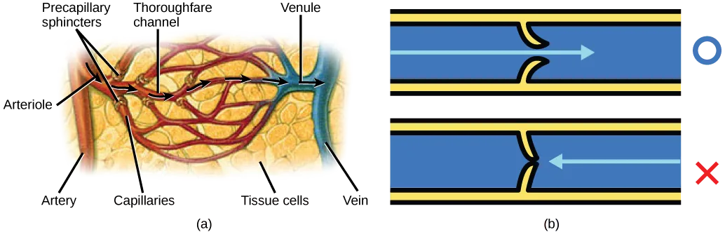
Blood flow through the capillary beds is regulated depending on the body’s needs and is directed by nerve and hormone signals. For example, after a large meal, most of the blood is diverted to the stomach by vasodilation of vessels of the digestive system and vasoconstriction of other vessels. During exercise, blood is diverted to the skeletal muscles through vasodilation while blood to the digestive system would be lessened through vasoconstriction. The blood entering some capillary beds is controlled by small muscles, called precapillary sphincters, illustrated below. If the sphincters are open, the blood will flow into the associated branches of the capillary blood. If all of the sphincters are closed, then the blood will flow directly from the arteriole to the venule through the thoroughfare channel. These muscles allow the body to precisely control when capillary beds receive blood flow. At any given moment only about 5–10% of our capillary beds actually have blood flowing through them.
Extended description
Panel (a), reading top to bottom then left to right: at the top, one leader line labels Precapillary sphincters at a cluster of narrow openings where several capillaries branch from the arteriole, a second labels Thoroughfare channel, the wider vessel running straight across the middle of the capillary web, and a third labels Venule, where the vessels rejoin at the upper right. At the left, one leader line labels Arteriole, the vessel feeding the web from the artery. Along the bottom, leader lines label Artery at the far left, Capillaries at the branching web in the middle, Tissue cells at the shaded mass the capillaries wrap around, and Vein at the vessel leaving the venule at the right. Black arrows trace blood entering at the artery and leaving at the vein, either threading through the narrow capillaries or bypassing them straight through the thoroughfare channel. Panel (b) stacks two copies of the same curved vessel with a yellow valve at its center: in the upper copy, a blue arrow passes rightward through the valve, shown open, beside a blue circle; in the lower copy, a blue arrow points leftward into the same valve, now pinched shut, beside a red X.
Varicose veins are veins that become enlarged because the valves no longer close properly, allowing blood to flow backward. Varicose veins are often most prominent on the legs. Why do you think this is the case?
Show model answer
Did your answer mention:
Link to Learning
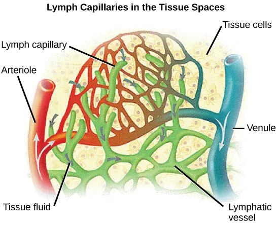
Proteins and other large solutes cannot leave the capillaries. The loss of the watery plasma creates a hyperosmotic solution within the capillaries, especially near the venules. This causes about 85% of the plasma that leaves the capillaries to eventually diffuse back into the capillaries near the venules. The remaining 15% of blood plasma drains out from the interstitial fluid into nearby lymphatic vessels, shown below. The fluid in the lymph is similar in composition to the interstitial fluid. The lymph fluid passes through lymph nodes before it returns to the heart via the superior vena cava. Lymph nodes are specialized organs that filter the lymph by percolation through a maze of connective tissue filled with white blood cells. The white blood cells remove infectious agents, such as bacteria and viruses, to clean the lymph before it returns to the bloodstream. After it is cleaned, the lymph returns to the heart by the action of smooth muscle pumping, skeletal muscle action, and one-way valves joining the returning blood near the junction of the venae cavae entering the right atrium of the heart.
Extended description
The image is titled ‘Lymph Capillaries in the Tissue Spaces.’ Reading top to bottom: at the upper left, one leader line labels Lymph capillary, pointing to the green branching network at the top of the illustration; at the upper right, one leader line labels Tissue cells, the pale stippled background the vessels run through. At the left, one leader line labels Arteriole, the red vessel entering from that side; at the right, one leader line labels Venule, the blue vessel leaving on that side; small pale arrows on both vessels show blood entering at the arteriole and leaving at the venule. At the lower left, one leader line labels Tissue fluid, the space between the reddish capillary bed and the green lymph network; at the lower right, one leader line labels Lymphatic vessel, the green vessel the lymph capillaries drain into. Small arrows within the tissue space show fluid moving outward from the capillary bed into the surrounding lymph capillaries.
Evolution Connection. Vertebrate Diversity in Blood Circulation.
Blood circulation has evolved differently in vertebrates and may show variation in different animals for the required amount of pressure, organ and vessel location, and organ size. Animals with long necks and those that live in cold environments have distinct blood pressure adaptations.
Long necked animals, such as giraffes, need to pump blood upward from the heart against gravity. The blood pressure required from the pumping of the left ventricle would be equivalent to 250 mm Hg (mm Hg = millimeters of mercury, a unit of pressure) to reach the height of a giraffe’s head, which is 2.5 meters higher than the heart. However, if checks and balances were not in place, this blood pressure would damage the giraffe’s brain, particularly if it was bending down to drink. These checks and balances include valves and feedback mechanisms that reduce the rate of cardiac output. Long-necked dinosaurs such as the sauropods had to pump blood even higher, up to ten meters above the heart. This would have required a blood pressure of more than 600 mm Hg, which could only have been achieved by an enormous heart. Evidence for such an enormous heart does not exist and mechanisms to reduce the blood pressure required include the slowing of metabolism as these animals grew larger. It is likely that they did not routinely feed on tree tops but grazed on the ground.
Living in cold water, whales need to maintain the temperature in their blood. This is achieved by the veins and arteries being close together so that heat exchange can occur. This mechanism is called a countercurrent heat exchanger. The blood vessels and the whole body are also protected by thick layers of blubber to prevent heat loss. In land animals that live in cold environments, thick fur and hibernation are used to retain heat and slow metabolism.
Blood Pressure
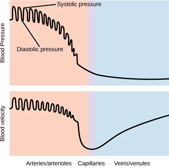
The pressure of the blood flow in the body is produced by the hydrostatic pressure of the fluid (blood) against the walls of the blood vessels. Fluid will move from areas of high to low hydrostatic pressures. In the arteries, the hydrostatic pressure near the heart is very high and blood flows to the arterioles where the rate of flow is slowed by the narrow openings of the arterioles. During systole, when new blood is entering the arteries, the artery walls stretch to accommodate the increase of pressure of the extra blood; during diastole, the walls return to normal because of their elastic properties. The blood pressure of the systole phase and the diastole phase, graphed below, gives the two pressure readings for blood pressure. For example, 120/80 indicates a reading of 120 mm Hg during the systole and 80 mm Hg during diastole. Throughout the cardiac cycle, the blood continues to empty into the arterioles at a relatively even rate. This resistance to blood flow is called peripheral resistance.
Extended description
Two line graphs stacked vertically share the same three x-axis zones, labeled left to right: Arteries/arterioles (shaded peach), Capillaries (a narrow pink band), and Veins/venules (shaded blue). The top graph’s y-axis is labeled Blood Pressure: within the arteries/arterioles zone the curve oscillates in a tight zigzag between an upper envelope labeled Systolic pressure and a lower envelope labeled Diastolic pressure, both sloping gently downward; the oscillation smooths out and the curve drops sharply through the capillaries zone, then levels off low and flat through the veins/venules zone. The bottom graph’s y-axis is labeled Blood velocity: the curve oscillates in the same tight zigzag at a high, roughly level value through the arteries/arterioles zone, drops sharply to its lowest point through the capillaries zone, then rises again through the veins/venules zone.
Blood Pressure Regulation
Cardiac output is the volume of blood pumped by the heart in one minute. It is calculated by multiplying the number of heart contractions that occur per minute (heart rate) times the stroke volume (the volume of blood pumped into the aorta per contraction of the left ventricle). Therefore, cardiac output can be increased by increasing heart rate, as when exercising. However, cardiac output can also be increased by increasing stroke volume, such as if the heart contracts with greater strength. Stroke volume can also be increased by speeding blood circulation through the body so that more blood enters the heart between contractions. During heavy exertion, the blood vessels relax and increase in diameter, offsetting the increased heart rate and ensuring adequate oxygenated blood gets to the muscles. Stress triggers a decrease in the diameter of the blood vessels, consequently increasing blood pressure. These changes can also be caused by nerve signals or hormones, and even standing up or lying down can have a great effect on blood pressure.
Summary
Blood primarily moves through the body by the rhythmic movement of smooth muscle in the vessel wall and by the action of the skeletal muscle as the body moves. Blood is prevented from flowing backward in the veins by one-way valves. Blood flow through the capillary beds is controlled by precapillary sphincters to increase and decrease flow depending on the body’s needs and is directed by nerve and hormone signals. Lymph vessels take fluid that has leaked out of the blood to the lymph nodes where it is cleaned before returning to the heart. During systole, blood enters the arteries, and the artery walls stretch to accommodate the extra blood. During diastole, the artery walls return to normal. The blood pressure of the systole phase and the diastole phase gives the two pressure readings for blood pressure.
Key terms
- blood pressure (BP) — pressure of blood in the arteries that helps to push blood through the body.
- cardiac output — the volume of blood pumped by the heart in one minute as a product of heart rate multiplied by stroke volume.
- lymph node — specialized organ that contains a large number of macrophages that clean the lymph before the fluid is returned to the heart.
- peripheral resistance — resistance of the artery and blood vessel walls to the pressure placed on them by the force of the heart pumping.
- precapillary sphincter — small muscle that controls blood circulation in the capillary beds.
- stroke volume — the volume of blood pumped into the aorta per contraction of the left ventricle.
Practice
Describe the system of blood flow through the body
A specialized organ that contains a large number of macrophages that clean the lymph before the fluid is returned to the heart is called a ________.
This structure sits along the lymphatic vessels and filters out bacteria and viruses using white blood cells before the fluid rejoins the bloodstream.A small muscle that controls blood circulation in the capillary beds is called a ________.
When these rings of smooth muscle close, blood bypasses the capillary bed entirely through the thoroughfare channel.Blood is prevented from flowing backward in the veins by ________.
Veins move blood against gravity, and this structure keeps it from sliding backward between heartbeats.Describe how blood pressure is regulated
High blood pressure would be a result of ________.
Cardiac output and peripheral resistance both push blood pressure in the same direction — find the combination that raises it instead of lowering it.How does blood pressure change during heavy exercise?
Show model answer
Did your answer mention:
The pressure of blood in the arteries that helps to push blood through the body is called ________.
Its two readings, such as 120 over 80, come from systole and diastole.The volume of blood pumped by the heart in one minute, calculated as heart rate multiplied by stroke volume, is called ________.
Increasing either how fast or how forcefully the heart beats raises this quantity.Resistance of the artery and blood vessel walls to the pressure placed on them by the force of the heart pumping is called ________.
Narrower vessels raise this resistance; wider ones lower it.The volume of blood pumped into the aorta per contraction of the left ventricle is called the ________.
Multiplying this quantity by heart rate gives cardiac output.This section is adapted from Biology 2e, Section 40.4: Blood Flow and Blood Pressure Regulation by Mary Ann Clark, Jung Choi, Matthew Douglas, and OpenStax, © OpenStax, licensed under CC BY-NC-SA 4.0. Access the original for free at openstax.org. Changes: the Evolution Connection’s opening sentence reads “Animals with long necks” where the source prints “Animals with longs necks” (reported as a source defect); figures re-encoded as WebP; two figures re-kinded from the manifest’s file-extension “photo” guess to “diagram” (Figure_40_04_02 and Figure_40_04_03 are each a hand-drawn illustration/graph, not a captured photograph); a longdesc added to all three figures — the precapillary-sphincter/valve Visual Connection illustration, the lymph-capillary illustration, and the blood-pressure/velocity graph — transcribing each drawing’s own printed labels, leader lines, and axis zones in reading order, without stating which statement is true or false; the note wrapping the precapillary-sphincter/valve Visual Connection rendered as its figure followed by a self-check, kept in the body in the “How Blood Flows Through the Body” section — the note copy and the <exercise> copy print identical question wording, so no adjudication was needed; the interactive note rendered as a Link to Learning callout, keeping the module’s own openstax.org/l/circulation redirect URL; the end-of-section Review Question and Critical Thinking Question adapted into the closing interactive Practice block (multiple choice and self-check respectively); rubric checkpoints added to each self-check (including the body Visual Connection), decomposing its model answer into check-off clauses with no new claims; all six glossary terms added as key-term recall items (lymph node, precapillary sphincter, blood pressure, cardiac output, peripheral resistance, stroke volume); one cloze recall item added from the section summary (“one-way valves”), since the module carries no comparison table to draw a sortbins from. One number is corrected with a visible Source note: about 20 liters of the day’s cardiac output is filtered at the capillaries, not “over 1,500” (erratum 448).