Excretion Systems
By the end of this section, you will be able to:
- Explain how vacuoles, present in microorganisms, work to excrete waste
- Describe the way in which flame cells and nephridia in worms perform excretory functions and maintain osmotic balance
- Explain how insects use Malpighian tubules to excrete wastes and maintain osmotic balance
Microorganisms and invertebrate animals use more primitive and simple mechanisms to get rid of their metabolic wastes than the mammalian system of kidney and urinary function. Three excretory systems evolved in organisms before complex kidneys: vacuoles, flame cells, and Malpighian tubules.
Contractile Vacuoles in Microorganisms
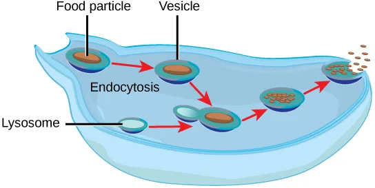
The most fundamental feature of life is the presence of a cell. In other words, a cell is the simplest functional unit of a life. Bacteria are unicellular, prokaryotic organisms that have some of the least complex life processes in place; however, prokaryotes such as bacteria do not contain membrane-bound vacuoles. The cells of microorganisms like bacteria, protozoa, and fungi are bound by cell membranes and use them to interact with the environment. Some cells, including some leucocytes in humans, are able to engulf food by endocytosis—the formation of vesicles by involution of the cell membrane within the cells. The same vesicles are able to interact and exchange metabolites with the intracellular environment. In some unicellular eukaryotic organisms such as the amoeba, shown below, cellular wastes and excess water are excreted by exocytosis, when the contractile vacuoles merge with the cell membrane and expel wastes into the environment. Contractile vacuoles (CV) should not be confused with vacuoles, which store food or water.
Extended description
Reading left to right: at the cell’s upper left edge, a scalloped pseudopod has just enclosed an orange particle labeled Food particle inside a blue-rimmed sac labeled Vesicle — the step labeled Endocytosis. A first red arrow carries this vesicle right to a second, unchanged vesicle; a second red arrow carries it down toward the lower half of the cell, where a third red arrow brings in the pale, unlabeled sac pointed to by the label Lysosome, fusing with it. A fourth red arrow carries the fused vesicle up and to the right, its contents now shown broken into small speckled fragments as digestion proceeds. A fifth red arrow carries this vesicle to the cell’s upper right edge, where the fragments have separated into loose brown specks shown passing through the membrane to the outside, completing exocytosis.
Flame Cells of Planaria and Nephridia of Worms
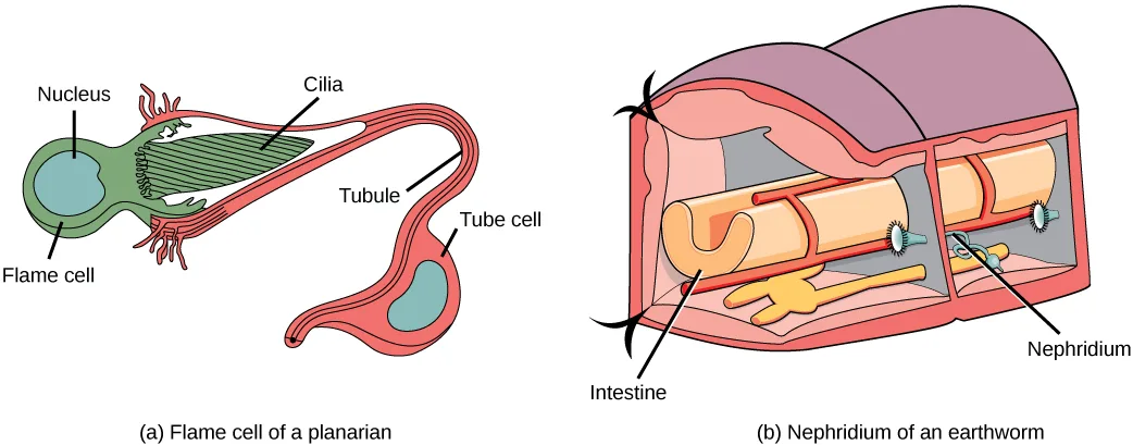
As multicellular systems evolved to have organ systems that divided the metabolic needs of the body, individual organs evolved to perform the excretory function. Planaria are flatworms that live in freshwater. Their excretory system consists of two tubules connected to a highly branched duct system. The cells in the tubules are called flame cells (or protonephridia) because they have a cluster of cilia that looks like a flickering flame when viewed under the microscope, as illustrated below. The cilia propel waste matter down the tubules and out of the body through excretory pores that open on the body surface; cilia also draw water from the interstitial fluid, allowing for filtration. Any valuable metabolites are recovered by reabsorption. Flame cells are found in flatworms, including parasitic tapeworms and free-living planaria. They also maintain the organism’s osmotic balance.
Extended description
Panel (a), left: a green, bulb-shaped structure labeled Flame cell holds a blue oval labeled Nucleus at its wide end; from its narrow end a red, comb-like tuft labeled Cilia projects into the opening of a red tube labeled Tubule, which narrows, then winds down to a second, smaller red bulb labeled Tube cell (itself holding an unlabeled nucleus) before continuing past it. Panel (b), right: a cutaway of a segmented earthworm body, its walls dividing the cavity into chambers; a coiled reddish tube labeled Nephridium runs between two small ciliated funnel-openings, one in each of two segment walls, passing alongside the paler Intestine that runs the length of the body.
Earthworms (annelids) have slightly more evolved excretory structures called nephridia, illustrated above. A pair of nephridia is present on each segment of the earthworm. They are similar to flame cells in that they have a tubule with cilia. Excretion occurs through a pore called the nephridiopore. They are more evolved than the flame cells in that they have a system for tubular reabsorption by a capillary network before excretion.
Malpighian Tubules of Insects
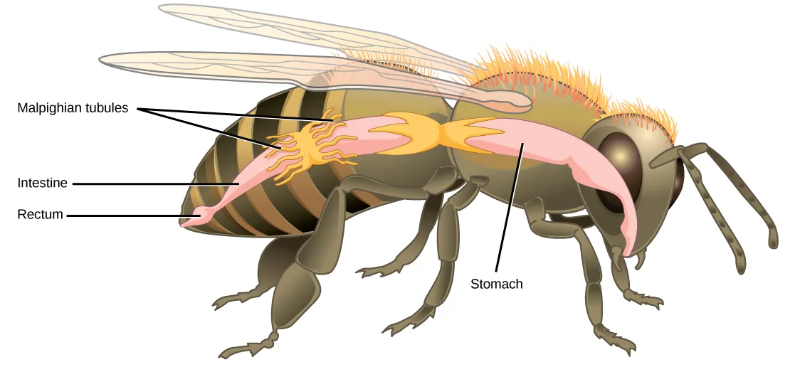
Malpighian tubules are found lining the gut of some species of arthropods, such as the bee illustrated below. They are usually found in pairs and the number of tubules varies with the species of insect. Malpighian tubules are convoluted, which increases their surface area, and they are lined with microvilli for reabsorption and maintenance of osmotic balance. Malpighian tubules work cooperatively with specialized glands in the wall of the rectum. Body fluids are not filtered as in the case of nephridia; urine is produced by tubular secretion mechanisms by the cells lining the Malpighian tubules that are bathed in hemolymph (a mixture of blood and interstitial fluid that is found in insects and other arthropods as well as most mollusks). Metabolic wastes like uric acid freely diffuse into the tubules. There are exchange pumps lining the tubules, which actively transport H⁺ ions into the cell and K⁺ or Na⁺ ions out; water passively follows to form urine. The secretion of ions alters the osmotic pressure which draws water, electrolytes, and nitrogenous waste (uric acid) into the tubules. Water and electrolytes are reabsorbed when these organisms are faced with low-water environments, and uric acid is excreted as a thick paste or powder. Not dissolving wastes in water helps these organisms to conserve water; this is especially important for life in dry environments.
Extended description
Three printed labels are stacked top to bottom at the left edge, each with a leader line running rightward into the abdomen: Malpighian tubules, with two leader lines pointing into the cluster of thread-like yellow tubules over the abdomen; Intestine, pointing to the pink tube running the length of the lower abdomen; and Rectum, pointing to the small pale pouch at the abdomen’s rear tip. A fourth label, Stomach, sits alone at the lower right with a leader line rising to the large pink bulge in the thorax, ahead of the abdomen.
Link to Learning
Watch a dissected cockroach, including a close-up look at its Malpighian tubules.
Summary
Many systems have evolved for excreting wastes that are simpler than the kidney and urinary systems of vertebrate animals. The simplest system is that of contractile vacuoles present in microorganisms. Flame cells and nephridia in worms perform excretory functions and maintain osmotic balance. Some insects have evolved Malpighian tubules to excrete wastes and maintain osmotic balance.
Key terms
- flame cell — (also, protonephridia) excretory cell found in flatworms.
- Malpighian tubule — excretory tubules found in arthropods.
- microvilli — cellular processes that increase the surface area of cells.
- nephridia — excretory structures found in annelids.
- nephridiopore — pore found at the end of nephridia.
Practice
Explain how vacuoles, present in microorganisms, work to excrete waste
Contractile vacuoles in microorganisms:
Check whether the vacuoles do more than one job, and where they come from according to the section — one option covers each, and only one option combines them.Explain two different excretory systems other than the kidneys.
Show model answer
Did your answer mention:
The simplest excretory system is that of ________ present in microorganisms.
This structure merges with the cell membrane during exocytosis to expel wastes, as shown in the amoeba illustration above.Describe the way in which flame cells and nephridia in worms perform excretory functions and maintain osmotic balance
Flame cells are primitive excretory organs found in ________.
This animal’s excretory system is described earlier in the section as two tubules connected to a highly branched duct system.Why might specialized organs have evolved for excretion of wastes?
Show model answer
Did your answer mention:
An excretory cell found in flatworms is called a(n) ________.
It’s named for how its tuft of beating cilia looks when viewed under a microscope.Excretory structures found in annelids, such as earthworms, are called ________.
Each segment of the earthworm has a pair of these, evolved beyond flame cells to add tubular reabsorption by a capillary network.The pore through which a nephridium releases waste to the exterior is called the ________.
Its name combines the structure it drains with the word for an opening to the outside.Explain how insects use Malpighian tubules to excrete wastes and maintain osmotic balance
Active transport of K⁺ in Malpighian tubules ensures that:
Check whether the section supports the first option on its own, the second on its own, or supports them at the same time.Excretory structures that line the gut of some arthropods and are convoluted to increase their surface area are called ________.
They work with specialized glands in the wall of the rectum and are usually found in pairs.Cellular processes that increase the surface area of cells are called ________.
The lining of the Malpighian tubules is covered in these projections, which aid reabsorption and osmotic balance.This section is adapted from Biology 2e, Section 41.3: Excretion Systems by Mary Ann Clark, Jung Choi, Matthew Douglas, and OpenStax, © OpenStax, licensed under CC BY-NC-SA 4.0. Access the original for free at openstax.org. Changes: figures re-encoded as WebP; all three figures re-kinded from the manifest’s file-extension “photo” guess to “diagram” (Figure_41_02_01, Figure_41_02_02, and Figure_41_02_03 are each a hand-drawn line illustration, not a captured photograph); a longdesc added to every diagram — the amoeba endocytosis/exocytosis sequence, the two-panel flame-cell/nephridium illustration, and the bee’s Malpighian-tubule anatomy — transcribing each drawing’s own printed labels and leader lines, in reading order; ion charges (H⁺, K⁺, Na⁺) set in Unicode where the source prints “H+”, “K+”, “Na+”; in-text pointers to figures (“Figure 41.9” through “Figure 41.11”) replaced with “shown below” or “illustrated above,” since Hugo does not number figures; the interactive note rendered as a Link to Learning callout, keeping the module’s own openstax.org/l/malpighian redirect URL; the end-of-section Review Questions and Critical Thinking Questions adapted into the closing interactive Practice block (multiple choice and self-check respectively); the Critical Thinking solution to “explain two different excretory systems other than the kidneys” — a numbered “(1)…(2)…(3)…(4)…” list in the source, printed here as sentences in order with the numbering removed — is kept in full (all four systems, though the question asks for two), since it makes no new claim beyond the source’s own text; rubric checkpoints added to each self-check, decomposing its model answer (the source solution) into check-off clauses with no new claims; five key-term recall items added from the glossary (flame cell, nephridia, nephridiopore, Malpighian tubule, microvilli), covering every glossary term in the section; one summary-sourced cloze text-recall item added (contractile vacuoles), since the section’s glossary carries no term for that system.