Hormonal Control of Osmoregulatory Functions
By the end of this section, you will be able to:
- Explain how hormonal cues help the kidneys synchronize the osmotic needs of the body
- Describe how hormones like epinephrine, norepinephrine, renin-angiotensin, aldosterone, anti-diuretic hormone, and atrial natriuretic peptide help regulate waste elimination, maintain correct osmolarity, and perform other osmoregulatory functions
While the kidneys operate to maintain osmotic balance and blood pressure in the body, they also act in concert with hormones. Hormones are small molecules that act as messengers within the body. Hormones are typically secreted from one cell and travel in the bloodstream to affect a target cell in another portion of the body. Different regions of the nephron bear specialized cells that have receptors to respond to chemical messengers and hormones. The table below summarizes the hormones that control the osmoregulatory functions.
Hormones That Affect Osmoregulation
| Hormone | Where produced | Function |
|---|---|---|
| Epinephrine and Norepinephrine | Adrenal medulla | Can decrease kidney function temporarily by vasoconstriction |
| Renin | Kidney nephrons | Increases blood pressure by acting on angiotensinogen |
| Angiotensin | Liver | Angiotensin II affects multiple processes and increases blood pressure |
| Aldosterone | Adrenal cortex | Prevents loss of sodium and water |
| Anti-diuretic hormone (vasopressin) | Hypothalamus (stored in the posterior pituitary) | Prevents water loss |
| Atrial natriuretic peptide | Heart atrium | Decreases blood pressure by acting as a vasodilator and increasing glomerular filtration rate; decreases sodium reabsorption in kidneys |
Epinephrine and Norepinephrine
Epinephrine and norepinephrine are synthesized by the adrenal medulla. They are the flight/fight hormones that are released when the body is under extreme stress. During stress, much of the body’s energy is used to combat imminent danger. Kidney function is halted temporarily by epinephrine and norepinephrine. These hormones function by acting directly on the smooth muscles of blood vessels to constrict them. Once the afferent arterioles are constricted, blood flow into the nephrons stops. These hormones go one step further and trigger the renin-angiotensin-aldosterone system.
Renin-Angiotensin-Aldosterone
The renin-angiotensin-aldosterone system, illustrated below, proceeds through several steps to produce angiotensin II, which acts to stabilize blood pressure and volume. Renin (secreted by a part of the juxtaglomerular complex) is produced by the granular cells of the afferent and efferent arterioles. Thus, the kidneys control blood pressure and volume directly. Renin acts on angiotensinogen, which is made in the liver and converts it to angiotensin I. Angiotensin converting enzyme (ACE) converts angiotensin I to angiotensin II. Angiotensin II raises blood pressure by constricting blood vessels. It also triggers the release of the mineralocorticoid aldosterone from the adrenal cortex, which in turn stimulates the renal tubules to reabsorb more sodium. Angiotensin II also triggers the release of anti-diuretic hormone (ADH) from the hypothalamus, leading to water retention in the kidneys. It acts directly on the nephrons and decreases glomerular filtration rate. Medically, blood pressure can be controlled by drugs that inhibit ACE (called ACE inhibitors).
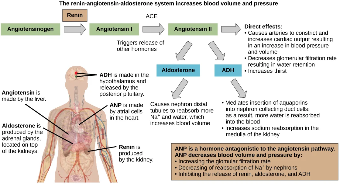
Extended description
A title across the top reads: The renin-angiotensin-aldosterone system increases blood volume and pressure. Below it, a chain of three green boxes runs left to right, joined by three gray arrows: Angiotensinogen, then an arrow (a smaller tan box labeled Renin sits directly above this arrow) to Angiotensin I, then an arrow labeled ACE to Angiotensin II, then a third arrow to an unboxed heading, Direct effects:, followed by three bullets — causes arteries to constrict and increases cardiac output, resulting in an increase in blood pressure and volume; decreases glomerular filtration rate, resulting in water retention; increases thirst. Under the caption Triggers release of other hormones, two more gray arrows point down from Angiotensin II to two blue boxes, Aldosterone and ADH. A further arrow below Aldosterone points to unbulleted text: causes nephron distal tubules to reabsorb more Na⁺ and water, which increases blood volume. A further arrow below ADH points to a two-bullet list: mediates insertion of aquaporins into nephron collecting duct cells, so more water is reabsorbed into the blood; increases sodium reabsorption in the medulla of the kidney. Beneath both, a wide tan-bordered box reads: ANP is a hormone antagonistic to the angiotensin pathway. ANP decreases blood volume and pressure by: — followed by three bullets — increasing the glomerular filtration rate; decreasing reabsorption of Na⁺ by nephrons; inhibiting the release of renin, aldosterone, and ADH. To the left of the flow chart, a torso-and-head illustration carries five bold labels with six leader lines running to the gland or organ that produces the named hormone. Near the top, the label ADH is made in the hypothalamus and released by the posterior pituitary sends two lines to two marked points in the head, one for the hypothalamus and one for the posterior pituitary. Below it, side by side, one line reads Angiotensin is made by the liver, leading to the liver, and another reads ANP is made by atrial cells in the heart, leading to the heart. Below those, again side by side, one line reads Aldosterone is produced by the adrenal glands, located on top of the kidneys, leading to the adrenal gland, and another reads Renin is produced by the kidney, leading to the kidney.
Mineralocorticoids
Mineralocorticoids are hormones synthesized by the adrenal cortex that affect osmotic balance. Aldosterone is a mineralocorticoid that regulates sodium levels in the blood. Almost all of the sodium in the blood is reclaimed by the renal tubules under the influence of aldosterone. Because sodium is always reabsorbed by active transport and water follows sodium to maintain osmotic balance, aldosterone manages not only sodium levels but also the water levels in body fluids. In contrast, the aldosterone also stimulates potassium secretion concurrently with sodium reabsorption. In contrast, absence of aldosterone means that no sodium gets reabsorbed in the renal tubules and all of it gets excreted in the urine. In addition, the daily dietary potassium load is not secreted and the retention of K⁺ can cause a dangerous increase in plasma K⁺ concentration. Patients who have Addison’s disease have a failing adrenal cortex and cannot produce aldosterone. They lose sodium in their urine constantly, and if the supply is not replenished, the consequences can be fatal.
Antidiuretic Hormone
As previously discussed, antidiuretic hormone or ADH (also called vasopressin), as the name suggests, helps the body conserve water when body fluid volume, especially that of blood, is low. It is formed by the hypothalamus and is stored and released from the posterior pituitary. It acts by inserting aquaporins in the collecting ducts and promotes reabsorption of water. ADH also acts as a vasoconstrictor and increases blood pressure during hemorrhaging.
Atrial Natriuretic Peptide Hormone
The atrial natriuretic peptide (ANP) lowers blood pressure by acting as a vasodilator. It is released by cells in the atrium of the heart in response to high blood pressure and in patients with sleep apnea. ANP affects salt release, and because water passively follows salt to maintain osmotic balance, it also has a diuretic effect. ANP also prevents sodium reabsorption by the renal tubules, decreasing water reabsorption (thus acting as a diuretic) and lowering blood pressure. Its actions suppress the actions of aldosterone, ADH, and renin.
Summary
Hormonal cues help the kidneys synchronize the osmotic needs of the body. Hormones like epinephrine, norepinephrine, renin-angiotensin, aldosterone, anti-diuretic hormone, and atrial natriuretic peptide help regulate the needs of the body as well as the communication between the different organ systems.
Key terms
- angiotensin converting enzyme (ACE) — enzyme that converts angiotensin I to angiotensin II.
- angiotensin I — product in the renin-angiotensin-aldosterone pathway.
- angiotensin II — molecule that affects different organs to increase blood pressure.
- anti-diuretic hormone (ADH) — hormone that prevents the loss of water.
- renin-angiotensin-aldosterone — biochemical pathway that activates angiotensin II, which increases blood pressure.
- vasodilator — compound that increases the diameter of blood vessels.
- vasopressin — another name for anti-diuretic hormone.
Practice
Explain how hormonal cues help the kidneys synchronize the osmotic needs of the body
Describe how hormones regulate blood pressure, blood volume, and kidney function.
Show model answer
Did your answer mention:
How does the renin-angiotensin-aldosterone mechanism function? Why is it controlled by the kidneys?
Show model answer
Did your answer mention:
Hormonal cues help the kidneys synchronize the ________ of the body.
This section’s opening sentence and its Summary both name the same two-word balance — of water and solutes — that hormonal signals keep the kidneys tracking.The biochemical pathway that activates angiotensin II, increasing blood pressure, is called the ________.
This is the same multi-hormone cascade whose first two steps convert a liver protein into two numbered forms before a mineralocorticoid and a water-conserving hormone take over.Describe how hormones like epinephrine, norepinephrine, renin-angiotensin, aldosterone, anti-diuretic hormone, and atrial natriuretic peptide help regulate waste elimination, maintain correct osmolarity, and perform other osmoregulatory functions
Renin is made by ________.
Compare the table’s ‘Where produced’ entry for renin with the prose that follows it — do the granular cells, the kidneys, and the nephrons name three different sites, or the same site described three ways?Patients with Addison’s disease ________.
Addison’s disease means a failing adrenal cortex that can no longer produce aldosterone — think about what aldosterone normally reclaims for the body, and what happens once that reclaiming stops.Which hormone elicits the “fight or flight” response?
This hormone, made alongside a closely related one by the adrenal medulla, is also what constricts the afferent arterioles and sets off the renin-angiotensin-aldosterone cascade during extreme stress.The enzyme that converts angiotensin I to angiotensin II is called ________.
The module later notes that blood pressure can be controlled by drugs that block this very enzyme — its full name literally describes the conversion it performs.A compound that increases the diameter of blood vessels is called a ________.
Atrial natriuretic peptide has this effect on blood vessels; the opposite effect would narrow them rather than widen them.The hormone that prevents the loss of water is called ________.
This hormone is formed in the hypothalamus but stored in and released from the posterior pituitary, and it acts by inserting aquaporins into the collecting ducts.This section is adapted from Biology 2e, Section 41.5: Hormonal Control of Osmoregulatory Functions by Mary Ann Clark, Jung Choi, Matthew Douglas, and OpenStax, © OpenStax, licensed under CC BY-NC-SA 4.0. Access the original for free at openstax.org. Changes: the figure re-encoded as WebP, re-kinded from the manifest’s file-extension “photo” guess to “diagram” (it is a hand-drawn flow chart and body illustration, not a captured photograph); its source alt, a letter-spaced screen-reader spelling (“A D H,” “A C E,” “A N P,” “angiotensin I I”), rewritten as a plain description, with the full walk-through moved into a longdesc counting every box, arrow, and leader-line label; the in-text pointer to the table (“Table 41.1 summarizes…”) replaced with “The table below summarizes…,” and the figure pointer (“illustrated in Figure 41.15”) replaced with “illustrated below,” since Hugo does not number tables or figures; the “Hormones That Affect Osmoregulation” table transcribed as a Markdown table with its spanning header kept as a bold lead-in line — its columns name a hormone, its production site, and its function rather than categories, so it is not rendered as a sortbins exercise; ion charges (K⁺, Na⁺) set in Unicode; the Renin Review Question (fs-idm77856336) keyed D, all of the above rather than the source-printed key A — the module’s own table row (“Renin — Where produced: Kidney nephrons”) and its prose (“Renin … is produced by the granular cells of the afferent and efferent arterioles”) both make options B (the kidneys) and C (the nephrons) true in addition to A, so only “all of the above” is consistent with the module’s own text; this is a disclosed departure from the source key, reported as a source defect; the end-of-section Review Questions and Critical Thinking Questions adapted into the closing interactive Practice block (multiple choice and self-check respectively), all five keyed exercises used; rubric checkpoints added to each self-check, decomposing its model answer (the source solution) into check-off clauses with no new claims; four key-term recall items added from the glossary (renin-angiotensin-aldosterone, angiotensin converting enzyme, vasodilator, anti-diuretic hormone with accept ADH/vasopressin), plus one summary-sourced cloze item blanking the section’s synchronizing “osmotic needs,” covering four of the section’s seven glossary terms — the other three (angiotensin I, angiotensin II, vasopressin) appear only in the Key terms list and the prose, each as a bolded defining term of its own paragraph or table row.