Adaptive Immune Response
By the end of this section, you will be able to:
- Explain adaptive immunity
- Compare and contrast adaptive and innate immunity
- Describe cell-mediated immune response and humoral immune response
- Describe immune tolerance
The adaptive, or acquired, immune response takes days or even weeks to become established—much longer than the innate response; however, adaptive immunity is more specific to pathogens and has memory. Adaptive immunity is an immunity that occurs after exposure to an antigen either from a pathogen or a vaccination. This part of the immune system is activated when the innate immune response is insufficient to control an infection. In fact, without information from the innate immune system, the adaptive response could not be mobilized. There are two types of adaptive responses: the cell-mediated immune response, which is carried out by T cells, and the humoral immune response, which is controlled by activated B cells and antibodies. Activated T cells and B cells that are specific to molecular structures on the pathogen proliferate and attack the invading pathogen. Their attack can kill pathogens directly or secrete antibodies that enhance the phagocytosis of pathogens and disrupt the infection. Adaptive immunity also involves a memory to provide the host with long-term protection from reinfection with the same type of pathogen; on reexposure, this memory will facilitate an efficient and quick response.
Antigen-presenting Cells
Unlike NK cells of the innate immune system, B cells (B lymphocytes) are a type of white blood cell that gives rise to antibodies, whereas T cells (T lymphocytes) are a type of white blood cell that plays an important role in the immune response. T cells are a key component in the cell-mediated response—the specific immune response that utilizes T cells to neutralize cells that have been infected with viruses and certain bacteria. There are three types of T cells: cytotoxic, helper, and suppressor T cells. Cytotoxic T cells destroy virus-infected cells in the cell-mediated immune response, and helper T cells play a part in activating both the antibody and the cell-mediated immune responses. Suppressor T cells deactivate T cells and B cells when needed, and thus prevent the immune response from becoming too intense.
An antigen is a foreign or “non-self” macromolecule that reacts with cells of the immune system. Not all antigens will provoke a response. For instance, individuals produce innumerable “self” antigens and are constantly exposed to harmless foreign antigens, such as food proteins, pollen, or dust components. The suppression of immune responses to harmless macromolecules is highly regulated and typically prevents processes that could be damaging to the host, known as tolerance.
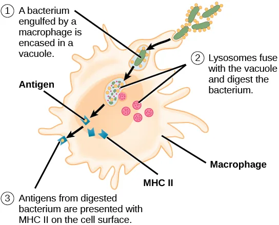
The innate immune system contains cells that detect potentially harmful antigens, and then inform the adaptive immune response about the presence of these antigens. An antigen-presenting cell (APC) is an immune cell that detects, engulfs, and informs the adaptive immune response about an infection. When a pathogen is detected, these APCs will phagocytose the pathogen and digest it to form many different fragments of the antigen. Antigen fragments will then be transported to the surface of the APC, where they will serve as an indicator to other immune cells. Dendritic cells are immune cells that process antigen material; they are present in the skin (Langerhans cells) and the lining of the nose, lungs, stomach, and intestines. Sometimes a dendritic cell presents on the surface of other cells to induce an immune response, thus functioning as an antigen-presenting cell. Macrophages also function as APCs. Before activation and differentiation, B cells can also function as APCs.
After phagocytosis by APCs, the phagocytic vesicle fuses with an intracellular lysosome forming phagolysosome. Within the phagolysosome, the components are broken down into fragments; the fragments are then loaded onto MHC class I or MHC class II molecules and are transported to the cell surface for antigen presentation, as shown below. Note that T lymphocytes cannot properly respond to the antigen unless it is processed and embedded in an MHC II molecule. APCs express MHC on their surfaces, and when combined with a foreign antigen, these complexes signal a “non-self” invader. Once the fragment of antigen is embedded in the MHC II molecule, the immune cell can respond. Helper T cells are one of the main lymphocytes that respond to antigen-presenting cells. Recall that all other nucleated cells of the body expressed MHC I molecules, which signal “healthy” or “normal.”
Link to Learning
Watch an animation of dendritic cells acting as sentinels in the body’s immune system, produced by Rockefeller University.
T and B Lymphocytes
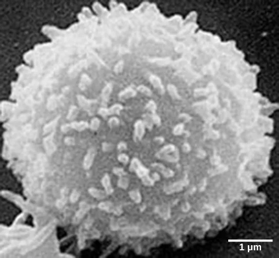
Lymphocytes in human circulating blood are approximately 80 to 90 percent T cells, shown below, and 10 to 20 percent B cells. Recall that the T cells are involved in the cell-mediated immune response, whereas B cells are part of the humoral immune response.
T cells encompass a heterogeneous population of cells with extremely diverse functions. Some T cells respond to APCs of the innate immune system, and indirectly induce immune responses by releasing cytokines. Other T cells stimulate B cells to prepare their own response. Another population of T cells detects APC signals and directly kills the infected cells. Other T cells are involved in suppressing inappropriate immune reactions to harmless or “self” antigens.
T and B cells exhibit a common theme of recognition/binding of specific antigens via a complementary receptor, followed by activation and self-amplification/maturation to specifically bind to the particular antigen of the infecting pathogen. T and B lymphocytes are also similar in that each cell only expresses one type of antigen receptor. Any individual may possess a population of T and B cells that together express a near limitless variety of antigen receptors that are capable of recognizing virtually any infecting pathogen. T and B cells are activated when they recognize small components of antigens, called epitopes, presented by APCs, shown below. Note that recognition occurs at a specific epitope rather than on the entire antigen; for this reason, epitopes are known as “antigenic determinants.” In the absence of information from APCs, T and B cells remain inactive, or naïve, and are unable to prepare an immune response. The requirement for information from the APCs of innate immunity to trigger B cell or T cell activation illustrates the essential nature of the innate immune response to the functioning of the entire immune system.

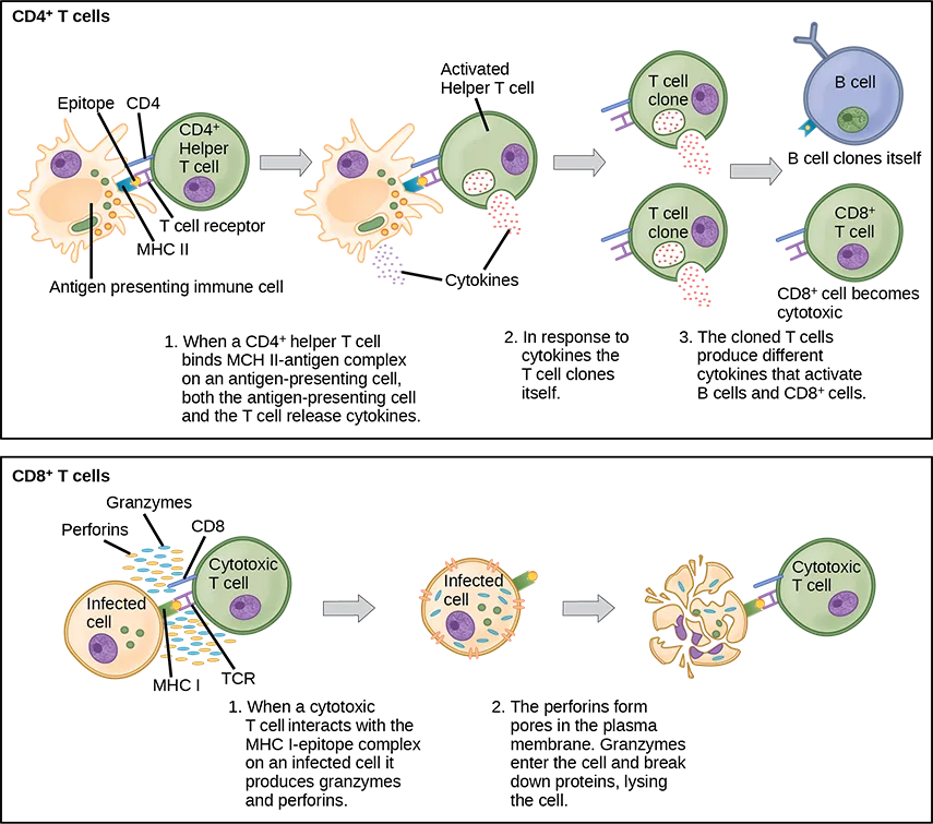
Naïve T cells can express one of two different molecules, CD4 or CD8, on their surface, shown below, and are accordingly classified as CD4⁺ or CD8⁺ cells. These molecules are important because they regulate how a T cell will interact with and respond to an APC. Naïve CD4⁺ cells bind APCs via their antigen-embedded MHC II molecules and are stimulated to become helper T (TH) lymphocytes, cells that go on to stimulate B cells (or cytotoxic T cells) directly or secrete cytokines to inform more and various target cells about the pathogenic threat. In contrast, CD8⁺ cells engage antigen-embedded MHC I molecules on APCs and are stimulated to become cytotoxic T lymphocytes (CTLs), which directly kill infected cells by apoptosis and emit cytokines to amplify the immune response. The two populations of T cells have different mechanisms of immune protection, but both bind MHC molecules via their antigen receptors called T cell receptors (TCRs). The CD4 or CD8 surface molecules differentiate whether the TCR will engage an MHC II or an MHC I molecule. Because they assist in binding specificity, the CD4 and CD8 molecules are described as coreceptors.
Extended description
Two boxed flow charts, each a left-to-right sequence of illustrations linked by gray arrows, with a numbered caption below each step. Top box, labeled CD4⁺ T cells: an antigen-presenting immune cell displays an epitope on an MHC II molecule; a CD4⁺ helper T cell binds it via its T cell receptor and CD4, and both cells release cytokines — caption 1. An arrow leads to the activated helper T cell, still releasing cytokines — caption 2. A second arrow leads to two identical T cell clones. Two more arrows lead from the clones to their targets: a B cell that clones itself, and a CD8⁺ T cell that becomes cytotoxic — caption 3. Bottom box, labeled CD8⁺ T cells: a cytotoxic T cell, bearing CD8, granzymes, and perforins, binds the MHC I–epitope complex on an infected cell — caption 1. An arrow leads to the infected cell as perforins open pores in its membrane and granzymes enter — caption 2. A second arrow leads to the lysed, fragmented cell beside the cytotoxic T cell.
Which of the following statements about T cells is false?
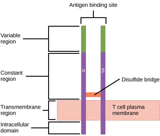
Re-read the paragraph on MHC I and MHC II directly above the figure — it names exactly which molecule appears on which cells, and only one of the four options gets that scope backward.Consider the innumerable possible antigens that an individual will be exposed to during a lifetime. The mammalian adaptive immune system is adept in responding appropriately to each antigen. Mammals have an enormous diversity of T cell populations, resulting from the diversity of TCRs. Each TCR consists of two polypeptide chains that span the T cell membrane, shown below; the chains are linked by a disulfide bridge. Each polypeptide chain is comprised of a constant domain and a variable domain: a domain, in this sense, is a specific region of a protein that may be regulatory or structural. The intracellular domain is involved in intracellular signaling. A single T cell will express thousands of identical copies of one specific TCR variant on its cell surface. The specificity of the adaptive immune system occurs because it synthesizes millions of different T cell populations, each expressing a TCR that differs in its variable domain. This TCR diversity is achieved by the mutation and recombination of genes that encode these receptors in stem cell precursors of T cells. The binding between an antigen-displaying MHC molecule and a complementary TCR “match” indicates that the adaptive immune system needs to activate and produce that specific T cell because its structure is appropriate to recognize and destroy the invading pathogen.
Extended description
A bracket labeled Antigen binding site spans the top of two parallel columns, marked α and β, representing the receptor’s two polypeptide chains. Reading down each column: a green segment labeled Variable region, then a longer purple segment labeled Constant region, crossing a pink band labeled T cell plasma membrane where the columns are labeled Transmembrane region, and ending in a short purple tail below the membrane labeled Intracellular domain. An orange Disulfide bridge connects the α and β columns partway down the constant region.
Helper T Lymphocytes
The TH lymphocytes function indirectly to identify potential pathogens for other cells of the immune system. These cells are important for extracellular infections, such as those caused by certain bacteria, helminths, and protozoa. TH lymphocytes recognize specific antigens displayed in the MHC II complexes of APCs. There are two major populations of TH cells: TH1 and TH2. TH1 cells secrete cytokines to enhance the activities of macrophages and other T cells. TH1 cells activate the action of cytotoxic T cells, as well as macrophages. TH2 cells stimulate naïve B cells to destroy foreign invaders via antibody secretion. Whether a TH1 or a TH2 immune response develops depends on the specific types of cytokines secreted by cells of the innate immune system, which in turn depends on the nature of the invading pathogen.
The TH1-mediated response involves macrophages and is associated with inflammation. Recall the frontline defenses of macrophages involved in the innate immune response. Some intracellular bacteria, such as Mycobacterium tuberculosis, have evolved to multiply in macrophages after they have been engulfed. These pathogens evade attempts by macrophages to destroy and digest the pathogen. When M. tuberculosis infection occurs, macrophages can stimulate naïve T cells to become TH1 cells. These stimulated T cells secrete specific cytokines that send feedback to the macrophage to stimulate its digestive capabilities and allow it to destroy the colonizing M. tuberculosis. In the same manner, TH1-activated macrophages also become better suited to ingest and kill tumor cells. In summary; TH1 responses are directed toward intracellular invaders while TH2 responses are aimed at those that are extracellular.
B Lymphocytes
When stimulated by the TH2 pathway, naïve B cells differentiate into antibody-secreting plasma cells. A plasma cell is an immune cell that secretes antibodies; these cells arise from B cells that were stimulated by antigens. Similar to T cells, naïve B cells initially are coated in thousands of B cell receptors (BCRs), which are membrane-bound forms of Ig (immunoglobulin, or an antibody). The B cell receptor has two heavy chains and two light chains connected by disulfide linkages. Each chain has a constant and a variable region; the latter is involved in antigen binding. Two other membrane proteins, Ig alpha and Ig beta, are involved in signaling. The receptors of any particular B cell, shown below, are all the same, but the hundreds of millions of different B cells in an individual have distinct recognition domains that contribute to extensive diversity in the types of molecular structures to which they can bind. In this state, B cells function as APCs. They bind and engulf foreign antigens via their BCRs and then display processed antigens in the context of MHC II molecules to TH2 cells. When a TH2 cell detects that a B cell is bound to a relevant antigen, it secretes specific cytokines that induce the B cell to proliferate rapidly, which makes thousands of identical (clonal) copies of it, and then it synthesizes and secretes antibodies with the same antigen recognition pattern as the BCRs. The activation of B cells corresponding to one specific BCR variant and the dramatic proliferation of that variant is known as clonal selection. This phenomenon drastically, but briefly, changes the proportions of BCR variants expressed by the immune system, and shifts the balance toward BCRs specific to the infecting pathogen.
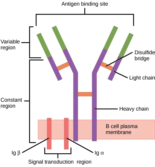
Extended description
Bracket labels along the left, top to bottom, read Antigen binding site (spanning the top of both forked arms), Variable region (the green segment of each light and heavy chain above the bend), Constant region (the purple segment of each chain below the bend, continuing down both stems), and Signal transduction region (two red proteins embedded in the plasma membrane, with short projections into the cytoplasm). On the right, leader lines label Disulfide bridge (the orange bars joining each light chain to its heavy chain, and joining the two heavy-chain stems to each other), Light chain (the shorter, upper subunit of each arm), and Heavy chain (the longer subunit running from each arm down into the membrane); the two red signal-transduction proteins in the membrane are labeled Ig beta on the left stem and Ig alpha on the right.
T and B cells differ in one fundamental way: whereas T cells bind antigens that have been digested and embedded in MHC molecules by APCs, B cells function as APCs that bind intact antigens that have not been processed. Although T and B cells both react with molecules that are termed “antigens,” these lymphocytes actually respond to very different types of molecules. B cells must be able to bind intact antigens because they secrete antibodies that must recognize the pathogen directly, rather than digested remnants of the pathogen. Bacterial carbohydrate and lipid molecules can activate B cells independently from the T cells.
Cytotoxic T Lymphocytes
CTLs, a subclass of T cells, function to clear infections directly. The cell-mediated part of the adaptive immune system consists of CTLs that attack and destroy infected cells. CTLs are particularly important in protecting against viral infections; this is because viruses replicate within cells where they are shielded from extracellular contact with circulating antibodies. When APCs phagocytize pathogens and present MHC I-embedded antigens to naïve CD8⁺ T cells that express complementary TCRs, the CD8⁺ T cells become activated to proliferate according to clonal selection. These resulting CTLs then identify non-APCs displaying the same MHC I-embedded antigens (for example, viral proteins)—for example, the CTLs identify infected host cells.
Intracellularly, infected cells typically die after the infecting pathogen replicates to a sufficient concentration and lyses the cell, as many viruses do. CTLs attempt to identify and destroy infected cells before the pathogen can replicate and escape, thereby halting the progression of intracellular infections. CTLs also support NK lymphocytes to destroy early cancers. Cytokines secreted by the TH1 response that stimulates macrophages also stimulate CTLs and enhance their ability to identify and destroy infected cells and tumors.
CTLs sense MHC I-embedded antigens by directly interacting with infected cells via their TCRs. Binding of TCRs with antigens activates CTLs to release perforin and granzyme, degradative enzymes that will induce apoptosis of the infected cell. Recall that this is a similar destruction mechanism to that used by NK cells. In this process, the CTL does not become infected and is not harmed by the secretion of perforin and granzymes. In fact, the functions of NK cells and CTLs are complementary and maximize the removal of infected cells, shown below. If the NK cell cannot identify the “missing self” pattern of down-regulated MHC I molecules, then the CTL can identify it by the complex of MHC I with foreign antigens, which signals “altered self.” Similarly, if the CTL cannot detect antigen-embedded MHC I because the receptors are depleted from the cell surface, NK cells will destroy the cell instead. CTLs also emit cytokines, such as interferons, that alter surface protein expression in other infected cells, such that the infected cells can be easily identified and destroyed. Moreover, these interferons can also prevent virally infected cells from releasing virus particles.
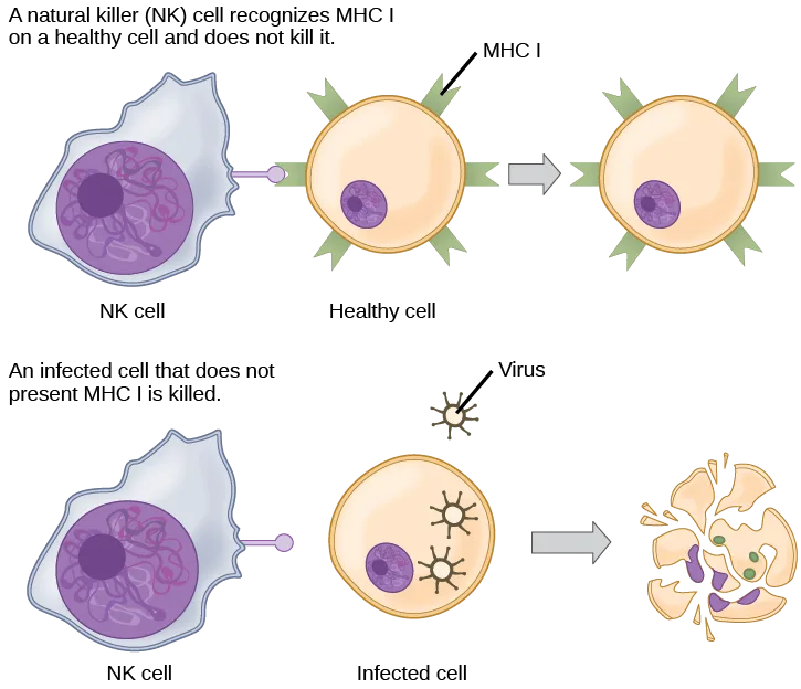
Extended description
Top row, left to right: an NK cell approaches a healthy cell whose surface is studded with MHC I markers; an arrow leads to the same healthy cell afterward, unchanged. Bottom row, left to right: an NK cell approaches an infected cell, marked by two small virus particles and displaying no MHC I; an arrow leads to that cell afterward, burst into fragments.
Based on what you know about MHC receptors, why do you think an organ transplanted from an incompatible donor to a recipient will be rejected?
Show model answer
Did your answer mention:
Plasma cells and CTLs are collectively called effector cells: they represent differentiated versions of their naïve counterparts, and they are involved in bringing about the immune defense of killing pathogens and infected host cells.
Mucosal Surfaces and Immune Tolerance
The innate and adaptive immune responses discussed thus far comprise the systemic immune system (affecting the whole body), which is distinct from the mucosal immune system. Mucosal immunity is formed by mucosa-associated lymphoid tissue, which functions independently of the systemic immune system, and which has its own innate and adaptive components. Mucosa-associated lymphoid tissue (MALT), shown below, is a collection of lymphatic tissue that combines with epithelial tissue lining the mucosa throughout the body. This tissue functions as the immune barrier and response in areas of the body with direct contact to the external environment. The systemic and mucosal immune systems use many of the same cell types. Foreign particles that make their way to MALT are taken up by absorptive epithelial cells called M cells and delivered to APCs located directly below the mucosal tissue. M cells function in the transport described, and are located in the Peyer’s patch, a lymphoid nodule. APCs of the mucosal immune system are primarily dendritic cells, with B cells and macrophages having minor roles. Processed antigens displayed on APCs are detected by T cells in the MALT and at various mucosal induction sites, such as the tonsils, adenoids, appendix, or the mesenteric lymph nodes of the intestine. Activated T cells then migrate through the lymphatic system and into the circulatory system to mucosal sites of infection.
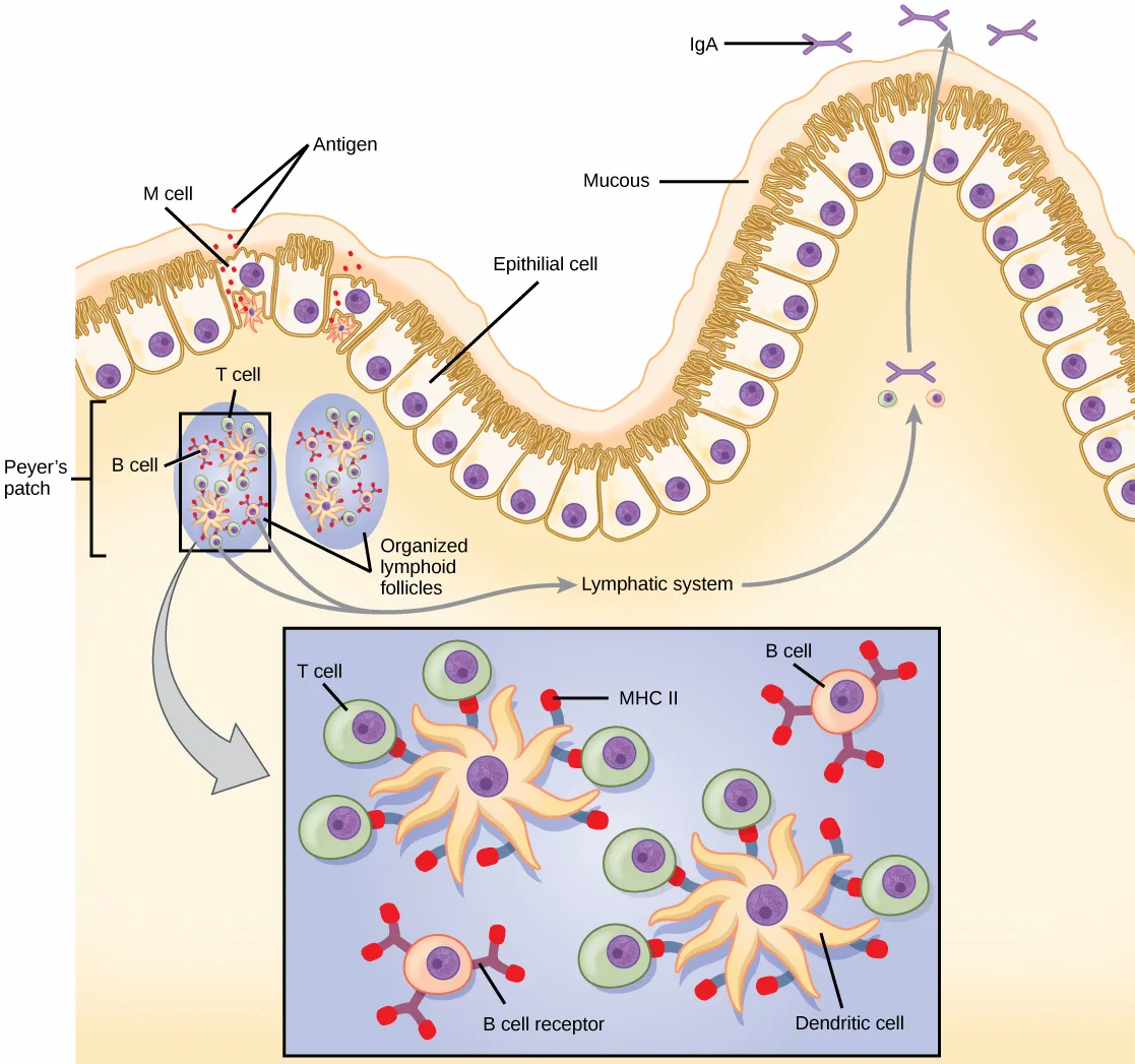
Extended description
Reading left to right across the intestinal lining: an M cell, shaped like an upside-down U, sits among column-shaped epithelial cells (labeled epithelial cell — the artwork’s own label misspells this ‘Epithilial cell’) whose surface cilia and mucous coat are labeled Mucous. Small dots labeled Antigen are taken up by the M cell into the pocket formed by its underside. Below the epithelium, a bracket labels the Peyer’s patch, containing two blue ovals labeled Organized lymphoid follicles, each holding T cell and B cell labels. One gray arrow leads down from the follicles into a boxed inset below; a second, curving gray arrow leads from the follicles, past a label reading Lymphatic system, up through the epithelium at the right, where Y-shaped antibodies labeled IgA are released into the intestinal lumen at the surface. The inset magnifies one follicle: star-shaped dendritic cells display red MHC II tags to green T cells and to pink B cells, one of which is labeled at its Y-shaped B cell receptor.
MALT is a crucial component of a functional immune system because mucosal surfaces, such as the nasal passages, are the first tissues onto which inhaled or ingested pathogens are deposited. The mucosal tissue includes the mouth, pharynx, and esophagus, and the gastrointestinal, respiratory, and urogenital tracts.
The immune system has to be regulated to prevent wasteful, unnecessary responses to harmless substances, and more importantly so that it does not attack “self.” The acquired ability to prevent an unnecessary or harmful immune response to a detected foreign substance known not to cause disease is described as immune tolerance. Immune tolerance is crucial for maintaining mucosal homeostasis given the tremendous number of foreign substances (such as food proteins) that APCs of the oral cavity, pharynx, and gastrointestinal mucosa encounter. Immune tolerance is brought about by specialized APCs in the liver, lymph nodes, small intestine, and lung that present harmless antigens to an exceptionally diverse population of regulatory T (Treg) cells, specialized lymphocytes that suppress local inflammation and inhibit the secretion of stimulatory immune factors. The combined result of Treg cells is to prevent immunologic activation and inflammation in undesired tissue compartments and to allow the immune system to focus on pathogens instead. In addition to promoting immune tolerance of harmless antigens, other subsets of Treg cells are involved in the prevention of the autoimmune response, which is an inappropriate immune response to host cells or self-antigens. Another Treg class suppresses immune responses to harmful pathogens after the infection has cleared to minimize host cell damage induced by inflammation and cell lysis.
Immunological Memory
The adaptive immune system possesses a memory component that allows for an efficient and dramatic response upon reinvasion of the same pathogen. Memory is handled by the adaptive immune system with little reliance on cues from the innate response. During the adaptive immune response to a pathogen that has not been encountered before, called a primary response, plasma cells secreting antibodies and differentiated T cells increase, then plateau over time. As B and T cells mature into effector cells, a subset of the naïve populations differentiates into B and T memory cells with the same antigen specificities, shown below.
A memory cell is an antigen-specific B or T lymphocyte that does not differentiate into effector cells during the primary immune response, but that can immediately become effector cells upon reexposure to the same pathogen. During the primary immune response, memory cells do not respond to antigens and do not contribute to host defenses. As the infection is cleared and pathogenic stimuli subside, the effectors are no longer needed, and they undergo apoptosis. In contrast, the memory cells persist in the circulation.
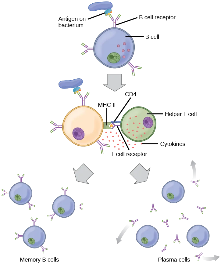
Extended description
Top panel: a bacterium’s antigen binds a B cell receptor on a labeled B cell. An arrow leads down to a middle panel where the B cell, now presenting the antigen on MHC II, contacts a helper T cell whose T cell receptor and CD4 recognize the complex; both cells release cytokines. Two arrows lead down from this panel to a bottom row of two panels: on the left, three labeled memory B cells; on the right, three labeled plasma cells shown secreting antibodies.
The Rh antigen is found on Rh-positive red blood cells. During pregnancy, Rh-negative people can usually carry an Rh-positive fetus to term without difficulty. However, a second Rh-positive fetus may trigger an immune attack that causes hemolytic disease of the newborn. Why do you think hemolytic disease is only a problem during the second or subsequent pregnancies?
Show model answer
Did your answer mention:
If the pathogen is never encountered again during the individual’s lifetime, B and T memory cells will circulate for a few years or even several decades and will gradually die off, having never functioned as effector cells. However, if the host is reexposed to the same pathogen type, circulating memory cells will immediately differentiate into plasma cells and CTLs without input from APCs or TH cells. One reason the adaptive immune response is delayed is because it takes time for naïve B and T cells with the appropriate antigen specificities to be identified and activated. Upon reinfection, this step is skipped, and the result is a more rapid production of immune defenses. Memory B cells that differentiate into plasma cells output tens to hundreds-fold greater antibody amounts than were secreted during the primary response, as the graph below illustrates. This rapid and dramatic antibody response may stop the infection before it can even become established, and the individual may not realize they had been exposed.
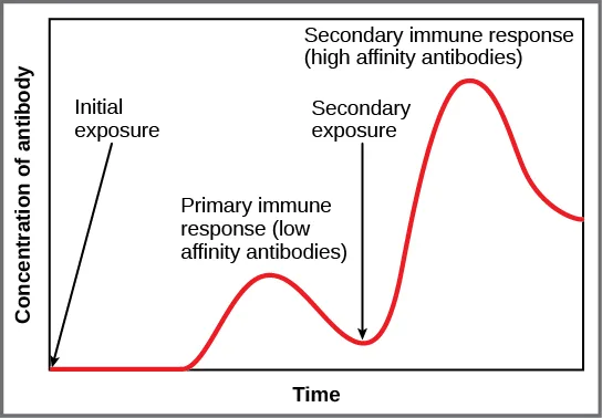
Extended description
The curve starts flat at zero before the initial exposure, arrowed at the far left. After a delay, it climbs to a low peak labeled the primary immune response (low affinity antibodies), then falls back toward the baseline. At the point labeled secondary exposure, the curve climbs again, faster and to roughly three times the height of the first peak, labeled the secondary immune response (high affinity antibodies), before gradually declining.
Vaccination is based on the knowledge that exposure to noninfectious antigens, derived from known pathogens, generates a mild primary immune response. The immune response to vaccination may not be perceived by the host as illness but still confers immune memory. When exposed to the corresponding pathogen to which an individual was vaccinated, the reaction is similar to a secondary exposure. Because each reinfection generates more memory cells and increased resistance to the pathogen, and because some memory cells die, certain vaccine courses involve one or more booster vaccinations to mimic repeat exposures: for instance, tetanus boosters are necessary every ten years because the memory cells only live that long.
Mucosal Immune Memory
A subset of T and B cells of the mucosal immune system differentiates into memory cells just as in the systemic immune system. Upon reinvasion of the same pathogen type, a pronounced immune response occurs at the mucosal site where the original pathogen deposited, but a collective defense is also organized within interconnected or adjacent mucosal tissue. For instance, the immune memory of an infection in the oral cavity would also elicit a response in the pharynx if the oral cavity was exposed to the same pathogen.
Career Connection. Vaccinologist.
Vaccination (or immunization) involves the delivery, usually by injection, shown below, of noninfectious antigen(s) derived from known pathogens. Other components, called adjuvants, are delivered in parallel to help stimulate the immune response. Immunological memory is the reason vaccines work. Ideally, the effect of vaccination is to elicit immunological memory, and thus resistance to specific pathogens without the individual having to experience an infection.

Vaccinologists are involved in the process of vaccine development from the initial idea to the availability of the completed vaccine. This process can take decades, can cost millions of dollars, and can involve many obstacles along the way. For instance, injected vaccines stimulate the systemic immune system, eliciting humoral and cell-mediated immunity, but have little effect on the mucosal response, which presents a challenge because many pathogens are deposited and replicate in mucosal compartments, and the injection does not provide the most efficient immune memory for these disease agents. For this reason, vaccinologists are actively involved in developing new vaccines that are applied via intranasal, aerosol, oral, or transcutaneous (absorbed through the skin) delivery methods. Importantly, mucosal-administered vaccines elicit both mucosal and systemic immunity and produce the same level of disease resistance as injected vaccines.

Currently, a version of intranasal influenza vaccine is available, and the polio and typhoid vaccines can be administered orally, shown above. Similarly, the measles and rubella vaccines are being adapted to aerosol delivery using inhalation devices. Eventually, transgenic plants may be engineered to produce vaccine antigens that can be eaten to confer disease resistance. Other vaccines may be adapted to rectal or vaginal application to elicit immune responses in rectal, genitourinary, or reproductive mucosa. Finally, vaccine antigens may be adapted to transdermal application in which the skin is lightly scraped and microneedles are used to pierce the outermost layer. In addition to mobilizing the mucosal immune response, this new generation of vaccines may end the anxiety associated with injections and, in turn, improve patient participation.
Primary Centers of the Immune System
Although the immune system is characterized by circulating cells throughout the body, the regulation, maturation, and intercommunication of immune factors occur at specific sites. The blood circulates immune cells, proteins, and other factors through the body. Approximately 0.1 percent of all cells in the blood are leukocytes, which encompass monocytes (the precursor of macrophages) and lymphocytes. The majority of cells in the blood are erythrocytes (red blood cells). Lymph is a watery fluid that bathes tissues and organs with protective white blood cells and does not contain erythrocytes. Cells of the immune system can travel between the distinct lymphatic and blood circulatory systems, which are separated by interstitial space, by a process called extravasation (passing through to surrounding tissue).
The cells of the immune system originate from hematopoietic stem cells in the bone marrow. Cytokines stimulate these stem cells to differentiate into immune cells. B cell maturation occurs in the bone marrow, whereas naïve T cells transit from the bone marrow to the thymus for maturation. In the thymus, immature T cells that express TCRs complementary to self-antigens are destroyed. This process helps prevent autoimmune responses.
On maturation, T and B lymphocytes circulate to various destinations. Lymph nodes scattered throughout the body, shown below, house large populations of T and B cells, dendritic cells, and macrophages. Lymph gathers antigens as it drains from tissues. These antigens then are filtered through lymph nodes before the lymph is returned to circulation. APCs in the lymph nodes capture and process antigens and inform nearby lymphocytes about potential pathogens.
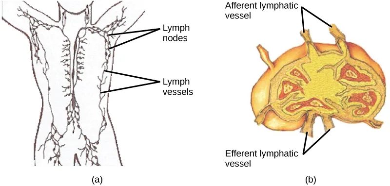
Extended description
Panel (a), a black-and-white outline of the upper body and neck, has two leader lines from the label Lymph nodes pointing to clusters in the neck and lower torso, and two leader lines from the label Lymph vessels pointing to the branching channels along the spine and into the limbs. Panel (b), a colored oval cross-section of one lymph node, has two leader lines from the label Afferent lymphatic vessel entering at the top, and two leader lines from the label Efferent lymphatic vessel leaving at the bottom; red and orange lobed areas fill the node’s interior.
The spleen houses B and T cells, macrophages, dendritic cells, and NK cells. The spleen, shown below, is the site where APCs that have trapped foreign particles in the blood can communicate with lymphocytes. Antibodies are synthesized and secreted by activated plasma cells in the spleen, and the spleen filters foreign substances and antibody-complexed pathogens from the blood. Functionally, the spleen is to the blood as lymph nodes are to the lymph.
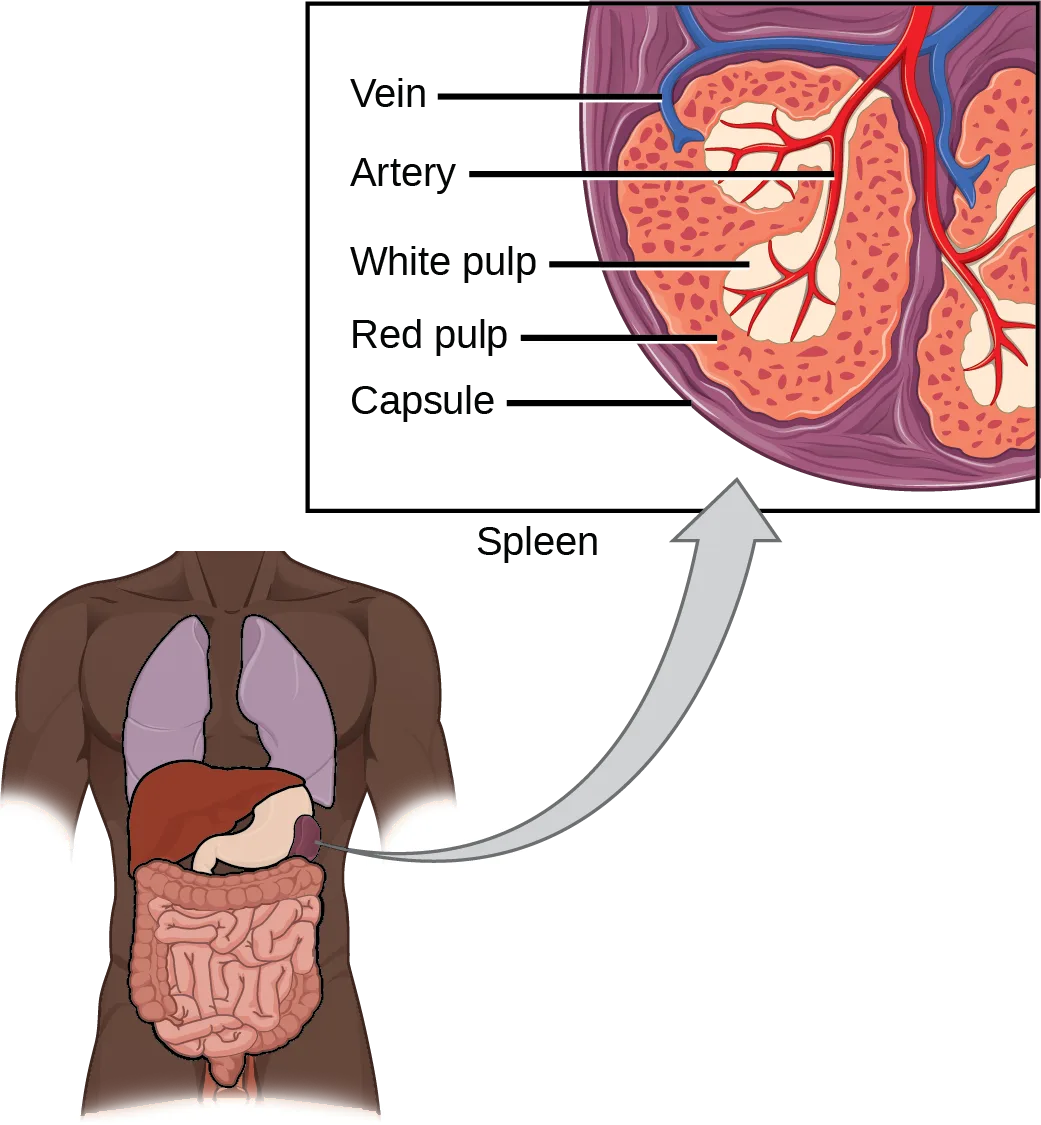
Extended description
The lower panel is a brown body outline showing the lungs, liver, stomach, and intestines, with the spleen — a small dark oval beside the stomach — connected by a gray arrow up to the boxed inset above. In the inset cross-section, five leader lines, top to bottom, label Vein (a blue vessel entering one lobe), Artery (a red vessel branching into the tissue), White pulp (pale oval nodules scattered through the tissue), Red pulp (the surrounding red-brown tissue), and Capsule (the purple outer membrane enclosing the whole cross-section).
Summary
The adaptive immune response is a slower-acting, longer-lasting, and more specific response than the innate response. However, the adaptive response requires information from the innate immune system to function. APCs display antigens via MHC molecules to complementary naïve T cells. In response, the T cells differentiate and proliferate, becoming TH cells or CTLs. TH cells stimulate B cells that have engulfed and presented pathogen-derived antigens. B cells differentiate into plasma cells that secrete antibodies, whereas CTLs induce apoptosis in intracellularly infected or cancerous cells. Memory cells persist after a primary exposure to a pathogen. If reexposure occurs, memory cells differentiate into effector cells without input from the innate immune system. The mucosal immune system is largely independent from the systemic immune system but functions in a parallel fashion to protect the extensive mucosal surfaces of the body.
Key terms
- adaptive immunity — immunity that has memory and occurs after exposure to an antigen either from a pathogen or a vaccination.
- antigen — foreign or “non-self” protein that triggers the immune response.
- antigen-presenting cell (APC) — immune cell that detects, engulfs, and informs the adaptive immune response about an infection by presenting the processed antigen on the cell surface.
- autoimmune response — inappropriate immune response to host cells or self-antigens.
- cell-mediated immune response — adaptive immune response that is carried out by T cells.
- clonal selection — activation of B cells corresponding to one specific BCR variant and the dramatic proliferation of that variant.
- cytotoxic T lymphocyte (CTL) — adaptive immune cell that directly kills infected cells via perforin and granzymes, and releases cytokines to enhance the immune response.
- dendritic cell — immune cell that processes antigen material and presents it on the surface of other cells to induce an immune response.
- effector cell — lymphocyte that has differentiated, such as a B cell, plasma cell, or cytotoxic T lymphocyte.
- epitope — small component of an antigen that is specifically recognized by antibodies, B cells, and T cells; the antigenic determinant.
- helper T lymphocyte (TH) — cell of the adaptive immune system that binds APCs via MHC II molecules and stimulates B cells or secretes cytokines to initiate the immune response.
- humoral immune response — adaptive immune response that is controlled by activated B cells and antibodies.
- immune tolerance — acquired ability to prevent an unnecessary or harmful immune response to a detected foreign body known not to cause disease or to self-antigens.
- lymph — watery fluid that bathes tissues and organs with protective white blood cells and does not contain erythrocytes.
- mucosa-associated lymphoid tissue (MALT) — collection of lymphatic tissue that combines with epithelial tissue lining the mucosa throughout the body.
- memory cell — antigen-specific B or T lymphocyte that does not differentiate into effector cells during the primary immune response but that can immediately become an effector cell upon reexposure to the same pathogen.
- plasma cell — immune cell that secretes antibodies; these cells arise from B cells that were stimulated by antigens.
- regulatory T (Treg) cell — specialized lymphocyte that suppresses local inflammation and inhibits the secretion of cytokines, antibodies, and other stimulatory immune factors; involved in immune tolerance.
Practice
Explain adaptive immunity
Immunity that has memory and occurs after exposure to an antigen either from a pathogen or a vaccination is called ________.
This is the immune system’s slower, second line of defense — the one this whole section describes, as opposed to the system that acts within minutes using genetically fixed defenses.Explain the difference between an epitope and an antigen.
Show model answer
Did your answer mention:
What is a naïve B or T cell?
Show model answer
Did your answer mention:
In mammalian adaptive immune systems, T cell receptors are extraordinarily diverse. What function of the immune system results from this diversity, and how is this diversity achieved?
Show model answer
Did your answer mention:
Foreign particles circulating in the blood are filtered by the ________.
The section draws a direct parallel between two organs: one filters lymph, the other filters this different body fluid. Which of the four options is the one described as being to the blood as lymph nodes are to the lymph?Compare and contrast adaptive and innate immunity
The adaptive immune response is a slower-acting, longer-lasting, and more specific response than the ________ response.
This section opens by comparing adaptive immunity to the body’s other, faster-acting line of defense.Which of the following is both a phagocyte and an antigen-presenting cell?
This cell type both engulfs pathogens directly, the way a phagocyte does, and displays the resulting antigen fragments on its own surface, the way an antigen-presenting cell does — which single option does both jobs?An immune cell that detects, engulfs, and processes an antigen, then displays fragments of it on its own surface to inform the adaptive immune response about an infection, is called a(n) ________.
Macrophages, dendritic cells, and (before activation) B cells can all serve this same role in bridging the innate and adaptive immune responses.What “self” pattern is identified by NK cells?
NK cells recognize a normal cell by checking for MHC I; the four options each pair “self” with a different qualifier — think about which one names a molecule that has been lost entirely, rather than one that looks foreign, unusual, or unrecognizable.Why is the immune response after reinfection much faster than the adaptive immune response after the initial infection?
Show model answer
Did your answer mention:
Describe cell-mediated immune response and humoral immune response
The adaptive immune response that is carried out by T cells is called the ________.
This is the T-cell arm of adaptive immunity, distinct from the one carried out by activated B cells and antibodies.The adaptive immune response that is controlled by activated B cells and antibodies is called the ________.
This is the B-cell-and-antibody arm of adaptive immunity, distinct from the one carried out directly by T cells.Which immune cells bind MHC molecules on APCs via CD8 coreceptors on their cell surfaces?
This is the CD8⁺ population of T cells, not the CD4⁺ population — check which named cell type is defined by that coreceptor rather than by the one helper T cells use.How does the TH1 response differ from the TH2 response?
Show model answer
Did your answer mention:
How do B and T cells differ with respect to antigens that they bind?
Show model answer
Did your answer mention:
Upon reexposure to a pathogen, a memory B cell can differentiate to which cell type?
A memory B cell skips the naive stage entirely and differentiates directly into the antibody-secreting cell type this section names in the paragraph about clonal selection — which option is that cell type, rather than a T-cell population?Describe immune tolerance
The acquired ability to prevent an unnecessary or destructive immune reaction to a harmless foreign particle, such as a food protein, is called ________.
This term describes the immune system deliberately NOT reacting to something harmless — think about which of the four options names a state of restraint rather than a state of reacting too much or attacking the wrong target.An inappropriate immune response to host cells or self-antigens is called the ________.
Some subsets of regulatory T cells specifically work to prevent the immune system from turning against its own tissue in this way.A specialized lymphocyte that suppresses local inflammation and inhibits the secretion of stimulatory immune factors, helping to maintain immune tolerance, is called a(n) ________.
APCs in the liver, lymph nodes, small intestine, and lung present harmless antigens to this diverse population of lymphocytes.A collection of lymphatic tissue that combines with epithelial tissue lining the mucosa throughout the body is called ________.
M cells in the Peyer’s patch deliver antigens to APCs of this tissue, which functions largely independently of the systemic immune system.This section is adapted from Biology 2e, Section 42.2: Adaptive Immune Response by Mary Ann Clark, Jung Choi, Matthew Douglas, and OpenStax, © OpenStax, licensed under CC BY-NC-SA 4.0. Access the original for free at openstax.org. Changes: figures re-encoded as WebP; six figures re-kinded from the manifest’s file-extension “photo” guess to “diagram” (Figure_42_02_01, Figure_42_02_05, Figure_42_02_06, Figure_42_02_08, Figure_42_02_10, and Figure_42_02_13ab are each a hand-drawn illustration or a line graph, not a captured photograph); a longdesc added to every diagram whose meaning is not carried by its caption alone — the T-cell-receptor, B-cell-receptor, MALT topology, antibody-titer graph, lymph-node, and spleen illustrations, plus the T-cell-activation and B-cell-activation Visual Connection figures — transcribing each drawing’s labels, panels, and arrows in reading order without stating either Visual Connection’s answer; CD4/CD8 superscript “+” markers set as Unicode ⁺ throughout prose, options, hints, and figure text, matching the ion-charge convention used elsewhere in this book; the MALT figure’s own printed label “Epithilial cell” is a source artwork typo for “epithelial cell,” corrected in the longdesc and reported as a source defect (the artwork itself is unchanged); the interactive note rendered as a Link to Learning callout with descriptive link text in place of the source’s bare “animation,” keeping the module’s own openstax.org/l/immune_system redirect URL; the career note rendered as a Career Connection callout with its own figures nested inside, keeping its title in italics; the three body Visual Connections (the T-cell false-statement question beside Figure_B42_02_04, the incompatible-donor-transplant question beside Figure_42_02_07, and the Rh-negative-pregnancy question beside Figure_42_02_09) kept in the body immediately after their figures — the note copy and the <exercise> copy of each print identical question and option wording, except the Rh-pregnancy item, whose note copy opens “A pregnant Rh-negative person can usually carry…” while its <exercise> copy opens “During pregnancy, Rh-negative people can usually carry…”; the page uses the <exercise> copy’s wording per house style, and both are quoted under Source defects for the parent to adjudicate; the six Review Questions and six Critical Thinking Questions adapted into the closing interactive Practice block (multiple choice and self-check respectively), each used once; rubric checkpoints added to each self-check, decomposing its model answer (the source solution) into check-off clauses with no new claims, spelling TH1/TH2 as plain “TH1”/“TH2” in the checkpoint lines since checkpoints render as plain text rather than through the markdown/HTML pipeline that renders the model answer above them; seven key-term recall items added from the glossary (adaptive immunity, antigen-presenting cell, cell-mediated immune response, humoral immune response, autoimmune response, regulatory T cell, and mucosa-associated lymphoid tissue), covering seven of the section’s eighteen glossary terms — the other eleven (antigen, clonal selection, cytotoxic T lymphocyte, dendritic cell, effector cell, epitope, helper T lymphocyte, immune tolerance, lymph, memory cell, plasma cell) are not keyed as recall items (several appear inside Practice as multiple-choice options or selfcheck answers), several as the bolded defining term of their own paragraph; one cloze recall item added from the section summary (adaptive immunity’s speed and specificity relative to the innate response); no sortbins was built — the section carries no comparison table with columns naming categories.