Hormonal Control of Human Reproduction
By the end of this section, you will be able to:
- Describe the roles of male and female reproductive hormones
- Discuss the interplay of the ovarian and menstrual cycles
- Describe the process of menopause
The human male and female reproductive cycles are controlled by the interaction of hormones from the hypothalamus and anterior pituitary with hormones from reproductive tissues and organs. In both sexes, the hypothalamus monitors and causes the release of hormones from the pituitary gland. When the reproductive hormone is required, the hypothalamus sends a gonadotropin-releasing hormone (GnRH) to the anterior pituitary. This causes the release of follicle stimulating hormone (FSH) and luteinizing hormone (LH) from the anterior pituitary into the blood. Note that the body must reach puberty in order for the adrenals to release the hormones that must be present for GnRH to be produced. Although FSH and LH are named after their functions in female reproduction, they are produced in both sexes and play important roles in controlling reproduction. Other hormones have specific functions in the male and female reproductive systems.
Male Hormones
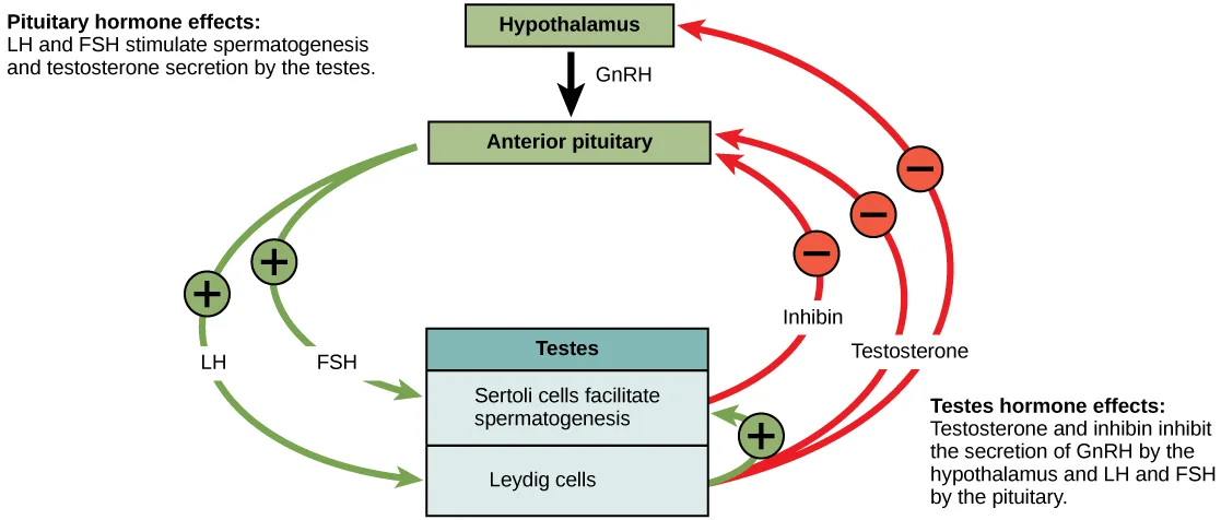
At the onset of puberty, the hypothalamus causes the release of FSH and LH into the male system for the first time. FSH enters the testes and stimulates the Sertoli cells to begin facilitating spermatogenesis using negative feedback, as illustrated below. LH also enters the testes and stimulates the interstitial cells of Leydig to make and release testosterone into the testes and the blood.
Testosterone, the hormone responsible for the secondary sexual characteristics that develop in the male during adolescence, stimulates spermatogenesis. These secondary sex characteristics include a deepening of the voice, the growth of facial, axillary, and pubic hair, and the beginnings of the sex drive.
Extended description
Reading top to bottom: a text box at upper left reads ‘Pituitary hormone effects: LH and FSH stimulate spermatogenesis and testosterone secretion by the testes.’ A green Hypothalamus box sits below it, connected by a black downward arrow labeled GnRH to a green Anterior pituitary box. Two green curved arrows leave the Anterior pituitary box and run down to a Testes box (blue header, with a Sertoli cells facilitate spermatogenesis row above a Leydig cells row); each arrow crosses a green plus-sign circle, labeled LH and FSH. A third green plus-sign circle sits on a short arrow inside the Testes box, running from the Leydig cells row up to the Sertoli cells row. Two red curved arrows run from the Testes box up and around the right side: the Inhibin arrow ends at the Anterior pituitary, and the Testosterone arrow forks into two branches, one ending at the Anterior pituitary and one at the Hypothalamus, so three arrowheads point at the boxes they inhibit and each of the three branches crosses its own red minus-sign circle. A text box at lower right reads ‘Testes hormone effects: Testosterone and inhibin inhibit the secretion of GnRH by the hypothalamus and LH and FSH by the pituitary.’
A negative feedback system occurs in the male with rising levels of testosterone acting on the hypothalamus and anterior pituitary to inhibit the release of GnRH, FSH, and LH. The Sertoli cells produce the hormone inhibin, which is released into the blood when the sperm count is too high. This inhibits the release of GnRH and FSH, which will cause spermatogenesis to slow down. If the sperm count reaches 20 million/ml, the Sertoli cells cease the release of inhibin, and the sperm count increases.
Female Hormones
The control of reproduction in females is more complex. As with the male, the anterior pituitary hormones cause the release of the hormones FSH and LH. In addition, estrogens and progesterone are released from the developing follicles. Estradiol is the reproductive hormone in females that assists in endometrial regrowth, ovulation, and calcium absorption; it is also responsible for the secondary sexual characteristics of females. These include breast development, flaring of the hips, and a shorter period necessary for bone maturation. Progesterone assists in endometrial regrowth and inhibition of FSH and LH release.
In females, FSH stimulates development of egg cells, called ova, which develop in structures called follicles. Follicle cells produce the hormone inhibin, which inhibits FSH production. LH also plays a role in the development of ova, induction of ovulation, and stimulation of estradiol, the most potent and prevalent form of estrogen, and progesterone production by the ovaries. Estradiol and progesterone are steroid hormones that prepare the body for pregnancy. Estradiol produces secondary sex characteristics in females, while both estradiol and progesterone regulate the menstrual cycle.
The Ovarian Cycle and the Menstrual Cycle
The ovarian cycle governs the preparation of endocrine tissues and release of eggs, while the menstrual cycle governs the preparation and maintenance of the uterine lining. These cycles occur concurrently and are coordinated over a 22–32 day cycle, with an average length of 28 days.
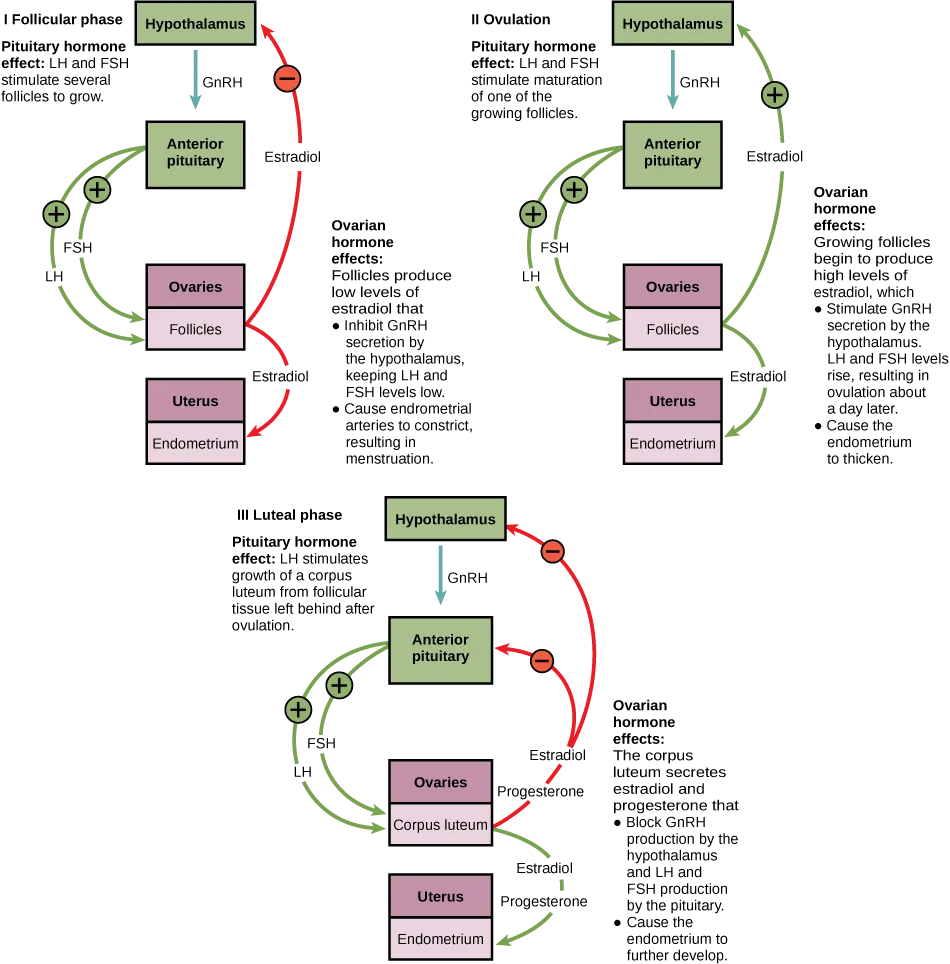
The first half of the ovarian cycle is the follicular phase shown below. Slowly rising levels of FSH and LH cause the growth of follicles on the surface of the ovary. This process prepares the egg for ovulation. As the follicles grow, they begin releasing estrogens and a low level of progesterone. At this stage, progesterone helps the thickening of the endometrium to make it receptive to implantation of the fertilized egg. If the fertilization of the egg happens, it takes place in the fallopian tube. The trip of a fertilized egg through the fallopian tube takes about seven days. At this stage of development, called the morula, there are 16-32 cells. (Source note: the source says 30-60; the morula is the 16- to 32-cell stage (Sadler, Langman’s Medical Embryology, 14th ed., chapter 3).) If pregnancy implantation does not occur, the lining is sloughed off. After about five days, estrogen levels rise and the menstrual cycle enters the proliferative phase. The endometrium begins to regrow, replacing the blood vessels and glands that deteriorated during the end of the last cycle.
Extended description
Three panels, top left, top right, and bottom: I Follicular phase, II Ovulation, and III Luteal phase. Each panel repeats the same four boxes top to bottom — a green Hypothalamus box, a green Anterior pituitary box, a mauve Ovaries box holding a pink sub-box, and a mauve Uterus box holding a pink Endometrium sub-box — plus its own ‘Pituitary hormone effect’ caption and its own bulleted ‘Ovarian hormone effects’ caption. In panel I the Ovaries sub-box reads Follicles; a teal arrow labeled GnRH runs from Hypothalamus down to Anterior pituitary, two green arrows labeled FSH and LH run from Anterior pituitary to Ovaries, and two arrows labeled Estradiol leave Follicles — one running up to Hypothalamus through a red minus-sign circle, one running down to Endometrium — with the caption noting the follicles’ low estradiol inhibits GnRH, keeping LH and FSH low, and constricts endometrial arteries, causing menstruation. Panel II repeats the same boxes and arrows, the Ovaries sub-box still reading Follicles, except the Estradiol arrow to Hypothalamus now crosses a green plus-sign circle, with the caption noting the growing follicles’ high estradiol now stimulates GnRH secretion, raising LH and FSH toward ovulation about a day later, and thickens the endometrium. In panel III the Ovaries sub-box reads Corpus luteum in place of Follicles; the teal GnRH arrow is unchanged, and two red arrows labeled Estradiol and Progesterone leave the Corpus luteum and run up to Anterior pituitary and Hypothalamus, each crossing a red minus-sign circle, while a green pair labeled Estradiol and Progesterone runs down to Endometrium — with the caption noting the corpus luteum’s estradiol and progesterone block GnRH, LH, and FSH production and cause the endometrium to further develop.
Which of the following statements about hormone regulation of the ovarian reproductive cycle is false?
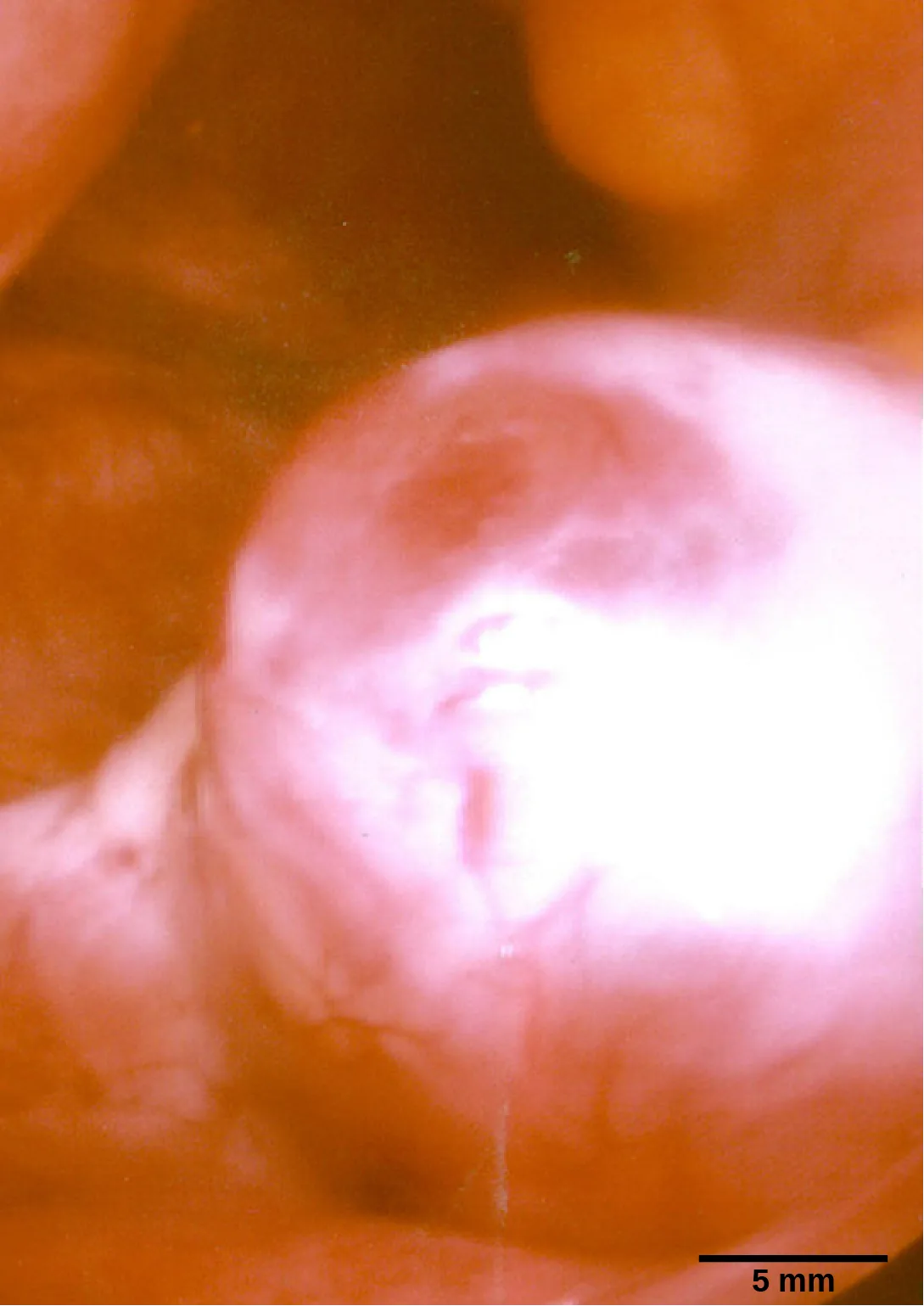
The diagram’s three panels each show where estradiol and progesterone are made at that stage — check which structure the follicular-phase and ovulation panels credit with producing them, before the corpus luteum ever forms.Just prior to the middle of the cycle (approximately day 14), the high level of estrogen causes FSH and especially LH to rise rapidly, then fall. The spike in LH causes ovulation: the most mature follicle, like that shown below, ruptures and releases its egg. The follicles that did not rupture degenerate and their eggs are lost. The level of estrogen decreases when the extra follicles degenerate.
Following ovulation, the ovarian cycle enters its luteal phase, illustrated above, and the menstrual cycle enters its secretory phase, both of which run from about day 15 to 28. The luteal and secretory phases refer to changes in the ruptured follicle. The cells in the follicle undergo physical changes and produce a structure called a corpus luteum. The corpus luteum produces estrogen and progesterone. The progesterone facilitates the regrowth of the uterine lining and inhibits the release of further FSH and LH. The uterus is being prepared to accept a fertilized egg, should it occur during this cycle. The inhibition of FSH and LH prevents any further eggs and follicles from developing, while the progesterone is elevated. The level of estrogen produced by the corpus luteum increases to a steady level for the next few days.
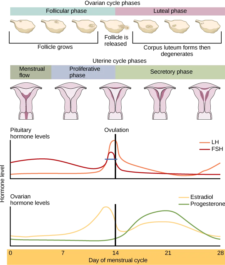
If no fertilized egg is implanted into the uterus, the corpus luteum degenerates and the levels of estrogen and progesterone decrease. The endometrium begins to degenerate as the progesterone levels drop, initiating the next menstrual cycle. The decrease in progesterone also allows the hypothalamus to send GnRH to the anterior pituitary, releasing FSH and LH and starting the cycles again. The figure below visually compares the ovarian and uterine cycles as well as the commensurate hormone levels.
Extended description
From top to bottom: an ‘Ovarian cycle phases’ bar split into a Follicular phase (left) and a Luteal phase (right), above seven small ovary icons captioned, left to right, Follicle grows (three icons), Follicle is released (one icon), and Corpus luteum forms then degenerates (three icons). Below it, a ‘Uterine cycle phases’ bar split into Menstrual flow, Proliferative phase, and Secretory phase, above five small uterus icons whose pink lining grows progressively thicker left to right. Below that, two line graphs share a ‘Day of menstrual cycle’ x-axis marked 0, 7, 14, 21, and 28, with a vertical black line at day 14 labeled Ovulation running through both. The upper graph, ‘Pituitary hormone levels,’ plots two curves: FSH (red) rises to a small early hump before day 7, dips, then rises again to a peak at day 14 alongside LH (orange), which stays lower until a taller, narrower peak also at day 14; both curves fall back to a low level after day 14; FSH stays low and flat to day 28, while LH rises again slightly from about day 24 to day 28. The lower graph, ‘Ovarian hormone levels,’ plots Estradiol (yellow), which rises to a peak just before day 14, drops sharply after day 14, then rises again to a lower, broader second peak around day 21 before declining toward day 28, and Progesterone (green), which stays near zero until after day 14, then rises to a peak around day 21 and falls back toward zero by day 28.
Which of the following statements about the menstrual cycle is false?
Follow the vertical line marking ovulation on the graph and compare what the two pituitary curves are doing right at that point against what the text says is happening a day or so later, once the corpus luteum has formed.Menopause
As people with ovarian reproductive systems approach their mid-40s to mid-50s, their ovaries begin to lose their sensitivity to FSH and LH. Menstrual periods become less frequent and finally cease; this is menopause. There are still eggs and potential follicles on the ovaries, but without the stimulation of FSH and LH, they will not produce a viable egg to be released. One outcome of this is the inability to have children.
The side effects of menopause include hot flashes, heavy sweating (especially at night), headaches, some hair loss, muscle pain, vaginal dryness, insomnia, depression, weight gain, and mood swings. Estrogen is involved in calcium metabolism and, without it, blood levels of calcium decrease. To replenish the blood, calcium is lost from bone which may decrease the bone density and lead to osteoporosis. Supplementation of estrogen in the form of hormone replacement therapy (HRT) can prevent bone loss, but the therapy can have negative side effects. While HRT is thought to give some protection from colon cancer, osteoporosis, heart disease, macular degeneration, and possibly depression, its negative side effects include increased risk of: stroke or heart attack, blood clots, breast cancer, ovarian cancer, endometrial cancer, gall bladder disease, and possibly dementia.
Career Connection. Reproductive Endocrinologist
A reproductive endocrinologist is a physician who treats a variety of hormonal disorders related to reproduction and infertility. The disorders include menstrual problems, infertility, pregnancy loss, sexual dysfunction, and menopause. Doctors may use fertility drugs, surgery, or assisted reproductive techniques (ART) in their therapy. ART involves the use of procedures to manipulate the egg or sperm to facilitate reproduction, such as in vitro fertilization.
Reproductive endocrinologists undergo extensive medical training, first in a four-year residency in obstetrics and gynecology, then in a three-year fellowship in reproductive endocrinology. To be board certified in this area, the physician must pass written and oral exams in both areas.
Summary
The male and female reproductive cycles are controlled by hormones released from the hypothalamus and anterior pituitary as well as hormones from reproductive tissues and organs. The hypothalamus monitors the need for the FSH and LH hormones made and released from the anterior pituitary. FSH and LH affect reproductive structures to cause the formation of sperm and the preparation of eggs for release and possible fertilization. In the male, FSH and LH stimulate Sertoli cells and interstitial cells of Leydig in the testes to facilitate sperm production. The Leydig cells produce testosterone, which also is responsible for the secondary sexual characteristics of males. In females, FSH and LH cause estrogen and progesterone to be produced. They regulate the female reproductive system which is divided into the ovarian cycle and the menstrual cycle. Menopause occurs when the ovaries lose their sensitivity to FSH and LH and the reproductive cycles slow to a stop.
Key terms
- estradiol — most potent form of estrogen that is responsible for the development of secondary female characteristics, maintenance of female reproductive organs, and preparation for pregnancy
- estrogen — reproductive hormone in females that assists in endometrial regrowth, ovulation, and calcium absorption
- follicle stimulating hormone (FSH) — reproductive hormone that causes sperm production in males and follicle development in females
- gonadotropin-releasing hormone (GnRH) — hormone from the hypothalamus that causes the release of FSH and LH from the anterior pituitary
- inhibin — hormone made by Sertoli cells; provides negative feedback to hypothalamus in control of FSH and GnRH release
- interstitial cell of Leydig — cell in seminiferous tubules that makes testosterone
- luteinizing hormone (LH) — reproductive hormone in both males and females, causes testosterone production in males and ovulation in females (Source note: the source glossary adds “and lactation”; this book attributes milk production to prolactin and milk release to oxytocin.)
- menopause — loss of reproductive capacity due to decreased sensitivity of the ovaries to FSH and LH
- menstrual cycle — cycle of the degradation and regrowth of the endometrium
- ovarian cycle — cycle of preparation of egg for ovulation and the conversion of the follicle to the corpus luteum
- ovulation — release of the egg by the most mature follicle
- progesterone — reproductive hormone in females; assists in endometrial regrowth and inhibition of FSH and LH release
- Sertoli cell — cell in seminiferous tubules that assists developing sperm and makes inhibin
- testosterone — reproductive hormone in men that assists in sperm production and promoting secondary sexual characteristics
Practice
Describe the roles of male and female reproductive hormones
Which hormone causes Leydig cells to make testosterone?
One pituitary gonadotropin targets the Sertoli cells and sperm production directly; the other targets the Leydig cells and steroid hormone production — match the cell type named in the question to the right gonadotropin.Which hormone causes FSH and LH to be released?
This signal comes from a gland one step upstream of the anterior pituitary — trace where in the signaling chain it originates, not where FSH and LH themselves are released.Which hormone signals ovulation?
Two of these are pituitary hormones that rise together near the middle of the cycle, but only one is the hormone whose sharp, rapid spike the text specifically ties to follicle rupture.If male reproductive pathways are not cyclical, how are they controlled?
Show model answer
Did your answer mention:
The hormone made by Sertoli cells that provides negative feedback to the hypothalamus in the control of FSH and GnRH release is called ________.
This hormone’s name describes what it does to FSH and GnRH release — the opposite of a hormone that promotes them.Discuss the interplay of the ovarian and menstrual cycles
Describe the events in the ovarian cycle leading up to ovulation.
Show model answer
Did your answer mention:
Which hormone causes the regrowth of the endometrial lining of the uterus?
Two of these hormones belong to the male reproductive system’s feedback loop and play no role in the uterine lining at all; between the remaining two, only one is a steroid hormone this section credits with regrowing the endometrium itself.The cycle of preparation of an egg for ovulation and the conversion of the follicle to the corpus luteum is called the ________.
This cycle is named for the organ where follicles grow and the corpus luteum forms — not for the uterine lining.The cycle of the degradation and regrowth of the endometrium is called the ________.
This cycle is named for the process the endometrium goes through each month, not for the ovary.Describe the process of menopause
The loss of reproductive capacity due to decreased sensitivity of the ovaries to FSH and LH is called ________.
This term names the permanent stopping point of the menstrual cycle, not a single irregular period.Menopause occurs when the ovaries lose their sensitivity to ________ and the reproductive cycles slow to a stop.
These are the two pituitary hormones this whole section has been naming together — the ones released after the hypothalamus sends GnRH to the anterior pituitary.Supplementation of estrogen in the form of ________ can prevent bone loss after menopause, though it can also have negative side effects.
This is not a fertility procedure — it is the general term the module uses for replacing the estrogen the ovaries stop making after menopause.This section is adapted from Biology 2e, Section 43.4: Hormonal Control of Human Reproduction by Mary Ann Clark, Jung Choi, Matthew Douglas, and OpenStax, © OpenStax, licensed under CC BY-NC-SA 4.0. Access the original for free at openstax.org. Changes: figures re-encoded as WebP; Figure_43_04_01 re-kinded from the manifest’s file-extension “photo” guess to “diagram” (it is a hand-drawn feedback-loop flow chart, not a captured photograph) and Figure_B43_04_04 re-kinded the other way, from the manifest’s “diagram” guess to “photo” (it is an actual light micrograph of a ruptured egg follicle); the three long, screen-reader-style source alts (Figure_43_04_01, Figure_B43_04_02, Figure_B43_04_03), each well over the 600-character alt limit, were rewritten as short plain-text alts describing what is drawn, with their full narratives moved into a longdesc transcribing every box, arrow, plus/minus circle, and caption in reading order — the two Visual Connection longdescs (Figure_B43_04_02, Figure_B43_04_03) transcribe only the printed labels and curve shapes and never state which of that item’s four statements is false; both notes wrapping a Visual Connection rendered as their figure followed by a multiple choice, kept in the body — the note copy and the <exercise> copy print identical question and option wording in both cases, so no adjudication was needed; the career note rendered as a Career Connection callout with its own title; exercise fs-idm71478992 (“Which hormone causes the regrowth of the endometrial lining of the uterus?”) keeps the module’s own key of progesterone, but its distractor “estrogen” is replaced with “inhibin” — reported as a source defect below; a duplicated word in the glossary definition of estradiol is corrected — see below; the end-of-section Review Questions and Critical Thinking Questions adapted into the closing interactive Practice block (multiple choice and self-check respectively), both Visual Connection items left in the body rather than duplicated in Practice; rubric checkpoints added to each self-check, decomposing its model answer (the source solution) into check-off clauses with no new claims; four key-term recall items added from the glossary (inhibin, ovarian cycle, menstrual cycle, menopause), covering four of the section’s fourteen glossary terms, plus one cloze item built from the section summary (blanking “FSH and LH”); one locally written multiple choice added to the thin Menopause objective group, built strictly from the section’s own sentence about hormone replacement therapy, disclosed here and in the ledger. Source defects: module m66674, exercise fs-idm71478992 — the module’s own text says “Estradiol is the reproductive hormone in females that assists in endometrial regrowth, ovulation, and calcium absorption” and “estrogen levels rise and the menstrual cycle enters the proliferative phase. The endometrium begins to regrow,” so the printed distractor “estrogen” is also true and not a valid wrong answer; the printed key, progesterone (supported by “The progesterone facilitates the regrowth of the uterine lining”), is kept, and the distractor is replaced with “inhibin,” which the module gives only the role of inhibiting FSH release. Glossary defect: module m66674, definition def-00001 (estradiol) reads “…responsible for the development of female secondary female characteristics…” — the word “female” is printed twice; corrected to “the development of secondary female characteristics.” Two claims are corrected with visible Source notes: the morula has 16 to 32 cells, not 30 to 60, and the LH key term no longer credits LH with lactation, which this book assigns to prolactin (errata 452–453).