Human Pregnancy and Birth
By the end of this section, you will be able to:
- Explain fetal development during the three trimesters of gestation
- Describe labor and delivery
- Compare the efficacy and duration of various types of contraception
- Discuss causes of infertility and the therapeutic options available
Pregnancy begins with the fertilization of an egg and continues through to the birth of the individual. The length of time of gestation varies among animals, but is very similar among the great apes: human gestation is 266 days, while chimpanzee gestation is 237 days, a gorilla’s is 257 days, and orangutan gestation is 260 days long. The fox has a 57-day gestation. Dogs and cats have similar gestations averaging 60 days. The longest gestation for a land mammal is an African elephant at 640 days. The longest gestations among marine mammals are the beluga and sperm whales at 460 days.
Human Gestation
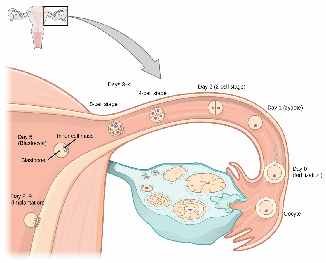
Twenty-four hours before fertilization, the egg has finished meiosis and becomes a mature oocyte. When fertilized (at conception) the egg becomes known as a zygote. The zygote travels through the oviduct to the uterus, shown below. The developing embryo must implant into the wall of the uterus within seven days, or it will deteriorate and die. The outer layers of the zygote (blastocyst) grow into the endometrium by digesting the endometrial cells, and wound healing of the endometrium closes up the blastocyst into the tissue. Another layer of the blastocyst, the chorion, begins releasing a hormone called human beta chorionic gonadotropin (β-HCG) which makes its way to the corpus luteum and keeps that structure active. This ensures adequate levels of progesterone that will maintain the endometrium of the uterus for the support of the developing embryo. Pregnancy tests determine the level of β-HCG in urine or serum. If the hormone is present, the test is positive.
Extended description
At upper left, a small inset drawing of the full female reproductive tract shows a box outlining one ovary and its fallopian tube, with a gray arrow curving down to the enlarged view below. Reading that enlarged view right to left, along the curved tube: a circle labeled Oocyte sits near the ovary, beside a cell labeled Day 0 (fertilization) that shows a sperm’s thread-like tail attached. Moving left, single circles are labeled Day 1 (zygote) and Day 2 (2-cell stage); then, under a Days 3-4 heading, a circle divided into four parts is labeled 4-cell stage and a denser cluster of cells is labeled 8-cell stage. The tube curves down into the uterus, where a larger circle is labeled Day 5 (Blastocyst); two leader lines from that circle label Inner cell mass, an off-center cluster, and Blastocoel, the fluid-filled center. At the lower left, embedded in the pink uterine wall, a small circle is labeled Day 8-9 (Implantation). Below the tube, an irregular teal shape (the ovary) holds several unlabeled round follicles.
The gestation period is divided into three equal periods or trimesters. During the first two to four weeks of the first trimester, nutrition and waste are handled by the endometrial lining through diffusion. As the trimester progresses, the outer layer of the embryo begins to merge with the endometrium, and the placenta forms. This organ takes over the nutrient and waste requirements of the embryo and fetus, with the parent’s blood passing nutrients to the placenta and removing waste from it. Chemicals from the fetus, such as bilirubin, are processed by the parent’s liver for elimination. Some of the parent’s immunoglobulins will pass through the placenta, providing passive immunity against some potential infections.
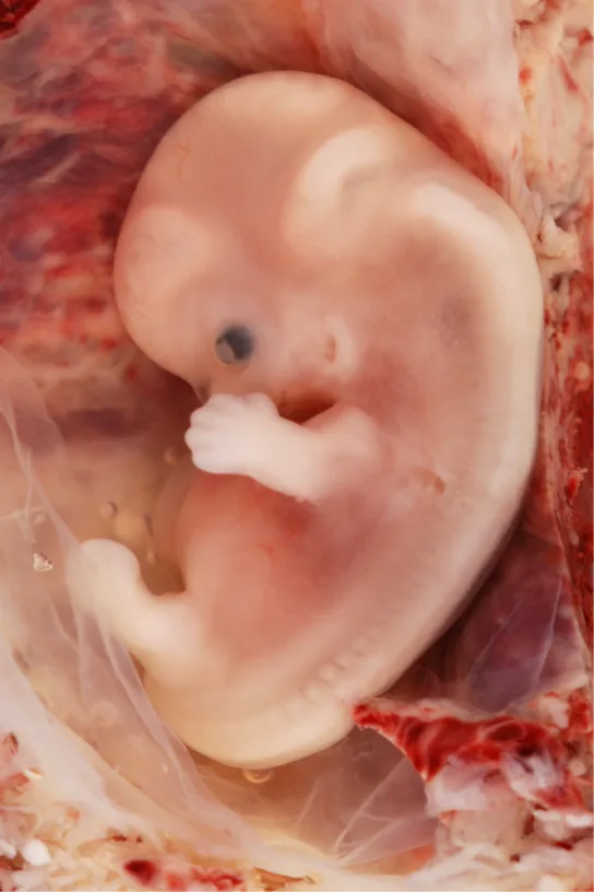
Internal organs and body structures begin to develop during the first trimester. By five weeks, limb buds, eyes, the heart, and liver have been basically formed. By eight weeks, the term fetus applies, and the body is essentially formed, as shown below. The individual is about five centimeters (two inches) in length and many of the organs, such as the lungs and liver, are not yet functioning. Exposure to any toxins is especially dangerous during the first trimester, as all of the body’s organs and structures are going through initial development. Anything that affects that development can have a severe effect on the fetus’ survival.
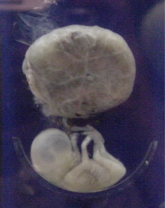
During the second trimester, the fetus grows to about 30 cm (12 inches), as shown below. It becomes active and the pregnant person usually feels the first movements. All organs and structures continue to develop. The placenta has taken over the functions of nutrition and waste and the production of estrogen and progesterone from the corpus luteum, which has degenerated. The placenta will continue functioning up through the delivery of the baby.
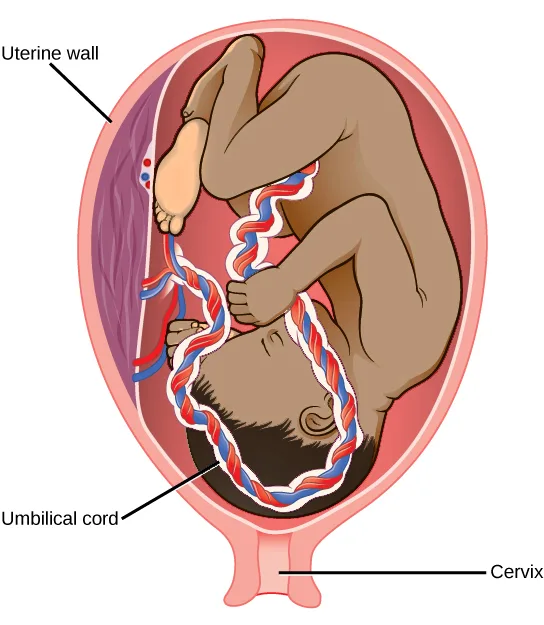
During the third trimester, the fetus grows to 3 to 4 kg (6 ½–8 ½ lbs.) and about 50 cm (19–20 inches) long, as illustrated below. This is the period of the most rapid growth during the pregnancy. Organ development continues to birth (and some systems, such as the nervous system and liver, continue to develop after birth). The pregnant person will be most uncomfortable during this trimester. They may urinate frequently due to pressure on the bladder from the fetus. There may also be intestinal blockage and circulatory problems, especially in the legs, where clots may form due to pressure from the fetus on returning veins as they enter the abdominal cavity.
Extended description
Three leader lines label the drawing. One near the upper left points to a purple-streaked patch of tissue on the uterus’s inner wall, labeled Uterine wall. One at the lower left points to the twisted red-and-blue-and-white cord running from the fetus’s belly up to that wall, labeled Umbilical cord. One at the lower right points to the narrow opening at the bottom of the uterus, labeled Cervix.
Link to Learning
Visit an interactive tool tracing the stages of human fetal development.
Labor and Birth
Labor is the physical efforts of expulsion of the fetus and the placenta from the uterus during birth (parturition). Toward the end of the third trimester, estrogen causes receptors on the uterine wall to develop and bind the hormone oxytocin. At this time, the baby reorients, facing forward and down with the back or crown of the head engaging the cervix (uterine opening). This causes the cervix to stretch and nerve impulses are sent to the hypothalamus, which signals for the release of oxytocin from the posterior pituitary. The oxytocin causes the smooth muscle in the uterine wall to contract. At the same time, the placenta releases prostaglandins into the uterus, increasing the contractions. A positive feedback relay occurs between the uterus, hypothalamus, and the posterior pituitary to assure an adequate supply of oxytocin. As more smooth muscle cells are recruited, the contractions increase in intensity and force.
There are three stages to labor. During stage one, the cervix thins and dilates. This is necessary for the baby and placenta to be expelled during birth. The cervix will eventually dilate to about 10 cm. During stage two, the baby is expelled from the uterus. The uterus contracts and the person giving birth pushes and compresses the abdominal muscles to aid the delivery. The last stage is the passage of the placenta after the baby has been born and the organ has completely disengaged from the uterine wall. If labor should stop before stage two is reached, synthetic oxytocin, known as Pitocin, can be administered to restart and maintain labor.
An alternative to labor and delivery is the surgical delivery of the baby through a procedure called a Caesarian section. This is major abdominal surgery and can lead to post-surgical complications for the person giving birth, but in some cases it may be the only way to safely deliver the baby.
The pregnant female’s mammary glands go through changes during the third trimester to prepare for lactation and breastfeeding. When the baby begins suckling at the breast, signals are sent to the hypothalamus causing the release of prolactin from the anterior pituitary. Prolactin causes the mammary glands to produce milk. Oxytocin is also released, promoting the release of the milk. The milk contains nutrients for the baby’s development and growth as well as immunoglobulins to protect the child from bacterial and viral infections.
Contraception and Birth Control
The prevention of a pregnancy comes under the terms contraception or birth control. Strictly speaking, contraception refers to preventing the sperm and egg from joining. Both terms are, however, frequently used interchangeably.
The table below lists common methods of contraception.
Contraceptive Methods
| Method | Examples | Failure Rate in Typical Use Over 12 Months |
|---|---|---|
| Barrier | male condom, female condom, sponge, cervical cap, diaphragm, spermicides | 15 to 24% |
| Hormonal | oral, patch, vaginal ring | 8% |
| injection | 3% | |
| implant | less than 1% | |
| Other | natural family planning | 12 to 25% |
| withdrawal | 27% | |
| sterilization | less than 1% |
The failure rates listed are not the ideal rates that could be realized, but the typical rates that occur. A failure rate is the number of pregnancies resulting from the method’s use over a twelve-month period. Barrier methods, such as condoms, cervical caps, and diaphragms, block sperm from entering the uterus, preventing fertilization. Spermicides are chemicals that are placed in the vagina that kill sperm. Sponges, which are saturated with spermicides, are placed in the vagina at the cervical opening. Combinations of spermicidal chemicals and barrier methods achieve lower failure rates than do the methods when used separately.
Nearly a quarter of the couples using barrier methods, natural family planning, or withdrawal can expect a failure of the method. Natural family planning is based on the monitoring of the menstrual cycle and having intercourse only during times when the egg is not available. During ovulation, a person’s body temperature may rise a degree Celsius and the cervical mucus may increase in volume and become more pliable. These changes give a general indication of when intercourse is more or less likely to result in fertilization. Withdrawal involves the removal of the penis from the vagina during intercourse, before ejaculation occurs. This is a risky method with a high failure rate due to the possible presence of sperm in the bulbourethral gland’s secretion, which may enter the vagina prior to removing the penis.
Hormonal methods use synthetic progesterone (sometimes in combination with estrogen), to inhibit the hypothalamus from releasing FSH or LH, and thus prevent an egg from being available for fertilization. The method of administering the hormone affects failure rate. The most reliable method, with a failure rate of less than 1 percent, is the implantation of the hormone under the skin. The same rate can be achieved through the sterilization procedures of vasectomy in the man or of tubal ligation in the woman, or by using an intrauterine device (IUD). IUDs are inserted into the uterus and establish an inflammatory condition that prevents fertilized eggs from implanting into the uterine wall.
Compliance with the contraceptive method is a strong contributor to the success or failure rate of any particular method. The only method that is completely effective at preventing conception is abstinence. The choice of contraceptive method depends on the goals of the woman or couple. Tubal ligation and vasectomy are considered permanent prevention, while other methods are reversible and provide short-term contraception.
Termination of an existing pregnancy can be spontaneous or voluntary. Spontaneous termination is a miscarriage and usually occurs very early in the pregnancy, usually within the first few weeks. This occurs when the fetus cannot develop properly and the gestation is naturally terminated. Voluntary termination of a pregnancy is an abortion. Laws regulating abortion vary between states and tend to view fetal viability as the criteria for allowing or preventing the procedure.
Infertility
Infertility is the inability to conceive a child or carry a child to birth. About 75 percent of causes of infertility can be identified; these include diseases, such as sexually transmitted diseases that can cause scarring of the reproductive tubes in males or females, or developmental problems frequently related to abnormal hormone levels in one of the individuals. Inadequate nutrition, especially starvation, can delay menstruation. Stress can also lead to infertility. Short-term stress can affect hormone levels, while long-term stress can delay puberty and cause less frequent menstrual cycles. Other factors that affect fertility include toxins (such as cadmium), tobacco smoking, marijuana use, gonadal injuries, and aging.
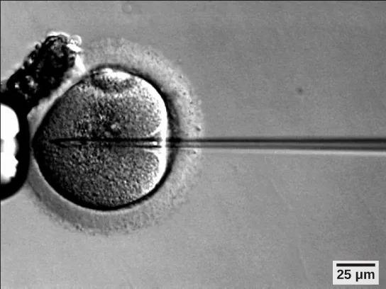
If infertility is identified, several assisted reproductive technologies (ART) are available to aid conception. A common type of ART is in vitro fertilization (IVF) where an egg and sperm are combined outside the body and then placed in the uterus. This technique was first successfully performed in mice by Anne McLaren and John Biggers, whose work is credited as among the most significant in reproductive medicine. Eggs are obtained from the woman after extensive hormonal treatments that prepare mature eggs for fertilization and prepare the uterus for implantation of the fertilized egg. Sperm are obtained, combined with the eggs, and supported through several cell divisions to ensure viability of the zygotes. When the embryos have reached the eight-cell stage, one or more is implanted into the gestational parent’s or carrier’s uterus. If fertilization is not accomplished by simple IVF, a procedure that injects the sperm into an egg can be used. This is called intracytoplasmic sperm injection (ICSI) and is shown below. IVF procedures produce a surplus of fertilized eggs and embryos that can be frozen and stored for future use. The procedures can also result in multiple births.
Summary
Human pregnancy begins with fertilization of an egg and proceeds through the three trimesters of gestation. The labor process has three stages (contractions, delivery of the fetus, expulsion of the placenta), each propelled by hormones. The first trimester lays down the basic structures of the body, including the limb buds, heart, eyes, and the liver. The second trimester continues the development of all of the organs and systems. The third trimester exhibits the greatest growth of the fetus and culminates in labor and delivery. Prevention of a pregnancy can be accomplished through a variety of methods including barriers, hormones, or other means. Assisted reproductive technologies may help individuals who have infertility problems.
Key terms
- contraception — (also, birth control) various means used to prevent pregnancy
- gestation — length of time for fetal development to birth
- human beta chorionic gonadotropin (β-HCG) — hormone produced by the chorion of the zygote that helps to maintain the corpus luteum and elevated levels of progesterone
- infertility — inability to conceive, carry, and deliver children
- morning sickness — condition in pregnant people during the first trimester; includes feelings of nausea
- placenta — organ that supports the diffusion of nutrients and waste between the pregnant person’s and fetus’ blood
Practice
Explain fetal development during the three trimesters of gestation
Nutrient and waste requirements for the developing fetus are handled during the first few weeks by:
The placenta hasn’t formed yet this early in the first trimester — think about how a molecule can cross a membrane when there is no vascular organ yet in place to carry it.Progesterone is made during the third trimester by the:
By the third trimester, one organ has taken over hormone production from the corpus luteum, which has already degenerated — check which structure the section says has assumed that role.Major organs begin to develop during which part of human gestation?
Limb buds, eyes, the heart, and the liver are named as basically formed by five weeks — that timing points to one specific period of gestation.Describe the major developments during each trimester of human gestation.
Show model answer
Did your answer mention:
The length of time for fetal development to birth is called ________.
This word also names the length of pregnancy in every mammal the section compares — human, chimpanzee, elephant, and whale each have their own value for it.The organ that supports the diffusion of nutrients and waste between the pregnant person’s and fetus’ blood is called the ________.
This organ forms partway through the first trimester and keeps performing this exchange function all the way through delivery.Describe labor and delivery
Which hormone is primarily responsible for the contractions during labor?
Trace which hormone the posterior pituitary releases once nerve impulses reach the hypothalamus from the stretching cervix, and which one the smooth muscle of the uterine wall responds to.Describe the stages of labor.
Show model answer
Did your answer mention:
The labor process has three stages (contractions, delivery of the fetus, expulsion of the placenta), each propelled by ________.
Oxytocin and prostaglandins are two examples of the broader chemical-messenger category this word names.Compare the efficacy and duration of various types of contraception
Which of these methods is regarded as permanent contraception?
Distinguish a method that can be reversed or simply stopped from one meant to end fertility for good.Which type of short term contraceptive method is generally more effective than others?
Compare the failure-rate range the table gives for each method category listed here, and pick the category with the lowest typical-use range.Various means used to prevent pregnancy are collectively called ________.
The section notes that, strictly speaking, this term refers only to preventing the sperm and egg from joining, though it is often used interchangeably with a broader synonym.Discuss causes of infertility and the therapeutic options available
The inability to conceive, carry, and deliver children is called ________.
About three-quarters of the causes of this condition can be identified, according to the section.Assisted reproductive technologies may help individuals who have which of the following?
This option names the inability to conceive a child or carry one to birth — check which of the four matches that meaning rather than a different reproductive process.A common type of assisted reproductive technology in which an egg and sperm are combined outside the body and then placed in the uterus is called ________.
Its Latin name literally means “in glass,” referring to where fertilization takes place.This section is adapted from Biology 2e, Section 43.5: Human Pregnancy and Birth by Mary Ann Clark, Jung Choi, Matthew Douglas, and OpenStax, © OpenStax, licensed under CC BY-NC-SA 4.0. Access the original for free at openstax.org. Changes: figures re-encoded as WebP with the source alt text edited to describe only what is visible; two figures re-kinded from the manifest’s file-extension “photo” guess to “diagram” (Figure_43_07_01, the day-by-day fertilization-to-implantation flow diagram, and Figure_43_07_04, the labeled third-trimester illustration, are each a hand-drawn or computer-rendered illustration, not a captured photograph); a longdesc added to the two diagrams whose printed labels are not carried by their one-line captions — the fertilization/implantation flow diagram (transcribing its day-by-day stage labels and the Inner cell mass/Blastocoel leader lines, traced right to left as the diagram itself progresses from ovary to uterus) and the third-trimester illustration (transcribing its three leader-line labels: Uterine wall, Umbilical cord, Cervix); in-text pointers to figures and the table (“Figure 43.18” through “Figure 43.22”, “Table 43.3”) replaced with “shown below,” “shown above,” “illustrated below,” or “the table below,” since Hugo does not number figures or tables; the interactive note rendered as a Link to Learning callout with descriptive link text, keeping the module’s own openstax.org/l/embryo_fetus redirect URL; the Contraceptive Methods table kept as a Markdown table in the body, with its merged Hormonal and Other category cells left blank on continuation rows in place of the source’s row-span; the end-of-section Review Questions and Critical Thinking Questions adapted into the closing interactive Practice block (multiple choice and self-check respectively); rubric checkpoints added to each self-check, decomposing its model answer (the source solution) into check-off clauses with no new claims; a cloze text-recall item added from the section summary (“each propelled by ________,” answer “hormones”) for the Describe labor and delivery group, which the source’s Review and Critical Thinking Questions do not otherwise cover; a select-the-term multiple choice built from the section summary’s closing sentence, and a locally written text-recall item built strictly from the section’s own sentence describing in vitro fertilization, added to the Discuss causes of infertility group, which the source exercise sets do not test at all; four key-term recall items added from the glossary (gestation, placenta, contraception, infertility), covering four of the section’s six glossary terms — human beta chorionic gonadotropin (β-HCG) and morning sickness appear only in the Key terms list (β-HCG also in the prose). Exercise fs-idp80601616 is keyed B, “diffusion through the endometrium,” on source authority; the module’s own sentence directly supports it (“During the first two to four weeks of the first trimester, nutrition and waste are handled by the endometrial lining through diffusion”).