Organogenesis and Vertebrate Axis Formation
By the end of this section, you will be able to:
- Describe the process of organogenesis
- Identify the anatomical axes formed in vertebrates
Gastrulation leads to the formation of the three germ layers that give rise, during further development, to the different organs in the animal body. This process is called organogenesis. Organogenesis is characterized by rapid and precise movements of the cells within the embryo.
Organogenesis
Organs form from the germ layers through the process of differentiation. During differentiation, the embryonic stem cells express specific sets of genes which will determine their ultimate cell type. For example, some cells in the ectoderm will express the genes specific to skin cells. As a result, these cells will differentiate into epidermal cells. The process of differentiation is regulated by cellular signaling cascades.
Scientists study organogenesis extensively in the lab in fruit flies (Drosophila) and the nematode Caenorhabditis elegans. Drosophila have segments along their bodies, and the patterning associated with the segment formation has allowed scientists to study which genes play important roles in organogenesis along the length of the embryo at different time points. The nematode C.elegans has roughly 1000 somatic cells and scientists have studied the fate of each of these cells during their development in the nematode life cycle. There is little variation in patterns of cell lineage between individuals, unlike in mammals where cell development from the embryo is dependent on cellular cues.
In vertebrates, one of the primary steps during organogenesis is the formation of the neural system. The ectoderm forms epithelial cells and tissues, and neuronal tissues. During the formation of the neural system, special signaling molecules called growth factors signal some cells at the edge of the ectoderm to become epidermis cells. The remaining cells in the center form the neural plate. If the signaling by growth factors were disrupted, then the entire ectoderm would differentiate into neural tissue.
The neural plate undergoes a series of cell movements where it rolls up and forms a tube called the neural tube, shown below. In further development, the neural tube will give rise to the brain and the spinal cord.
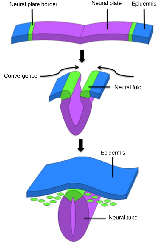
Extended description
Three panels, top to bottom, connected by two downward black block arrows. Panel 1: a flat layered sheet, colored (left to right) blue, a thin green stripe, wide purple, a thin green stripe, and blue again. Three leader lines label it: ‘Neural plate border’ points to the left green stripe (a matching green stripe sits at the right, unlabeled), ‘Neural plate’ points to the wide purple middle, and ‘Epidermis’ points to the right blue block. Panel 2: the same sheet has begun folding — its two edges have risen into a pair of flaps (each blue on the outside, green in the middle, purple at the base) tilting toward each other over a deepening purple groove. Two curved black arrows, labeled ‘Convergence’ at the left, curl inward from each flap toward the top center. A leader line labeled ‘Neural fold’ points to the raised edge of the right-hand flap. Panel 3: the sheet has closed into a wavy blue layer, labeled ‘Epidermis’, spanning the top; beneath it, a purple tube tapering to a point, labeled ‘Neural tube’ by a leader line, with a lighter purple line running down its center marking the lumen. Fourteen small green ovals — seven on each side, in two loosely staggered rows — sit tucked between the epidermis and the widening top of the tube.
The mesoderm that lies on either side of the vertebrate neural tube will develop into the various connective tissues of the animal body. A spatial pattern of gene expression reorganizes the mesoderm into groups of cells called somites with spaces between them. The somites shown below will further develop into the cells that form the vertebrae and ribs, the dermis of the dorsal skin, the skeletal muscles of the back, and the skeletal muscles of the body wall and limbs. The mesoderm also forms a structure called the notochord, which is rod-shaped and forms the central axis of the animal body.
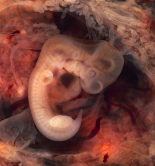
Vertebrate Axis Formation
Even as the germ layers form, the ball of cells still retains its spherical shape. However, animal bodies have lateral-medial (left-right), dorsal-ventral (back-belly), and anterior-posterior (head-feet) axes, shown below.
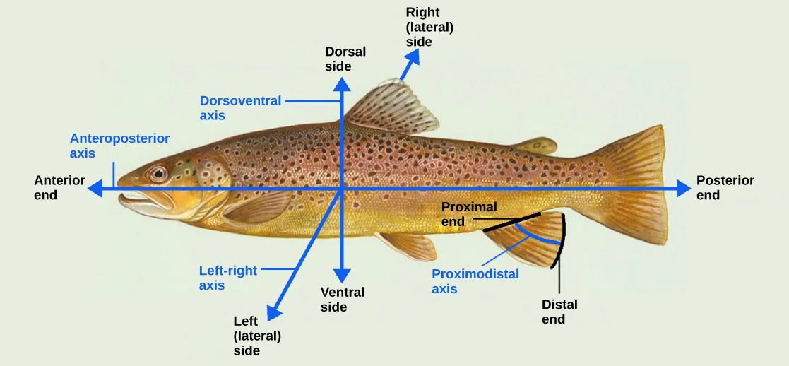
Extended description
A photographic side view of a trout, overlaid with three double-headed blue arrows crossing at its center and one short black-and-blue arrow at its lower fin. The horizontal arrow, labeled ‘Anteroposterior axis’, runs the length of the fish; its left arrowhead is labeled ‘Anterior end’ at the head and its right arrowhead ‘Posterior end’ at the tail. The vertical arrow, labeled ‘Dorsoventral axis’, runs from the fish’s back to its belly; its upper arrowhead is labeled ‘Dorsal side’ and its lower arrowhead ‘Ventral side’. A diagonal arrow, labeled ‘Left-right axis’, runs from lower left to upper right; its lower arrowhead is labeled ‘Left (lateral) side’ and its upper arrowhead, pointing toward the dorsal fin, is labeled ‘Right (lateral) side’. At the lower fin near the tail, a short arrow labeled ‘Proximodistal axis’ runs from where the fin meets the body, labeled ‘Proximal end’, to the fin’s outer edge, labeled ‘Distal end’.
How are these established? In one of the most seminal experiments ever to be carried out in developmental biology, Spemann and Mangold took dorsal cells from one embryo and transplanted them into the belly region of another embryo. They found that the transplanted embryo now had two notochords: one at the dorsal site from the original cells and another at the transplanted site. This suggested that the dorsal cells were genetically programmed to form the notochord and define the axis. Since then, researchers have identified many genes that are responsible for axis formation. Mutations in these genes lead to the loss of symmetry required for organism development.
Animal bodies have externally visible symmetry. However, the internal organs are not symmetric. For example, the heart is on the left side and the liver on the right. The formation of the central left-right axis is an important process during development. This internal asymmetry is established very early during development and involves many genes. Research is still ongoing to fully understand the developmental implications of these genes.
Summary
Organogenesis is the formation of organs from the germ layers. Each germ layer gives rise to specific tissue types. The first stage is the formation of the neural system in the ectoderm. The mesoderm gives rise to somites and the notochord. Formation of vertebrate axis is another important developmental stage.
Key terms
- neural tube — tube-like structure that forms from the ectoderm and gives rise to the brain and spinal cord.
- organogenesis — process of organ formation.
- somite — group of cells separated by small spaces that form from the mesoderm and give rise to connective tissue.
Practice
Describe the process of organogenesis
Which of the following gives rise to the skin cells?
Skin is made of epithelial tissue; the section names which germ layer forms epithelial cells and tissues as well as neuronal tissue.The ribs form from the ________.
This structure also gives rise to the vertebrae, the dermis of the dorsal skin, and the skeletal muscles of the back — one of four options names the segmented groups of mesoderm cells responsible for all of them.Explain how the different germ layers give rise to different tissue types.
Show model answer
Did your answer mention:
A tube-like structure that forms from the ectoderm and gives rise to the brain and spinal cord is called the ________.
This structure rolls up from the flat plate at the embryo’s dorsal midline, closing the epidermis over the top as it forms.The process of organ formation from the three germ layers is called ________.
It names the whole process by which the germ layers differentiate into organs — the process that skin-cell formation is one event within.A group of cells separated by small spaces that forms from the mesoderm and gives rise to connective tissue is called a ________.
Clusters of these cells are visible as segments along the length of a five-week human embryo, flanking the developing neural tube.Identify the anatomical axes formed in vertebrates
Explain the role of axis formation in development.
Show model answer
Did your answer mention:
Formation of vertebrate ________ is another important developmental stage.
This is the section’s own word for the line of symmetry it describes as lateral-medial, dorsal-ventral, and anterior-posterior.In the Spemann and Mangold transplant experiment, dorsal cells moved into the belly region of another embryo were found to be genetically programmed to form the ________, which defines the axis.
This rod-shaped structure, described earlier as forming the central axis of the animal body, appeared a second time at the transplant site.This section is adapted from Biology 2e, Section 43.7: Organogenesis and Vertebrate Axis Formation by Mary Ann Clark, Jung Choi, Matthew Douglas, and OpenStax, © OpenStax, licensed under CC BY-NC-SA 4.0. Access the original for free at openstax.org. Changes: figures re-encoded as WebP with the source alt text kept as printed; two figures (Figure_43_06_01, Figure_B43_06_03) re-kinded from the manifest’s file-extension “photo” guess to “diagram” — both are labeled line drawings, and the source’s own alt text opens “Illustration shows…” for each — while Figure_43_06_02 (an actual photograph of a five-week human embryo) is kept as the manifest’s “photo” guess; a longdesc added to Figure_43_06_01 (the three-panel neural-tube formation sequence, including a count of all fourteen green ovals in the third panel) and to Figure_B43_06_03 (the three-axis body-plan diagram, transcribing all twelve printed labels), since neither figure’s one-line caption carries its labels; Figure_43_06_02 gets no longdesc, since its caption already states everything the photograph shows; in-text pointers to figures (“Figure 43.28,” “Figure 43.29,” “Figure 43.30”) replaced with “shown below,” since Hugo does not number figures; the end-of-section Review Questions and Critical Thinking Questions adapted into the closing interactive Practice block (multiple choice and self-check respectively) — this module prints no Visual Connection item; rubric checkpoints added to each self-check, decomposing its model answer (the source solution) into check-off clauses with no new claims; five recall items added beyond the four end-of-section exercises — three glossary textin items (neural tube, organogenesis, somite), one cloze textin built from the section summary’s closing sentence (axis), and one locally written textin built strictly from the Spemann and Mangold paragraph (notochord), added because the module’s glossary and summary otherwise leave the “Identify the anatomical axes formed in vertebrates” objective without enough coverage on their own — disclosed here and in the source ledger; a grammar fix — the module’s “Mutations in these genes leads to the loss of symmetry” corrected to “lead” (the plural subject “genes” takes “lead,” matching the parallel sentence in this module’s own Critical Thinking solution, which prints “genes lead”) — reported as a source defect.