Types of Microorganisms
By the end of this section, you will be able to:
- List the various types of microorganisms and describe their defining characteristics
- Give examples of different types of cellular and viral microorganisms and infectious agents
- Describe the similarities and differences between archaea and bacteria
- Provide an overview of the field of microbiology
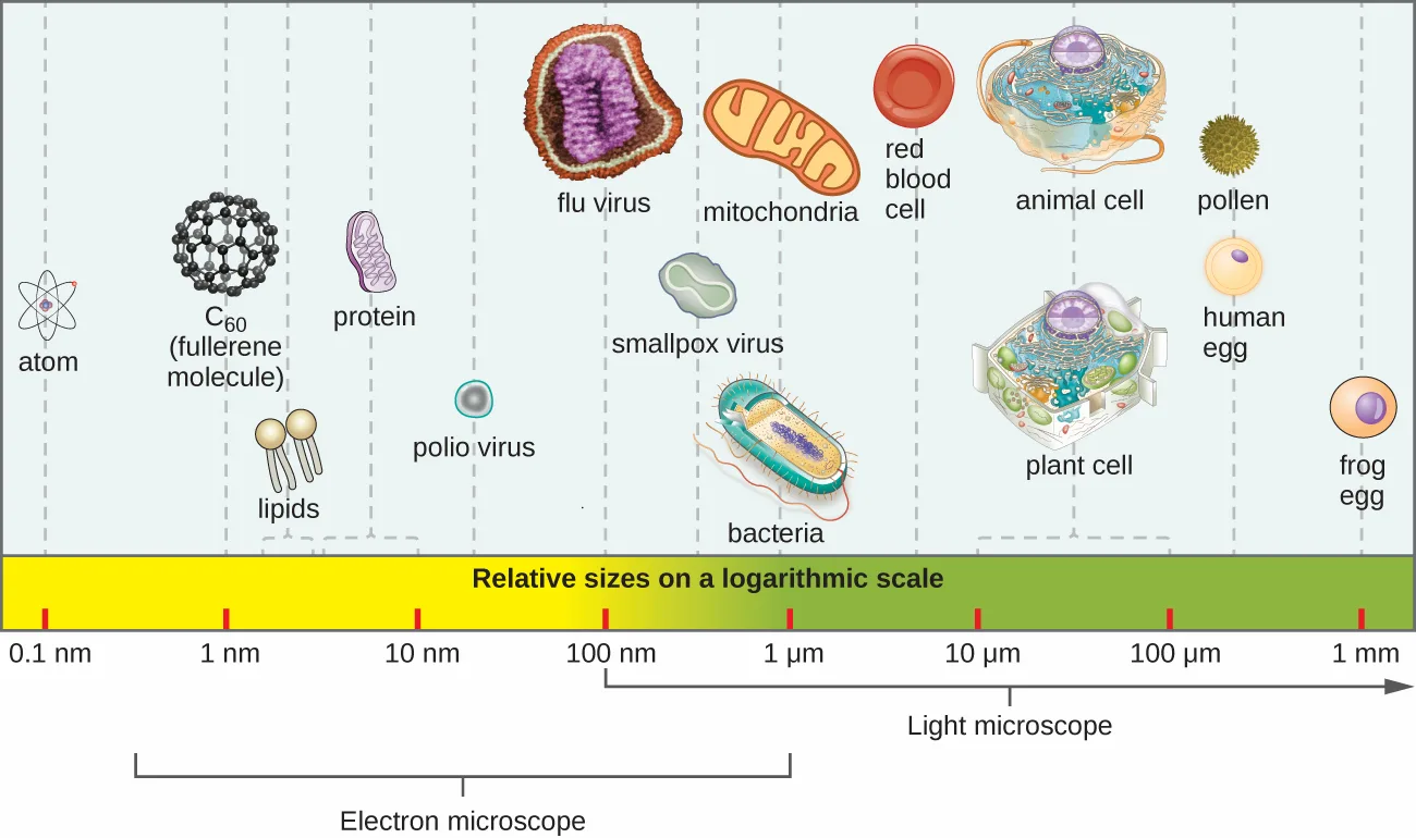
Most microbes are unicellular and small enough that they require artificial magnification to be seen. However, there are some unicellular microbes that are visible to the naked eye, and some multicellular organisms that are microscopic. An object must measure about 100 micrometers (µm) to be visible without a microscope, but most microorganisms are many times smaller than that. For some perspective, consider that a typical animal cell measures roughly 10 µm across but is still microscopic. Bacterial cells are typically about 1 µm, and viruses can be 10 times smaller than bacteria (see the figure below). See the table below for units of length used in microbiology.
Extended description
The scale is logarithmic, each labelled tick ten times the one before it, and runs from 0.1 nm to 1 mm. Reading it from left to right: an atom (about 0.1 nm); a C60 fullerene molecule (about 1 nm); lipids (2–5 nm) and a protein (5–10 nm); a polio virus (about 50 nm), a flu virus (about 100 nm), and a smallpox virus (about 500 nm), grouped together; a mitochondrion and a bacterial cell (about 1 µm); a red blood cell (just under 10 µm); a plant or animal cell (10–100 µm); a pollen grain and a human egg (about 0.1 mm); and, at the far right, a frog egg (about 1 mm). A bracket beneath the scale marks the electron-microscope range, from below 1 nm to about 1 µm; a second bracket marks the light-microscope range, from about 100 nm upward.
Units of Length Commonly Used in Microbiology
| Metric Unit | Meaning of Prefix | Metric Equivalent |
|---|---|---|
| meter (m) | — | 1 m = m |
| decimeter (dm) | 1/10 | 1 dm = 0.1 m = m |
| centimeter (cm) | 1/100 | 1 cm = 0.01 m = m |
| millimeter (mm) | 1/1000 | 1 mm = 0.001 m = m |
| micrometer (µm) | 1/1,000,000 | 1 µm = 0.000001 m = m |
| nanometer (nm) | 1/1,000,000,000 | 1 nm = 0.000000001 m = m |
Microorganisms differ from each other not only in size, but also in structure, habitat, metabolism, and many other characteristics. While we typically think of microorganisms as being unicellular, there are also many multicellular organisms that are too small to be seen without a microscope. Some microbes, such as viruses, are even acellular (not composed of cells).
Microorganisms are found in each of the three domains of life: Archaea, Bacteria, and Eukarya. Microbes within the domains Bacteria and Archaea are all prokaryotes (their cells lack a nucleus), whereas microbes in the domain Eukarya are eukaryotes (their cells have a nucleus). Some microorganisms, such as viruses, do not fall within any of the three domains of life. In this section, we will briefly introduce each of the broad groups of microbes. Later chapters will go into greater depth about the diverse species within each group.
Link to Learning
How big is a bacterium or a virus compared to other objects? Check out an interactive tool comparing the sizes of different microorganisms to get a feel for the scale involved.
Prokaryotic Microorganisms
Bacteria are found in nearly every habitat on earth, including within and on humans. Most bacteria are harmless or helpful, but some are pathogens, causing disease in humans and other animals. Bacteria are prokaryotic because their genetic material (DNA) is not housed within a true nucleus. Most bacteria have cell walls that contain peptidoglycan.
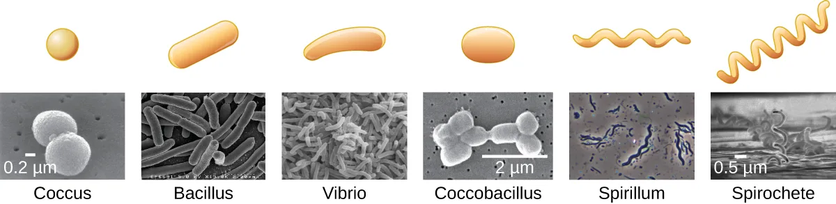
Bacteria are often described in terms of their general shape. Common shapes include spherical (coccus), rod-shaped (bacillus), or curved (spirillum, spirochete, or vibrio). The figure below shows examples of these shapes.
They have a wide range of metabolic capabilities and can grow in a variety of environments, using different combinations of nutrients. Some bacteria are photosynthetic, such as oxygenic cyanobacteria and anoxygenic green sulfur and green nonsulfur bacteria; these bacteria use energy derived from sunlight, and fix carbon dioxide for growth. Other types of bacteria are nonphotosynthetic, obtaining their energy from organic or inorganic compounds in their environment.
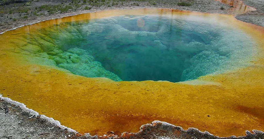
Archaea are also unicellular prokaryotic organisms. Archaea and bacteria have different evolutionary histories, as well as significant differences in genetics, metabolic pathways, and the composition of their cell walls and membranes. Unlike most bacteria, archaeal cell walls do not contain peptidoglycan, but their cell walls are often composed of a similar substance called pseudopeptidoglycan. Like bacteria, archaea are found in nearly every habitat on earth, even extreme environments that are very cold, very hot, very basic, or very acidic (see the photo below). Some archaea live in the human body, but none have been shown to be human pathogens.
Check Your Understanding
What are the two main types of prokaryotic organisms?
Prokaryotes fall into exactly two domains discussed in this section.Sort each characteristic of bacteria and archaea to the group it describes.
Bacteria
Archaea
Eukaryotic Microorganisms
The domain Eukarya contains all eukaryotes, including uni- or multicellular eukaryotes such as protists, fungi, plants, and animals. The major defining characteristic of eukaryotes is that their cells contain a nucleus.
Protists
Protists are an informal grouping of eukaryotes that are not plants, animals, or fungi. Some algae are protists and others are bacteria; all protozoa are examples of protists.
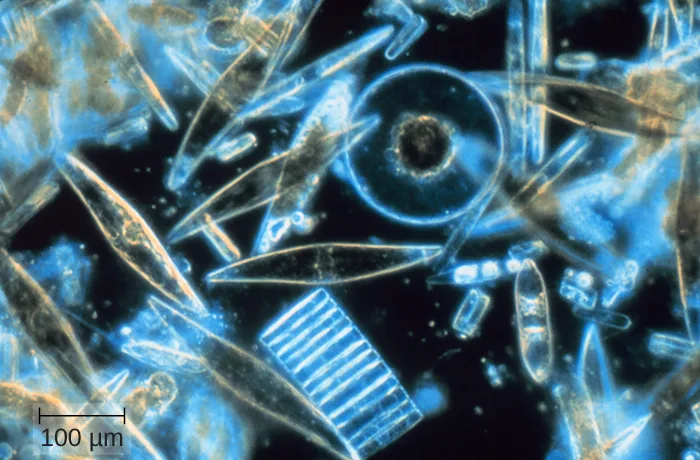
Algae (singular: alga) are mostly made up of protists that can be either unicellular or multicellular and vary widely in size, appearance, and habitat (see the micrograph below). Algal protists are surrounded by cell walls made of cellulose, a type of carbohydrate. Algae are photosynthetic organisms that extract energy from the sun and release oxygen and carbohydrates into their environment. Cyanobacteria, a type of bacteria, is also considered an algae, but these organisms are bacterial prokaryotes and therefore have a peptidoglycan-based cell wall, unlike the cellulose-based cell wall of the algal protists. Because other organisms can use the waste products of all algae for energy, algae are important parts of many ecosystems. Many consumer products contain ingredients derived from algae, such as carrageenan or alginic acid, which are found in some brands of ice cream, salad dressing, beverages, lipstick, and toothpaste. A derivative of algae also plays a prominent role in the microbiology laboratory. Agar, a gel derived from algae, can be mixed with various nutrients and used to grow microorganisms in a Petri dish. Algae are also being developed as a possible source for biofuels.
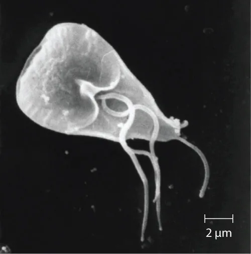
Protozoa (singular: protozoan) are protists that make up the backbone of many food webs by providing nutrients for other organisms. Protozoa are very diverse. Some protozoa move with help from hair-like structures called cilia or whip-like structures called flagella. Others extend part of their cell membrane and cytoplasm to propel themselves forward. These cytoplasmic extensions are called pseudopods (“false feet”). Some protozoa are photosynthetic; others feed on organic material. Some are free-living, whereas others are parasitic, only able to survive by extracting nutrients from a host organism. Most protozoa are harmless, but some are pathogens that can cause disease in animals or humans (see the micrograph below).
Fungi
Fungi (singular: fungus) are also eukaryotes. Some multicellular fungi, such as mushrooms, resemble plants, but they are actually quite different. Fungi are not photosynthetic, and their cell walls are usually made out of chitin rather than cellulose.
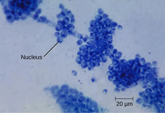
Unicellular fungi—yeasts—are included within the study of microbiology. There are more than 1000 known species. Yeasts are found in many different environments, from the deep sea to the human navel. Some yeasts have beneficial uses, such as causing bread to rise and beverages to ferment; but yeasts can also cause food to spoil. Some even cause diseases, such as vaginal yeast infections and oral thrush (see the micrograph below).
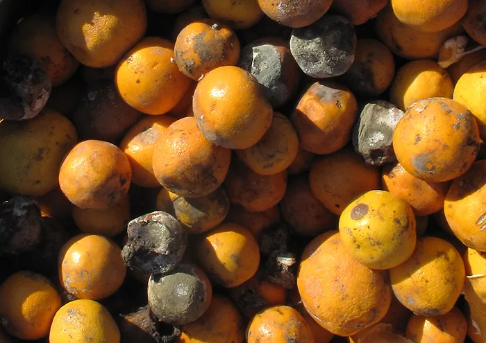
Other fungi of interest to microbiologists are multicellular organisms called molds. Molds are made up of long filaments that form visible colonies (see the photo below). Molds are found in many different environments, from soil to rotting food to dank bathroom corners. Molds play a critical role in the decomposition of dead plants and animals. Some molds can cause allergies, and others produce disease-causing metabolites called mycotoxins. Molds have been used to make pharmaceuticals, including penicillin, which is one of the most commonly prescribed antibiotics, and cyclosporine, used to prevent organ rejection following a transplant.
Check Your Understanding
Sort each organism type to the eukaryotic group it belongs to.
Protists
Fungi
Name some of the defining characteristics of protists and fungi.
Show model answer
Did your answer mention:
Helminths
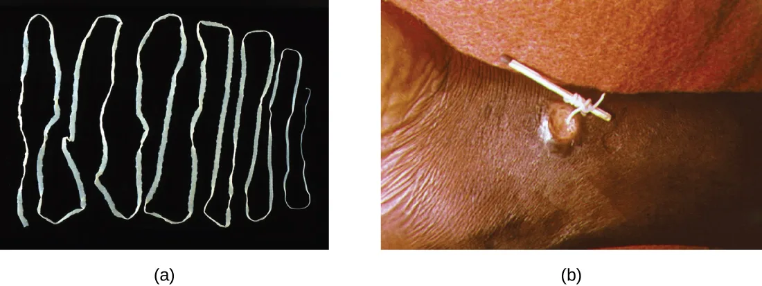
Multicellular parasitic worms called helminths are not technically microorganisms, as most are large enough to see without a microscope. However, these worms fall within the field of microbiology because diseases caused by helminths involve microscopic eggs and larvae. One example of a helminth is the guinea worm, or Dracunculus medinensis, which causes dizziness, vomiting, diarrhea, and painful ulcers on the legs and feet when the worm works its way out of the skin (see the photos below). Infection typically occurs after a person drinks water containing water fleas infected by guinea-worm larvae. In the mid-1980s, there were an estimated 3.5 million cases of guinea-worm disease, but the disease has been largely eradicated. In 2014, there were only 126 cases reported, thanks to the coordinated efforts of the World Health Organization (WHO) and other groups committed to improvements in drinking water sanitation. (C. Greenaway, “Dracunculiasis (Guinea Worm Disease),” Canadian Medical Association Journal 170 no. 4 (2004): 495–500; World Health Organization, “Dracunculiasis (Guinea-Worm Disease),” WHO, 2015, http://www.who.int/mediacentre/factsheets/fs359/en/, accessed October 2, 2015.)
Viruses
Viruses are acellular microorganisms, which means they are not composed of cells. Essentially, a virus consists of proteins and genetic material—either DNA or RNA, but never both—that are inert outside of a host organism. However, by incorporating themselves into a host cell, viruses are able to co-opt the host’s cellular mechanisms to multiply and infect other hosts.
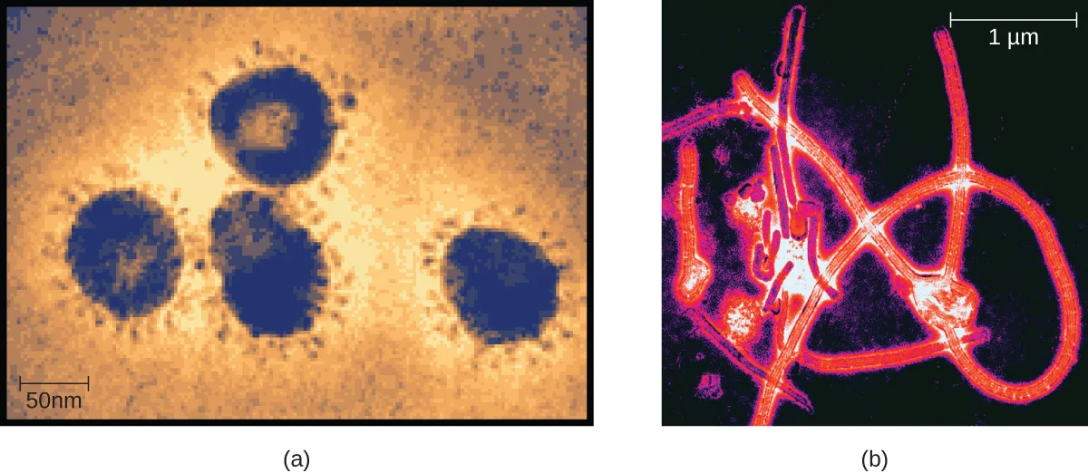
Viruses can infect all types of cells, from human cells to the cells of other microorganisms. In humans, viruses are responsible for numerous diseases, from the common cold to deadly Ebola (see the micrographs below). However, many viruses do not cause disease.
Check Your Understanding
Are helminths microorganisms? Explain why or why not.
Think about what makes something ‘micro’ and whether that applies to the adult worm versus its eggs and larvae.How are viruses different from other microorganisms?
Show model answer
Did your answer mention:
Microbiology as a Field of Study
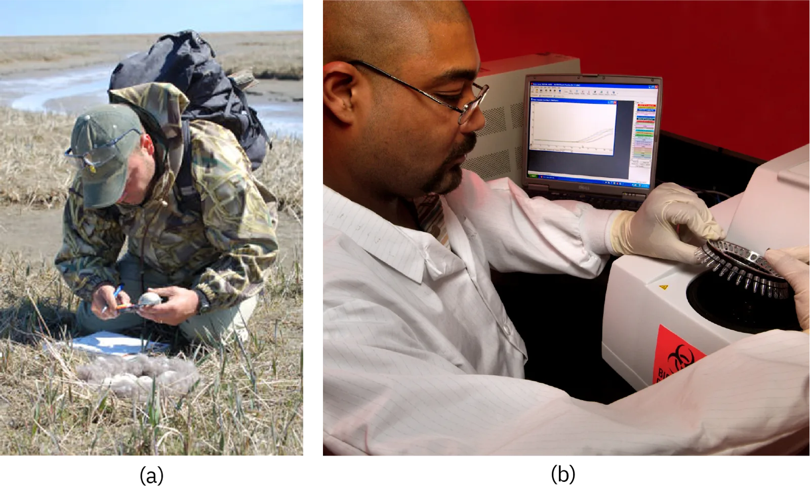
Microbiology is a broad term that encompasses the study of all different types of microorganisms. But in practice, microbiologists tend to specialize in one of several subfields. For example, bacteriology is the study of bacteria; mycology is the study of fungi; protozoology is the study of protozoa; parasitology is the study of helminths and other parasites; and virology is the study of viruses (see the photos below). Immunology, the study of the immune system, is often included in the study of microbiology because host–pathogen interactions are central to our understanding of infectious disease processes. Microbiologists can also specialize in certain areas of microbiology, such as clinical microbiology, environmental microbiology, applied microbiology, or food microbiology.
In this textbook, we are primarily concerned with clinical applications of microbiology, but since the various subfields of microbiology are highly interrelated, we will often discuss applications that are not strictly clinical.
Eye on Ethics. Bioethics in Microbiology
In the 1940s, the U.S. government was looking for a solution to a medical problem: the prevalence of sexually transmitted diseases (STDs) among soldiers. Several now-infamous government-funded studies used human subjects to research common STDs and treatments. In one such study, American researchers intentionally exposed more than 1300 human subjects in Guatemala to syphilis, gonorrhea, and chancroid to determine the ability of penicillin and other antibiotics to combat these diseases. Subjects of the study included Guatemalan soldiers, prisoners, prostitutes, and psychiatric patients—none of whom were informed that they were taking part in the study. Researchers exposed subjects to STDs by various methods, from facilitating intercourse with infected prostitutes to inoculating subjects with the bacteria known to cause the diseases. This latter method involved making a small wound on the subject’s genitals or elsewhere on the body, and then putting bacteria directly into the wound. (Kara Rogers, “Guatemala Syphilis Experiment: American Medical Research Project,” Encyclopaedia Britannica, http://www.britannica.com/event/Guatemala-syphilis-experiment, accessed June 24, 2015.) In 2011, a U.S. government commission tasked with investigating the experiment revealed that only some of the subjects were treated with penicillin, and 83 subjects died by 1953, likely as a result of the study. (Susan Donaldson James, “Syphilis Experiments Shock, But So Do Third-World Drug Trials,” ABC World News, August 30, 2011, http://abcnews.go.com/Health/guatemala-syphilis-experiments-shock-us-drug-trials-exploit/story?id=14414902, accessed June 24, 2015.)
Unfortunately, this is one of many horrific examples of microbiology experiments that have violated basic ethical standards. Even if this study had led to a life-saving medical breakthrough (it did not), few would argue that its methods were ethically sound or morally justifiable. But not every case is so clear cut. Professionals working in clinical settings are frequently confronted with ethical dilemmas, such as working with patients who decline a vaccine or life-saving blood transfusion. These are just two examples of life-and-death decisions that may intersect with the religious and philosophical beliefs of both the patient and the health-care professional.
No matter how noble the goal, microbiology studies and clinical practice must be guided by a certain set of ethical principles. Studies must be done with integrity. Patients and research subjects provide informed consent (not only agreeing to be treated or studied but demonstrating an understanding of the purpose of the study and any risks involved). Patients’ rights must be respected. Procedures must be approved by an institutional review board. When working with patients, accurate record-keeping, honest communication, and confidentiality are paramount. Animals used for research must be treated humanely, and all protocols must be approved by an institutional animal care and use committee. These are just a few of the ethical principles explored in the Eye on Ethics boxes throughout this book.
Clinical Focus. Resolution
Cora’s CSF samples show no signs of inflammation or infection, as would be expected with a viral infection. However, there is a high concentration of a particular protein, 14-3-3 protein, in her CSF. An electroencephalogram (EEG) of her brain function is also abnormal. The EEG resembles that of a patient with a neurodegenerative disease like Alzheimer’s or Huntington’s, but Cora’s rapid cognitive decline is not consistent with either of these. Instead, her doctor concludes that Cora has Creutzfeldt-Jakob disease (CJD), a type of transmissible spongiform encephalopathy (TSE).
CJD is an extremely rare disease, with only about 300 cases in the United States each year. It is not caused by a bacterium, fungus, or virus, but rather by prions—which do not fit neatly into any particular category of microbe. Like viruses, prions are not found on the tree of life because they are acellular. Prions are extremely small, about one-tenth the size of a typical virus. They contain no genetic material and are composed solely of a type of abnormal protein.
CJD can have several different causes. It can be acquired through exposure to the brain or nervous-system tissue of an infected person or animal. Consuming meat from an infected animal is one way such exposure can occur. There have also been rare cases of exposure to CJD through contact with contaminated surgical equipment (Greg Botelho, “Case of Creutzfeldt-Jakob Disease Confirmed in New Hampshire,” CNN, 2013, http://www.cnn.com/2013/09/20/health/creutzfeldt-jakob-brain-disease/) and from cornea and growth-hormone donors who unknowingly had CJD. (P. Rudge et al., “Iatrogenic CJD Due to Pituitary-Derived Growth Hormone With Genetically Determined Incubation Times of Up to 40 Years,” Brain 138 no. 11 (2015): 3386–3399; J.G. Heckmann et al., “Transmission of Creutzfeldt-Jakob Disease via a Corneal Transplant,” Journal of Neurology, Neurosurgery & Psychiatry 63 no. 3 (1997): 388–390.) In rare cases, the disease results from a specific genetic mutation that can sometimes be hereditary. However, in approximately 85% of patients with CJD, the cause of the disease is spontaneous (or sporadic) and has no identifiable cause. (National Institute of Neurological Disorders and Stroke, “Creutzfeldt-Jakob Disease Fact Sheet,” NIH, 2015, http://www.ninds.nih.gov/disorders/cjd/detail_cjd.htm#288133058.) Based on her symptoms and their rapid progression, Cora is diagnosed with sporadic CJD.
Unfortunately for Cora, CJD is a fatal disease for which there is no approved treatment. Approximately 90% of patients die within 1 year of diagnosis. (National Institute of Neurological Disorders and Stroke, “Creutzfeldt-Jakob Disease Fact Sheet,” NIH, 2015, http://www.ninds.nih.gov/disorders/cjd/detail_cjd.htm#288133058, accessed June 22, 2015.) Her doctors focus on limiting her pain and cognitive symptoms as her disease progresses. Eight months later, Cora dies. Her CJD diagnosis is confirmed with a brain autopsy.
The previous part of this case is in A Systematic Approach; the case began in What Our Ancestors Knew.
Summary
- Microorganisms are very diverse and are found in all three domains of life: Archaea, Bacteria, and Eukarya.
- Archaea and bacteria are classified as prokaryotes because they lack a cellular nucleus. Archaea differ from bacteria in evolutionary history, genetics, metabolic pathways, and cell wall and membrane composition.
- Archaea inhabit nearly every environment on earth, but no archaea have been identified as human pathogens.
- Eukaryotes studied in microbiology include algae, protozoa, fungi, and helminths.
- Algae are plant-like organisms that can be either unicellular or multicellular, and derive energy via photosynthesis.
- Protozoa are unicellular organisms with complex cell structures; most are motile.
- Microscopic fungi include molds and yeasts.
- Helminths are multicellular parasitic worms. They are included in the field of microbiology because their eggs and larvae are often microscopic.
- Viruses are acellular microorganisms that require a host to reproduce.
- The field of microbiology is extremely broad. Microbiologists typically specialize in one of many subfields, but all health professionals need a solid foundation in clinical microbiology.
Key terms
- acellular — not made of cells.
- bacteria — (singular: bacterium) any of various unicellular prokaryotic microorganisms typically (but not always) having cell walls that contain peptidoglycan.
- pathogen — a disease-causing microorganism.
- archaea — any of various unicellular prokaryotic microorganisms, typically having cell walls containing pseudopeptidoglycan.
- protists — informal name for diverse group of eukaryotic organisms, including unicellular, colonial, and multicellular types that lack specialized tissues.
- algae — (singular: alga) any of various unicellular and multicellular photosynthetic eukaryotic organisms; distinguished from plants by their lack of vascular tissues and organs.
- protozoan (plural: protozoa) — a unicellular eukaryotic organism, usually motile.
- fungi — (singular: fungus) any of various unicellular or multicellular eukaryotic organisms, typically having cell walls made out of chitin and lacking photosynthetic pigments, vascular tissues, and organs.
- mold — a multicellular fungus, typically made up of long filaments.
- helminth — a multicellular parasitic worm.
- virus — an acellular microorganism, consisting of proteins and genetic material (DNA or RNA), that can replicate itself by infecting a host cell.
- microbiology — the study of microorganisms.
- bacteriology — the study of bacteria.
- mycology — the study of fungi.
- protozoology — the study of protozoa.
- parasitology — the study of parasites.
- virology — the study of viruses.
- immunology — the study of the immune system.
Practice
List the various types of microorganisms and describe their defining characteristics
Which of the following types of microorganisms is photosynthetic?
Photosynthetic organisms use sunlight for energy; ask which option is a plant-like protist rather than a fungus, an acellular agent, or a worm.Which of the following is a type of fungal microorganism?
Fungi typically lack photosynthetic pigments and have chitin cell walls; ask which option is a unicellular fungus rather than a bacterium, protist, or alga.A ________ is a disease-causing microorganism.
This is the general term for any microorganism that causes disease, not a specific type of microbe.Name three structures that various protozoa use for locomotion.
Show model answer
Did your answer mention:
Give examples of different types of cellular and viral microorganisms and infectious agents
Which of the following is a prokaryotic microorganism?
Two of the three domains of life hold only prokaryotes; ask which option belongs to one of them rather than being a multicellular worm, a protist, or a fungus.Which of the following is acellular?
Acellular means not made of cells; three of the options are types of cellular organisms.Multicellular parasitic worms studied by microbiologists are called ___________.
This group includes parasitic worms whose eggs and larvae are often too small to see without a microscope.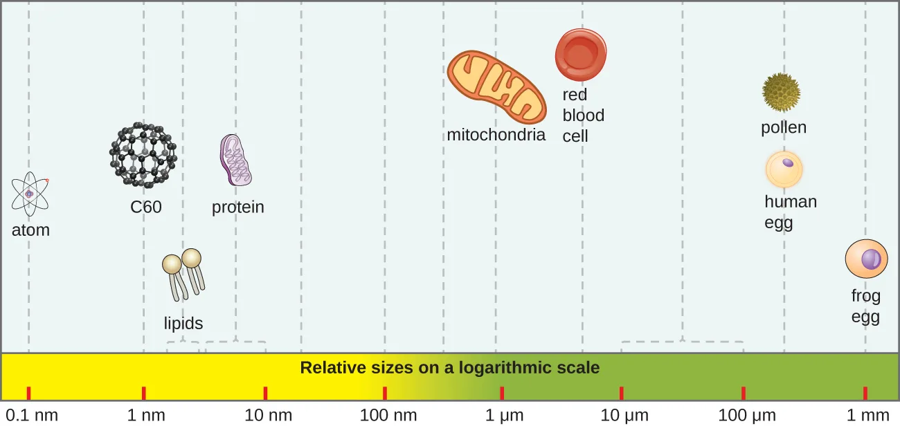
Sort each of these to the size range it falls into.
About 100 nm or smaller
About 1 µm
10–100 µm
Describe the actual and relative sizes of a virus, a bacterium, and a plant or animal cell.
Show model answer
Did your answer mention:
Sort each behavior to whether it describes a virus outside a host cell or inside one.
Outside a host cell
Inside a host cell
Describe the similarities and differences between archaea and bacteria
The cells of prokaryotic organisms lack a _______.
This is the membrane-bound structure that houses a eukaryotic cell’s genetic material but is absent from a prokaryotic cell.Archaea differ from bacteria in ________, genetics, metabolic pathways, and cell wall and membrane composition.
Think about each group’s distinct line of descent over time, separate from a difference in cell structure or chemistry.Archaea are found in nearly every habitat on Earth, including environments that are very cold, very hot, very basic, or very ________.
Think of the opposite of basic on the pH scale.Provide an overview of the field of microbiology
Which of the following is not a subfield of microbiology?
Three of these subfields study microorganisms specifically; one instead studies plants.The study of viruses is ___________.
This is the subfield of microbiology devoted specifically to viruses.Microbiologists typically specialize in one of many subfields, but all health professionals need a solid foundation in ________ microbiology.
This is the subfield most directly concerned with diagnosing and treating patients.This section is adapted from Microbiology, Section 1.3: Types of Microorganisms by Nina Parker, Mark Schneegurt, Anh-Hue Thi Tu, Philip Lister, Brian M. Forster, and OpenStax, © OpenStax, licensed under CC BY-NC-SA 4.0. Access the original for free at openstax.org. Changes: all 11 figures re-encoded as WebP; kind set explicitly on every figure after inspection, overriding the media manifest’s file-type guess on four of them (the sizes chart and the Art Connection chart are drawn diagrams despite being JPEGs; the shared bacterial-shapes reference figure is treated as a diagram; the field-work photos are photos despite being the chapter’s one PNG); the sizes-chart alt rewritten into a left-to-right walkthrough (correcting an apparent source typo, “a from egg,” to the pictured frog egg) with an added extended description, the field-work alt rewritten to describe what each photo shows (also correcting an apparent source typo, “centerfuge,” to centrifuge), the two-panel worm and virus alts tightened from “Figure a/Figure b” phrasing to parenthetical (a)/(b) form, and a caption written for the Art Connection chart since the source prints none; the Link to Learning, Eye on Ethics, and Clinical Focus notes rendered as callouts, with the Eye on Ethics and Clinical Focus footnote citations kept as parentheticals after the sentences they support; the source’s “Go back to the previous Clinical Focus box” link replaced with a plain sentence to this chapter’s preceding section; the Units of Length table transcribed as a Markdown table with its metric-equivalent exponents set as math; two of the six Check Your Understanding questions remain body self-checks with author-written rubrics from the same subsection’s text (“Name some of the defining characteristics of protists and fungi,” kept in prose because it would re-ask its sibling’s sort-into-bins bins, and “How are viruses different from other microorganisms,” kept in prose because the Practice block’s source Multiple Choice already grades the acellular fact); the other four are graded from the module’s own sentences, a table, or the module’s own paired terms rather than answered in prose, since the source prints no key for them; all five Multiple Choice and all four Fill in the Blank questions adapted into the closing Practice block; two of three Short Answer questions (“Name three structures that various protozoa use for locomotion” and “Describe the actual and relative sizes of a virus, a bacterium, and a plant or animal cell”) remain self-checks whose model answers and rubrics are author-written from this section’s own text, since the source prints no answer key for them and the module names no further locomotion structures or size-scale entries to build honest multiple-choice distractors from; one Short Answer question (“Describe the differences between bacteria and archaea”) omitted from Practice because it duplicates the body’s Check Your Understanding question already answered in place; both Critical Thinking questions (one paired with the Art Connection figure) are now graded from the module’s own size comparisons and its own contrast between a virus outside and inside a host cell, rather than answered in prose; three further Practice items author-written to round out a thin objective group — two clozes built from single sentences of the module’s own Summary and one cloze from a body sentence of the Archaea paragraph, none introducing a new claim; key terms compiled from the module’s 18 distinct defined terms and the book’s Glossary appendix, including the three (microbiology, parasitology, protozoology) recovered from a glossary formatting defect already on record (erratum 320) — none needed a sentence-derived substitute — and one further apparent glossary typo corrected on this page (“cell wells” to “cell walls” in the bacteria definition, reported for the errata file); a footnote citation’s misspelled “Encylopaedia Britannica” corrected to “Encyclopaedia Britannica” (also reported); the archaea Key terms bullet uses the appendix’s organism-sense entry rather than its domain-sense “Archaea” entry, matching how this module uses the word; 4 of the source’s body Check Your Understanding questions and both of its unkeyed Critical Thinking questions are graded from the module’s own sentences, tables, or figures rather than answered in prose; the source prints no key for them: “what are the two main types of prokaryotic organisms” is now a multiplechoice keyed by the module’s own two-domain framing, with distractors the module’s other paired eukaryotic groups; “name some of the defining characteristics of bacteria and archaea” is now a sort-into-bins item built from the module’s own cell-wall-composition and human-pathogen sentences; “name two types of protists and two types of fungi” is now a sort-into-bins item built from the module’s own named examples; “are helminths microorganisms” is now a multiplechoice keyed by the module’s own size sentence, with distractors the module’s descriptions of protists, viruses, and bacteria; “where would a virus, bacterium, animal cell, and a prion belong on this chart” is now a sort-into-bins item built from the module’s own size figures, keeping the mediafigure beside it; “contrast the behavior of a virus outside versus inside a cell” is now a sort-into-bins item built from the module’s own two-state description.