How We See the Invisible World
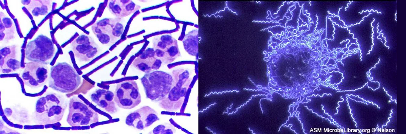
When we look at a rainbow, its colors span the full spectrum of light that the human eye can detect and differentiate. Each hue represents a different frequency of visible light, processed by our eyes and brains and rendered as red, orange, yellow, green, or one of the many other familiar colors that have always been a part of the human experience. But only recently have humans developed an understanding of the properties of light that allow us to see images in color.
Over the past several centuries, we have learned to manipulate light to peer into previously invisible worlds–those too small or too far away to be seen by the naked eye. Through a microscope, we can examine microbial cells, using various techniques to manipulate color, size, and contrast in ways that help us identify species and diagnose disease.
The two micrographs above illustrate how we can apply the properties of light to visualize and magnify images; but these stunning micrographs are just two examples of the numerous types of images we are now able to produce with different microscopic technologies. This chapter explores how various types of microscopes manipulate light in order to provide a window into the world of microorganisms. By understanding how various kinds of microscopes work, we can produce highly detailed images of microbes that can be useful for both research and clinical applications.
Sections
- The Properties of Light — the electromagnetic spectrum, wavelength, frequency, and energy; how light interacts with matter and is refracted by lenses; magnification, resolution, and numerical aperture; and fluorescence.
- Peering Into the Invisible World — the Janssens, Antonie van Leeuwenhoek, Robert Hooke, and the difference between simple and compound microscopes.
- Instruments of Microscopy — the parts of a brightfield microscope and total magnification; darkfield, phase-contrast, differential interference contrast, fluorescence, confocal, and two-photon microscopes; transmission and scanning electron microscopes; and scanning probe microscopes.
- Staining Microscopic Specimens — preparing and fixing specimens, simple and differential stains, and the Gram, acid-fast, capsule, endospore, and flagella stains, with the Clinical Focus case resolved.
This chapter is adapted from Microbiology, Chapter 2: How We See the Invisible World by Nina Parker, Mark Schneegurt, Anh-Hue Thi Tu, Philip Lister, Brian M. Forster, and OpenStax, © OpenStax, licensed under CC BY-NC-SA 4.0. Access the original for free at openstax.org. Each section page records its own changes from the source. Changes: the chapter-opening micrograph pair is the source’s, re-encoded for the web with its credit kept in the caption and its alt text rewritten to describe what the two micrographs show; the source’s reference to the figure by number is replaced with a describing phrase.