The Properties of Light
By the end of this section, you will be able to:
- Identify and define the characteristics of electromagnetic radiation (EMR) used in microscopy
- Explain how lenses are used in microscopy to manipulate visible and ultraviolet (UV) light
Clinical Focus. Part 1
Cindy, a 17-year-old counselor at a summer sports camp, scraped her knee playing basketball 2 weeks ago. At the time, she thought it was only a minor abrasion that would heal, like many others before it. Instead, the wound began to look like an insect bite and has continued to become increasingly painful and swollen.
The camp nurse examines the lesion and observes a large amount of pus oozing from the surface. Concerned that Cindy may have developed a potentially aggressive infection, she swabs the wound to collect a sample from the infection site. Then she cleans out the pus and dresses the wound, instructing Cindy to keep the area clean and to come back the next day. When Cindy leaves, the nurse sends the sample to the closest medical lab to be analyzed under a microscope.
- What are some things we can learn about these bacteria by looking at them under a microscope?
The case continues in Instruments of Microscopy.
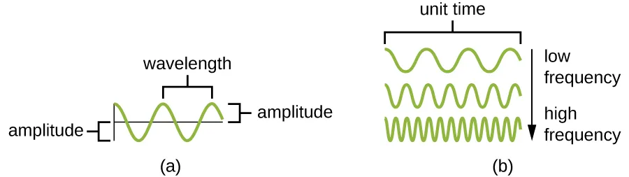
Visible light consists of electromagnetic waves that behave like other waves. Hence, many of the properties of light that are relevant to microscopy can be understood in terms of light’s behavior as a wave. An important property of light waves is the wavelength, or the distance between one peak of a wave and the next peak. The height of each peak (or depth of each trough) is called the amplitude. In contrast, the frequency of the wave is the rate of vibration of the wave, or the number of wavelengths within a specified time period (illustrated below).
Interactions of Light
Light waves interact with materials by being reflected, absorbed, or transmitted. Reflection occurs when a wave bounces off of a material. For example, a red piece of cloth may reflect red light to our eyes while absorbing other colors of light. Absorbance occurs when a material captures the energy of a light wave. In the case of glow-in-the-dark plastics, the energy from light can be absorbed and then later re-emitted as another form of phosphorescence. Transmission occurs when a wave travels through a material, like light through glass (the process of transmission is called transmittance). When a material allows a large proportion of light to be transmitted, it may do so because it is thinner, or more transparent (having more transparency and less opacity). The figure below illustrates the difference between transparency and opacity.
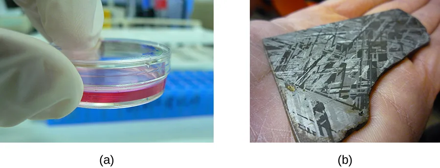
Light waves can also interact with each other by interference, creating complex patterns of motion. Dropping two pebbles into a puddle causes the waves on the puddle’s surface to interact, creating complex interference patterns. Light waves can interact in the same way.
In addition to interfering with each other, light waves can also interact with small objects or openings by bending or scattering. This is called diffraction. Diffraction is larger when the object is smaller relative to the wavelength of the light (the distance between two consecutive peaks of a light wave). Often, when waves diffract in different directions around an obstacle or opening, they will interfere with each other.
Check Your Understanding
If a light wave has a long wavelength, is it likely to have a low or high frequency?
Think about how many wave peaks pass a point per unit time when the peaks are spaced farther apart.If an object is transparent, does it reflect, absorb, or transmit light?
Transparency and opacity describe how much light passes through a material versus how much is blocked.Lenses and Refraction
In the context of microscopy, refraction is perhaps the most important behavior exhibited by light waves. Refraction occurs when light waves change direction as they enter a new medium (illustrated below). Different transparent materials transmit light at different speeds; thus, light can change speed when passing from one material to another. This change in speed usually also causes a change in direction (refraction), with the degree of change dependent on the angle of the incoming light.
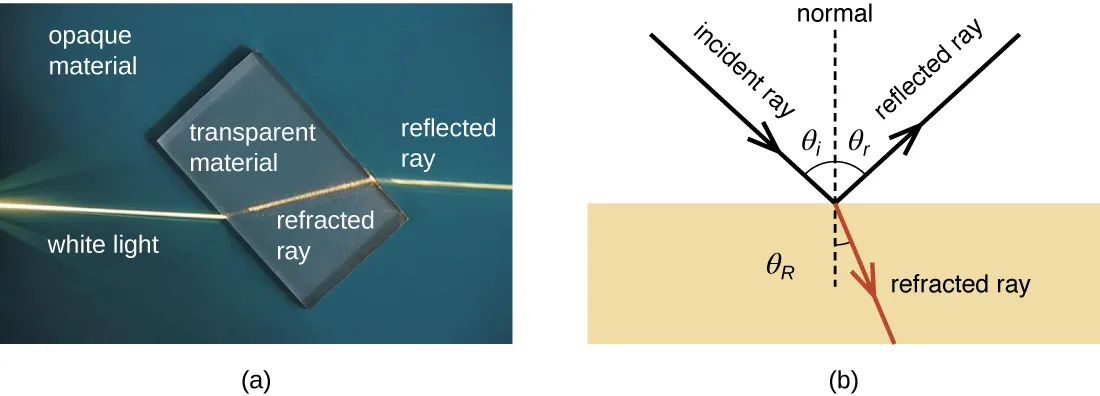
The extent to which a material slows transmission speed relative to empty space is called the refractive index of that material. Large differences between the refractive indices of two materials will result in a large amount of refraction when light passes from one material to the other. For example, light moves much more slowly through water than through air, so light entering water from air can change direction greatly. We say that the water has a higher refractive index than air (as shown in the photo below).

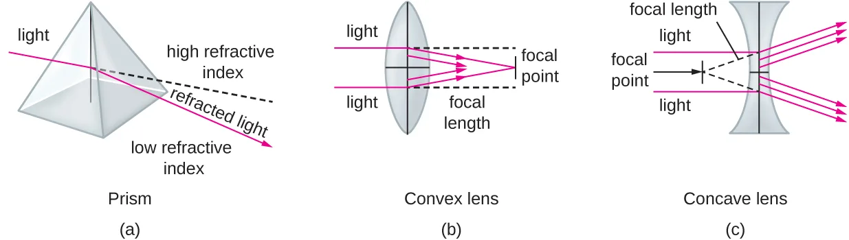
When light crosses a boundary into a material with a higher refractive index, its direction turns to be closer to perpendicular to the boundary (i.e., more toward a normal to that boundary; see the photo above). This is the principle behind lenses. We can think of a lens as an object with a curved boundary (or a collection of prisms) that collects all of the light that strikes it and refracts it so that it all meets at a single point called the image point (focus). A convex lens can be used to magnify because it can focus at closer range than the human eye, producing a larger image. Concave lenses and mirrors can also be used in microscopes to redirect the light path. The diagram below shows the focal point (the image point when light entering the lens is parallel) and the focal length (the distance to the focal point) for convex and concave lenses.
The human eye contains a lens that enables us to see images. This lens focuses the light reflecting off of objects in front of the eye onto the surface of the retina, which is like a screen in the back of the eye. Artificial lenses placed in front of the eye (contact lenses, glasses, or microscopic lenses) focus light before it is focused (again) by the lens of the eye, manipulating the image that ends up on the retina (e.g., by making it appear larger).
Images are commonly manipulated by controlling the distances between the object, the lens, and the screen, as well as the curvature of the lens. For example, for a given amount of curvature, when an object is closer to the lens, the focal points are farther from the lens. As a result, it is often necessary to manipulate these distances to create a focused image on a screen. Similarly, more curvature creates image points closer to the lens and a larger image when the image is in focus. This property is often described in terms of the focal distance, or distance to the focal point.
Check Your Understanding
Explain how a lens focuses light at the image point.
Think about what a lens does to light that strikes its curved boundary from many different points.Name some factors that affect the focal length of a lens.
This is about where images come into focus, not about how sharp or magnified they appear.Electromagnetic Spectrum and Color
Visible light is just one form of electromagnetic radiation (EMR), a type of energy that is all around us. Other forms of EMR include microwaves, X-rays, and radio waves, among others. The different types of EMR fall on the electromagnetic spectrum, which is defined in terms of wavelength and frequency. The spectrum of visible light occupies a relatively small range of frequencies between infrared and ultraviolet light (illustrated below).
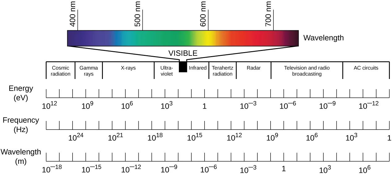
Extended description
Reading left to right, the labeled regions are cosmic radiation, gamma rays, X-rays, ultraviolet, a narrow visible band, infrared, terahertz radiation, radar, television and radio broadcasting, and AC circuits. Above the visible band, a colored strip is enlarged to show violet at about 400 nanometers shading through blue, green, yellow, and orange to red at about 700 nanometers. Below the regions, three number lines run in the same left-to-right order: energy in electron volts, from about 10¹² on the left down to about 10⁻¹² on the right; frequency in hertz, from more than 10²⁴ down to about 1; and wavelength in meters, from about 10⁻¹⁸ up to about 10⁶.
Whereas wavelength represents the distance between adjacent peaks of a light wave, frequency, in a simplified definition, represents the rate of oscillation. Waves with higher frequencies have shorter wavelengths and, therefore, have more oscillations per unit time than lower-frequency waves. Higher-frequency waves also contain more energy than lower-frequency waves. This energy is delivered as elementary particles called photons. Higher-frequency waves deliver more energetic photons than lower-frequency waves.
Photons with different energies interact differently with the retina. In the spectrum of visible light, each color corresponds to a particular frequency and wavelength (as shown in the figure above). The lowest frequency of visible light appears as the color red, whereas the highest appears as the color violet. When the retina receives visible light of many different frequencies, we perceive this as white light. However, white light can be separated into its component colors using refraction. If we pass white light through a prism, different colors will be refracted in different directions, creating a rainbow-like spectrum on a screen behind the prism. This separation of colors is called dispersion, and it occurs because, for a given material, the refractive index is different for different frequencies of light.
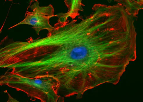
Certain materials can refract nonvisible forms of EMR and, in effect, transform them into visible light. Certain fluorescent dyes, for instance, absorb ultraviolet or blue light and then use the energy to emit photons of a different color, giving off light rather than simply vibrating. This occurs because the energy absorption causes electrons to jump to higher energy states, after which they then almost immediately fall back down to their ground states, emitting specific amounts of energy as photons. Not all of the energy is emitted in a given photon, so the emitted photons will be of lower energy and, thus, of lower frequency than the absorbed ones. Thus, a dye such as Texas red may be excited by orange light, but emit red light; or a dye such as fluorescein isothiocyanate (FITC) may absorb blue light and emit green light (illustrated below). (Source note: the source says Texas red is excited by blue light and that FITC absorbs invisible ultraviolet light. The manufacturer’s spectral data put Texas red’s excitation peak near 589 nm, in the orange, and FITC’s near 495 nm, in the visible blue; this page states the colors those measurements support. The paragraph’s point, that emitted light carries less energy than absorbed light, is unchanged.) In some materials, the photons may be emitted following a delay after absorption; in this case, the process is called phosphorescence. Glow-in-the-dark plastic works by using phosphorescent material.
Check Your Understanding
Which has a higher frequency: red light or green light?
Think about which end of the visible spectrum — red or violet — has the lowest frequency.Explain why dispersion occurs when white light passes through a prism.
Think about whether every frequency of light bends by the same amount when it enters a new medium.Why do fluorescent dyes emit a different color of light than they absorb?
Show model answer
Did your answer mention:
Magnification, Resolution, and Contrast
Microscopes magnify images and use the properties of light to create useful images of small objects. Magnification is defined as the ability of a lens to enlarge the image of an object when compared to the real object. For example, a magnification of 10× means that the image appears 10 times the size of the object as viewed with the naked eye.
Greater magnification typically improves our ability to see details of small objects, but magnification alone is not sufficient to make the most useful images. It is often useful to enhance the resolution of objects: the ability to tell that two separate points or objects are separate. A low-resolution image appears fuzzy, whereas a high-resolution image appears sharp. Two factors affect resolution. The first is wavelength. Shorter wavelengths are able to resolve smaller objects; thus, an electron microscope has a much higher resolution than a light microscope, since it uses an electron beam with a very short wavelength, as opposed to the long-wavelength visible light used by a light microscope. The second factor that affects resolution is numerical aperture, which is a measure of a lens’s ability to gather light. The higher the numerical aperture, the better the resolution.
Link to Learning
Read this article about numerical aperture to learn more about factors that can increase or decrease the numerical aperture of a lens.
Even when a microscope has high resolution, it can be difficult to distinguish small structures in many specimens because microorganisms are relatively transparent. It is often necessary to increase contrast to detect different structures in a specimen. Various types of microscopes use different features of light or electrons to increase contrast—visible differences between the parts of a specimen (see Instruments of Microscopy). Additionally, dyes that bind to some structures but not others can be used to improve the contrast between images of relatively transparent objects (see Staining Microscopic Specimens).
Check Your Understanding
Sort each phrase under the imaging property it describes.
Magnification
Resolution
Contrast
Name two factors that affect resolution.
One factor is a property of the light or electron beam itself; the other is a property of the lens.Summary
- Light waves interacting with materials may be reflected, absorbed, or transmitted, depending on the properties of the material.
- Light waves can interact with each other (interference) or be distorted by interactions with small objects or openings (diffraction).
- Refraction occurs when light waves change speed and direction as they pass from one medium to another. Differences in the refraction indices of two materials determine the magnitude of directional changes when light passes from one to the other.
- A lens is a medium with a curved surface that refracts and focuses light to produce an image.
- Visible light is part of the electromagnetic spectrum; light waves of different frequencies and wavelengths are distinguished as colors by the human eye.
- A prism can separate the colors of white light (dispersion) because different frequencies of light have different refractive indices for a given material.
- Fluorescent dyes and phosphorescent materials can effectively transform nonvisible electromagnetic radiation into visible light.
- The power of a microscope can be described in terms of its magnification and resolution.
- Resolution can be increased by shortening wavelength, increasing the numerical aperture of the lens, or using stains that enhance contrast.
Key terms
- wavelength — the distance between one peak of a wave and the next peak.
- amplitude — the height of a wave.
- frequency — the rate of vibration for a light wave or other electromagnetic wave.
- reflection — when light bounces back from a surface.
- absorbance — when a molecule captures energy from a photon and vibrates or stretches, using the energy.
- transmittance — the amount of light that passes through a medium.
- transparency — the property of allowing light to pass through.
- opacity — the property of absorbing or blocking light.
- interference — distortion of a light wave due to interaction with another wave.
- diffraction — the changing of direction (bending or spreading) that occurs when a light wave interacts with an opening or barrier.
- refraction — bending of light waves, which occurs when a light wave passes from one medium to another.
- refractive index — a measure of the magnitude of slowing of light waves by a particular medium.
- image point (focus) — a property of the lens and the distance of the object to the lens; the point at which an image is in focus (the image point is often called the focus).
- focal point — a property of the lens; the image point when light entering the lens is parallel (i.e., the object is an infinite distance from the lens).
- focal length — the distance from the lens to the image point when the object is at a definite distance from the lens (this is also the distance to the focal point).
- dispersion — the separation of light of different frequencies due to different degrees of refraction.
- fluorescent — the ability of certain materials to absorb energy and then immediately release that energy in the form of light.
- phosphorescence — the ability of certain materials to absorb energy and then release that energy as light after a delay.
- magnification — the power of a microscope (or lens) to produce an image that appears larger than the actual specimen, expressed as a factor of the actual size.
- resolution — the ability to distinguish between two points in an image.
- numerical aperture — a measure of a lens’s ability to gather light.
- contrast — visible differences between parts of a microscopic specimen.
Practice
Identify and define the characteristics of electromagnetic radiation (EMR) used in microscopy
Which of the following has the highest energy?
Shorter wavelengths correspond to higher frequencies, and a wave’s frequency is directly related to how much energy its photons carry.You place a specimen under the microscope and notice that parts of the specimen begin to emit light immediately. These materials can be described as _____________.
One of these two light-emitting processes releases its light right away after absorbing energy; the other releases it only after a delay.When you see light bend as it moves from air into water, you are observing _________.
This is the name for a change in a light wave’s direction when it passes from one medium into another.The height of a light wave’s peak — or the depth of its trough — is called its ________.
This wave property is distinct from wavelength (peak-to-peak distance) and frequency (rate of vibration).Looking at the electromagnetic spectrum figure above, which of these has the lowest energy — visible light, X-rays, ultraviolet rays, or infrared rays?
Energy rises with frequency and falls with wavelength; find which of the four regions sits farthest toward the long-wavelength, low-frequency end of the spectrum shown in the figure above.Explain how lenses are used in microscopy to manipulate visible and ultraviolet (UV) light
Which type of lens can be used to magnify an object because it can focus at closer range than the human eye, producing a larger image?
This lens type bulges outward at its center, in contrast with the lens type that curves inward.A lens is a medium with a ________ that refracts and focuses light to produce an image.
Think about the physical shape of the boundary that lets a lens bend light toward a single point, not what the lens is made of.A measure of a lens’s ability to gather light, where a higher value means better resolution, is called its ________.
This property of a lens is one of two factors that affect resolution; the other is a property of the light itself.This section is adapted from Microbiology, Section 2.1: The Properties of Light by Nina Parker, Mark Schneegurt, Anh-Hue Thi Tu, Philip Lister, Brian M. Forster, and OpenStax, © OpenStax, licensed under CC BY-NC-SA 4.0. Access the original for free at openstax.org. Changes: figures re-encoded as WebP, with kind="diagram" set on the wave, refraction, focal-length, and electromagnetic-spectrum figures (each drawn or containing a drawn panel) and kind="photo" set on the transparency/opacity, refractive-index, and fluorescence-micrograph figures, overriding the media manifest’s JPEG-based guess; source alts rewritten for all seven figures to describe what is visibly drawn or photographed, and a longdesc added for the electromagnetic-spectrum chart walking its regions and number lines in reading order; the refraction figure’s source alt, which names the diagram’s refracted-ray arrow “the reflected ray” a second time (duplicating the label already given to the first arrow), is corrected in the rewritten alt and reported as a source defect, as is the electromagnetic-spectrum figure’s alt, whose “10 superscript 24 Hzon the left” runs two words together; the source’s “10⨯” multiplication glyph is set as “10×” (U+00D7) per house notation; feature boxes rendered as callouts; of the ten Check Your Understanding bullets across four boxes, one (why fluorescent dyes emit a different color than they absorb) remains a body self-check with a model answer and rubric assembled from this section’s own preceding text, and the other nine are rendered as multiplechoice or sortbins, graded from this section’s own sentences (the magnification-vs-resolution and resolution-vs-contrast bullets share one sort-into-bins item, “Sort each phrase under the imaging property it describes”); the Clinical Focus box’s “Jump to the next Clinical Focus box” link replaced with a plain sentence linking to Section 2.3; the two cross-references to Sections 2.3 and 2.4 rendered as absolute links to those pages; the end-of-section Multiple Choice, Fill in the Blank, Short Answer, and Critical Thinking questions adapted into the closing interactive Practice block, sorted under the objective each supports; the Short Answer question “Explain how a prism separates white light into different colors” is not used in Practice because it duplicates, reworded, the Check Your Understanding bullet “Explain why dispersion occurs when white light passes through a prism,” which is graded in the body; the Critical Thinking item “In [the electromagnetic spectrum figure], which of the following has the lowest energy?” is rendered as a multiplechoice naming the figure by description and listing its four printed alternatives (visible light, X-rays, ultraviolet rays, infrared rays) as options, keyed by this section’s energy–frequency–wavelength sentences, even though the source prints no <solution> for it; 1 of the source’s unkeyed Critical Thinking questions and 9 Check Your Understanding questions are graded from the module’s own sentences or figure rather than answered in prose — the source prints no key for them; the first objective group is filled out with a body-sentence text-recall item for “amplitude” and the second objective group, whose module carries no Multiple Choice, Fill in the Blank, True/False, or Matching item testing it, is filled out with a select-the-term multiple choice built from a body sentence about convex lenses, a cloze built from this section’s own summary sentence about lenses, and a term-recall item built from the “numerical aperture” Key terms entry; key terms compiled from the module’s twenty-two defined terms and the book’s Glossary appendix (all twenty-two taken from the glossary; none from a defining sentence). The fluorochrome example sentence’s excitation colors are corrected from the source’s “blue” for Texas red and “invisible ultraviolet” for FITC to orange and blue, with a visible Source note (erratum 374).