Peering Into the Invisible World
By the end of this section, you will be able to:
- Describe historical developments and individual contributions that led to the invention and development of the microscope
- Compare and contrast the features of simple and compound microscopes
Some of the fundamental characteristics and functions of microscopes can be understood in the context of the history of their use. Italian scholar Girolamo Fracastoro is regarded as the first person to formally postulate that disease was spread by tiny invisible seminaria, or “seeds of the contagion.” In his book De Contagione (1546), he proposed that these seeds could attach themselves to certain objects (which he called fomes [cloth]) that supported their transfer from person to person. However, since the technology for seeing such tiny objects did not yet exist, the existence of the seminaria remained hypothetical for a little over a century—an invisible world waiting to be revealed.
Early Microscopes
Antonie van Leeuwenhoek, sometimes hailed as “the Father of Microbiology,” is typically credited as the first person to have created microscopes powerful enough to view microbes (see the images below). Born in the city of Delft in the Dutch Republic, van Leeuwenhoek began his career selling fabrics. However, he later became interested in lens making (perhaps to look at threads) and his innovative techniques produced microscopes that allowed him to observe microorganisms as no one had before. In 1674, he described his observations of single-celled organisms, whose existence was previously unknown, in a series of letters to the Royal Society of London. His report was initially met with skepticism, but his claims were soon verified and he became something of a celebrity in the scientific community.
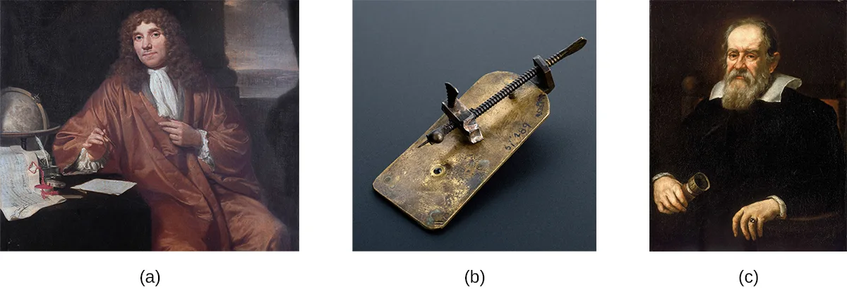
While van Leeuwenhoek is credited with the discovery of microorganisms, others before him had contributed to the development of the microscope. These included eyeglass makers in the Netherlands in the late 1500s, as well as the Italian astronomer Galileo Galilei, who used a compound microscope to examine insect parts (see the images above). Whereas van Leeuwenhoek used a simple microscope, in which light is passed through just one lens, Galileo’s compound microscope was more sophisticated, passing light through two sets of lenses.
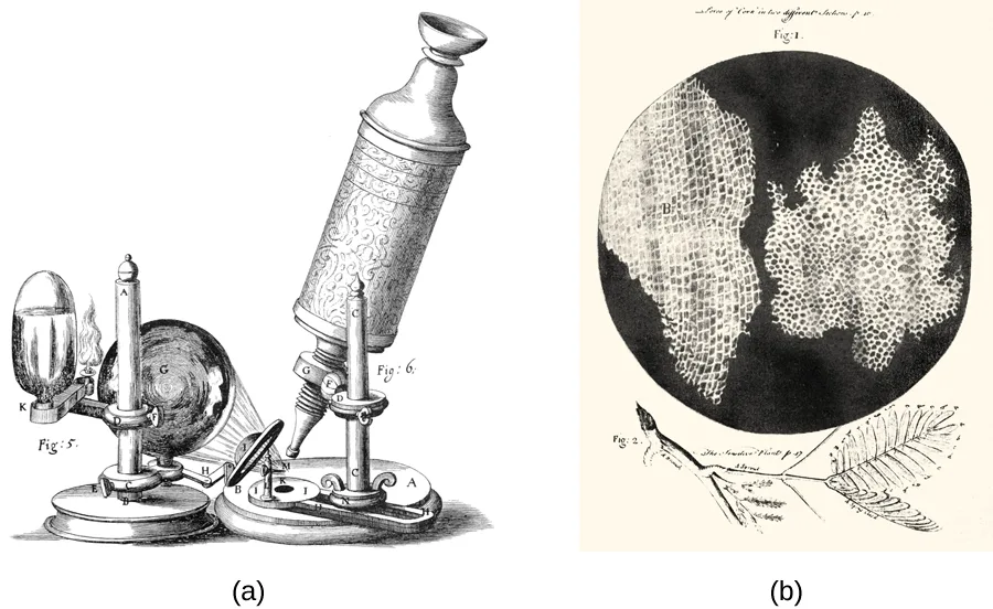
Van Leeuwenhoek’s contemporary, the Englishman Robert Hooke (1635–1703), also made important contributions to microscopy, publishing in his book Micrographia (1665) many observations using compound microscopes. Viewing a thin sample of cork through his microscope, he was the first to observe the structures that we now know as cells (see the images below). Hooke described these structures as resembling “Honey-comb,” and as “small Boxes or Bladders of Air,” noting that each “Cavern, Bubble, or Cell” is distinct from the others (in Latin, “cell” literally means “small room”). They likely appeared to Hooke to be filled with air because the cork cells were dead, with only the rigid cell walls providing the structure.
Extended description
Panel (a) shows the apparatus labeled with letters in Hooke’s original plate: an oil lamp and a water-filled glass sphere focus light onto a small lens near the base, which illuminates the specimen mounted below the tall cylindrical microscope body; a separate eyepiece tube stands beside it. Panel (b) shows the circular microscope field divided into two large, irregular honeycomb-like regions, each packed with small rectangular cells in rows, with a small drawing of a leafy plant stem beneath the circle.
Check Your Understanding
Explain the difference between simple and compound microscopes by sorting each phrase under the type of microscope it describes.
Simple microscope
Compound microscope
Compare and contrast the contributions of van Leeuwenhoek, Hooke, and Galileo to early microscopy.
Show model answer
Did your answer mention:
Micro Connection. Who Invented the Microscope?
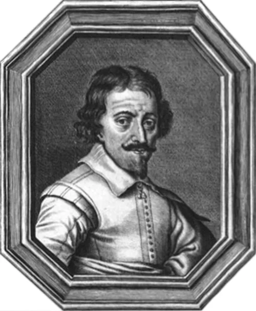
While Antonie van Leeuwenhoek and Robert Hooke generally receive much of the credit for early advances in microscopy, neither can claim to be the inventor of the microscope. Some argue that this designation should belong to Hans and Zaccharias Janssen, Dutch spectacle-makers who may have invented the telescope, the simple microscope, and the compound microscope during the late 1500s or early 1600s (see the portrait below). Unfortunately, little is known for sure about the Janssens, not even the exact dates of their births and deaths. The Janssens were secretive about their work and never published. It is also possible that the Janssens did not invent anything at all; their neighbor, Hans Lippershey, also developed microscopes and telescopes during the same time frame, and he is often credited with inventing the telescope. The historical records from the time are as fuzzy and imprecise as the images viewed through those early lenses, and any archived records have been lost over the centuries.
By contrast, van Leeuwenhoek and Hooke can thank ample documentation of their work for their respective legacies. Like Janssen, van Leeuwenhoek began his work in obscurity, leaving behind few records. However, his friend, the prominent physician Reinier de Graaf, wrote a letter to the editor of the Philosophical Transactions of the Royal Society of London calling attention to van Leeuwenhoek’s powerful microscopes. From 1673 onward, van Leeuwenhoek began regularly submitting letters to the Royal Society detailing his observations. In 1674, his report describing single-celled organisms produced controversy in the scientific community, but his observations were soon confirmed when the society sent a delegation to investigate his findings. He subsequently enjoyed considerable celebrity, at one point even entertaining a visit by the czar of Russia.
Similarly, Robert Hooke had his observations using microscopes published by the Royal Society in a book called Micrographia in 1665. The book became a bestseller and greatly increased interest in microscopy throughout much of Europe.
Summary
- Antonie van Leeuwenhoek is credited with the first observation of microbes, including protists and bacteria, with simple microscopes that he made.
- Robert Hooke was the first to describe what we now call cells.
- Simple microscopes have a single lens, while compound microscopes have multiple lenses.
Key terms
- compound microscope — a microscope that uses multiple lenses to focus light from the specimen.
- simple microscope — a type of microscope with only one lens to focus light from the specimen.
Practice
Describe historical developments and individual contributions that led to the invention and development of the microscope
Who was the first to describe “cells” in dead cork tissue?
This scientist published his observations of cork in his 1665 book Micrographia.Who is the probable inventor of the compound microscope?
This spectacle-maker, working with his father, may also have invented the telescope, though the historical evidence is inconclusive.Who is credited with the first observation of microbes, made using microscopes he built himself?
This scientist began his career selling fabrics before turning to lens making, and reported his observations to the Royal Society of London in 1674.Why is Antonie van Leeuwenhoek’s work much better known than that of Zaccharias Janssen?
Show model answer
Did your answer mention:
Why did the cork cells observed by Robert Hooke appear to be empty, as opposed to being full of other structures?
Show model answer
Did your answer mention:
Compare and contrast the features of simple and compound microscopes
A microscope that uses multiple lenses is called a ________ microscope.
This is the type of microscope Galileo used to examine insect parts, passing light through two sets of lenses.Which of the following distinguishes a compound microscope from a simple microscope, according to this section’s summary?
Recall how many lenses each type uses to focus light from the specimen.________ — a type of microscope with only one lens to focus light from the specimen.
This is the type of microscope Antonie van Leeuwenhoek used to observe microorganisms.This section is adapted from Microbiology, Section 2.2: Peering Into the Invisible World by Nina Parker, Mark Schneegurt, Anh-Hue Thi Tu, Philip Lister, Brian M. Forster, and OpenStax, © OpenStax, licensed under CC BY-NC-SA 4.0. Access the original for free at openstax.org. Changes: figures re-encoded as WebP; kind="photo" set on the van Leeuwenhoek/Galileo composite (an oil portrait, a photographed replica instrument, and a second oil portrait — all photographic reproductions, none drawn) and kind="diagram" set on the Hooke composite and the Janssen portrait (both are 17th-century engravings, genuinely drawn), overriding the media manifest’s JPEG-based photo guess for all three; the source alt text rewritten for all three figures to describe what is visibly shown rather than restate “Photo a/b/c is of…”; a longdesc added for the Hooke figure, walking each panel; the four figure cross-references inside running prose (“see [Figure 2.9]” twice, “[Figure 2.10]”, and “[Figure 2.11]”) replaced with plain descriptive phrases pointing at the adjacent figure; the Micro Connection box rendered as a callout, its figure kept at the end of the box in the source’s own document order even though it is referenced inline in the box’s first paragraph; of the two Check Your Understanding bullets, the first (the difference between simple and compound microscopes) is rendered as a sortbins graded from this section’s own sentences (1 Check Your Understanding question graded from the module’s own sentences rather than answered in prose; the source prints no key for it) and the second (comparing van Leeuwenhoek, Hooke, and Galileo) remains a body self-check with a model answer and rubric assembled from this section’s own preceding text; model answers for the section’s two Short Answer questions are written from this section’s text, because the source prints no answer key for either; key terms compiled from the module’s two defined terms and the book’s Glossary appendix (both taken from the glossary; none from a defining sentence); the end-of-section Multiple Choice, Fill in the Blank, and Short Answer questions adapted into the closing interactive Practice block; because this section’s five source exercises could not fill both objective groups to the book’s floor, three filler items were added and ledgered: one Multiple Choice built from this section’s own text (who is credited with the first observation of microbes), one Multiple Choice built from a single sentence of the section’s Summary (the lens-count distinction between simple and compound microscopes), and one term-recall text item built from the Key terms block (simple microscope); this section has no Clinical Focus box.