The Cell
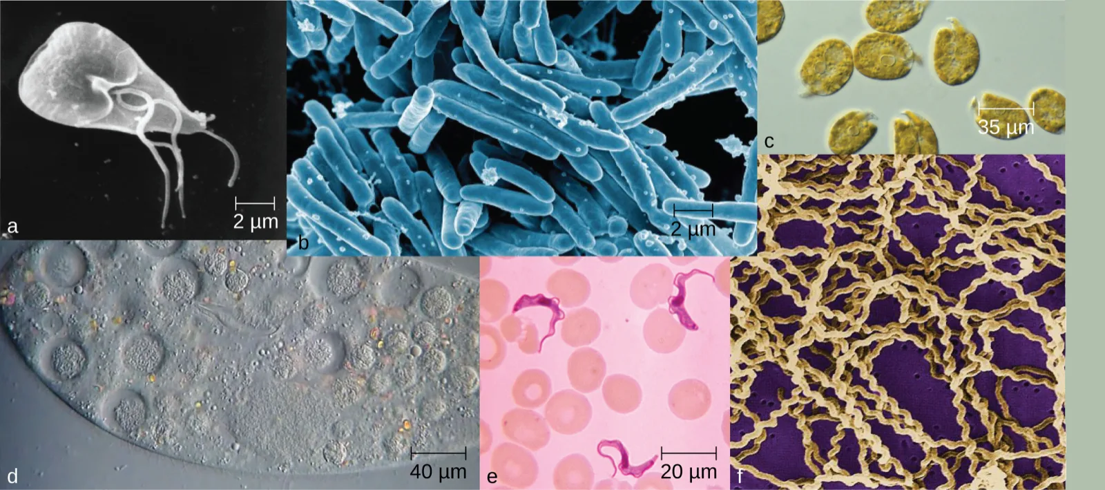
Life takes many forms, from giant redwood trees towering hundreds of feet in the air to the tiniest known microbes, which measure only a few billionths of a meter. Humans have long pondered life’s origins and debated the defining characteristics of life, but our understanding of these concepts has changed radically since the invention of the microscope. In the 17th century, observations of microscopic life led to the development of the cell theory: the idea that the fundamental unit of life is the cell, that all organisms contain at least one cell, and that cells only come from other cells.
Despite sharing certain characteristics, cells may vary significantly. The two main types of cells are prokaryotic cells (lacking a nucleus) and eukaryotic cells (containing a well-organized, membrane-bound nucleus). Each type of cell exhibits remarkable variety in structure, function, and metabolic activity, as the micrographs above suggest. This chapter will focus on the historical discoveries that have shaped our current understanding of microbes, including their origins and their role in human disease. We will then explore the distinguishing structures found in prokaryotic and eukaryotic cells.
Sections
- Spontaneous Generation — the theory of spontaneous generation, Redi’s and Needham’s and Spallanzani’s experiments, and Pasteur’s swan-neck flasks that disproved it.
- Foundations of Modern Cell Theory — Hooke, Schleiden, Schwann, Virchow, and Remak on cell theory; the endosymbiotic theory; Semmelweis, Snow, Pasteur, Lister, and Koch on the germ theory of disease.
- Unique Characteristics of Prokaryotic Cells — cell morphology and arrangement, the nucleoid, plasmids, ribosomes, inclusions, endospores, the plasma membrane and transport, cell walls, glycocalyces, flagella, fimbriae, and pili, and how archaea differ from bacteria.
- Unique Characteristics of Eukaryotic Cells — the nucleus, the endomembrane system, peroxisomes, the cytoskeleton, mitochondria and chloroplasts, the plasma membrane and endocytosis, cell walls, the extracellular matrix, flagella and cilia, mitosis and meiosis, and the Clinical Focus case resolved.
This chapter is adapted from Microbiology, Chapter 3: The Cell by Nina Parker, Mark Schneegurt, Anh-Hue Thi Tu, Philip Lister, Brian M. Forster, and OpenStax, © OpenStax, licensed under CC BY-NC-SA 4.0. Access the original for free at openstax.org. Each section page records its own changes from the source. Changes: the chapter-opening micrograph panel is the source’s, re-encoded for the web with its credit kept in the caption and its alt text rewritten to describe what each of the six panels shows (the source alt misspells “microbes” and guesses cell sizes the panels’ scale bars do not all support); the source’s reference to the figure by number is replaced with a describing phrase.