Foundations of Modern Cell Theory
By the end of this section, you will be able to:
- Explain the key points of cell theory and the individual contributions of Hooke, Schleiden, Schwann, Remak, and Virchow
- Explain the key points of endosymbiotic theory and cite the evidence that supports this concept
- Explain the contributions of Semmelweis, Snow, Pasteur, Lister, and Koch to the development of germ theory
While some scientists were arguing over the theory of spontaneous generation, other scientists were making discoveries leading to a better understanding of what we now call the cell theory. Modern cell theory has two basic tenets:
- All cells only come from other cells (the principle of biogenesis).
- Cells are the fundamental units of organisms.
Today, these tenets are fundamental to our understanding of life on earth. However, modern cell theory grew out of the collective work of many scientists.
The Origins of Cell Theory
The English scientist Robert Hooke first used the term “cells” in 1665 to describe the small chambers within cork that he observed under a microscope of his own design. To Hooke, thin sections of cork resembled “Honey-comb,” or “small Boxes or Bladders of Air.” He noted that each “Cavern, Bubble, or Cell” was distinct from the others (shown below). At the time, Hooke was not aware that the cork cells were long dead and, therefore, lacked the internal structures found within living cells.
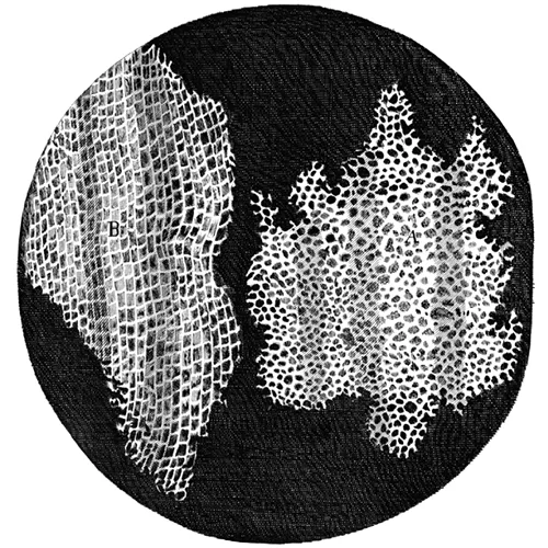
Despite Hooke’s early description of cells, their significance as the fundamental unit of life was not yet recognized. Nearly 200 years later, in 1838, Matthias Schleiden (1804–1881), a German botanist who made extensive microscopic observations of plant tissues, described them as being composed of cells. Visualizing plant cells was relatively easy because plant cells are clearly separated by their thick cell walls. Schleiden believed that cells formed through crystallization, rather than cell division.
Theodor Schwann (1810–1882), a noted German physiologist, made similar microscopic observations of animal tissue. In 1839, after a conversation with Schleiden, Schwann realized that similarities existed between plant and animal tissues. This laid the foundation for the idea that cells are the fundamental components of plants and animals.
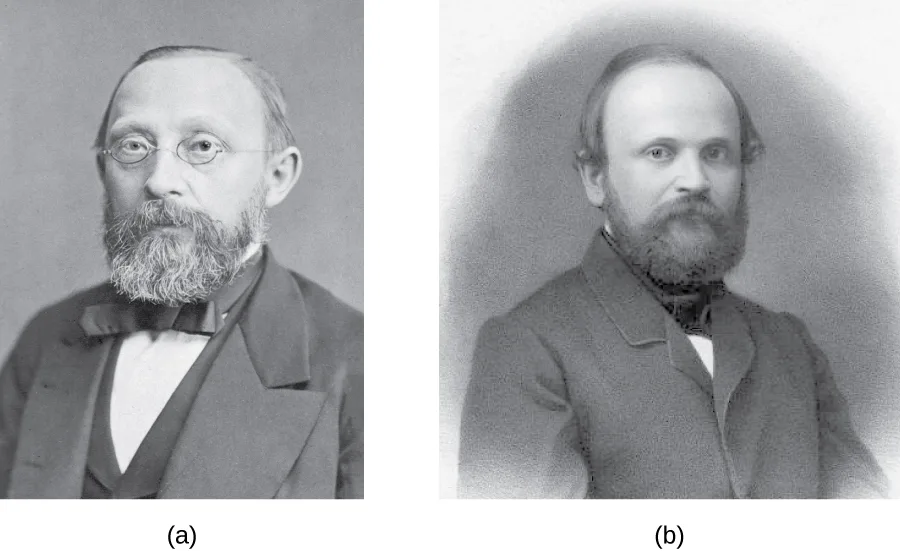
In the 1850s, two Polish scientists living in Germany pushed this idea further, culminating in what we recognize today as the modern cell theory. In 1852, Robert Remak (1815–1865), a prominent neurologist and embryologist, published convincing evidence that cells are derived from other cells as a result of cell division. However, this idea was questioned by many in the scientific community. Three years later, Rudolf Virchow (1821–1902), a well-respected pathologist, published an editorial essay entitled “Cellular Pathology,” which popularized the concept of cell theory using the Latin phrase omnis cellula a cellula (“all cells arise from cells”), which is essentially the second tenet of modern cell theory (M. Schultz, “Rudolph Virchow,” Emerging Infectious Diseases 14, no. 9 (2008): 1480–1481). Given the similarity of Virchow’s work to Remak’s, there is some controversy as to which scientist should receive credit for articulating cell theory. See the following Eye on Ethics feature for more about this controversy.
Eye on Ethics. Science and Plagiarism
Rudolf Virchow, a prominent, Polish-born, German scientist, is often remembered as the “Father of Pathology.” Well known for innovative approaches, he was one of the first to determine the causes of various diseases by examining their effects on tissues and organs. He was also among the first to use animals in his research and, as a result of his work, he was the first to name numerous diseases and created many other medical terms. Over the course of his career, he published more than 2,000 papers and headed various important medical facilities, including the Charité – Universitätsmedizin Berlin, a prominent Berlin hospital and medical school. But he is, perhaps, best remembered for his 1855 editorial essay titled “Cellular Pathology,” published in Archiv für Pathologische Anatomie und Physiologie, a journal that Virchow himself cofounded and still exists today.
Despite his significant scientific legacy, there is some controversy regarding this essay, in which Virchow proposed the central tenet of modern cell theory—that all cells arise from other cells. Robert Remak, a former colleague who worked in the same laboratory as Virchow at the University of Berlin, had published the same idea 3 years before. Though it appears Virchow was familiar with Remak’s work, he neglected to credit Remak’s ideas in his essay. When Remak wrote a letter to Virchow pointing out similarities between Virchow’s ideas and his own, Virchow was dismissive. In 1858, in the preface to one of his books, Virchow wrote that his 1855 publication was just an editorial piece, not a scientific paper, and thus there was no need to cite Remak’s work.
By today’s standards, Virchow’s editorial piece would certainly be considered an act of plagiarism, since he presented Remak’s ideas as his own. However, in the 19th century, standards for academic integrity were much less clear. Virchow’s strong reputation, coupled with the fact that Remak was a Jew in a somewhat anti-Semitic political climate, shielded him from any significant repercussions. Today, the process of peer review and the ease of access to the scientific literature help discourage plagiarism. Although scientists are still motivated to publish original ideas that advance scientific knowledge, those who would consider plagiarizing are well aware of the serious consequences.
In academia, plagiarism represents the theft of both individual thought and research—an offense that can destroy reputations and end careers (B. Kisch, “Forgotten Leaders in Modern Medicine, Valentin, Gouby, Remak, Auerbach,” Transactions of the American Philosophical Society 44 (1954): 139–317; H. Harris, The Birth of the Cell, New Haven, CT: Yale University Press, 2000, p. 133; C. Webster, ed., Biology, Medicine and Society 1840-1940, Cambridge, UK: Cambridge University Press, 1981, pp. 118–119; C. Zuchora-Walske, Key Discoveries in Life Science, Minneapolis, MN: Lerner Publishing, 2015, pp. 12–13).
Check Your Understanding
What are the key points of the cell theory?
Think back to the two tenets named at the very start of this section.What contributions did Rudolf Virchow and Robert Remak make to the development of the cell theory?
Show model answer
Did your answer mention:
Endosymbiotic Theory
As scientists were making progress toward understanding the role of cells in plant and animal tissues, others were examining the structures within the cells themselves. In 1831, Scottish botanist Robert Brown (1773–1858) was the first to describe observations of nuclei, which he observed in plant cells. Then, in the early 1880s, German botanist Andreas Schimper (1856–1901) was the first to describe the chloroplasts of plant cells, identifying their role in starch formation during photosynthesis and noting that they divided independent of the nucleus.
Based upon the chloroplasts’ ability to reproduce independently, Russian botanist Konstantin Mereschkowski (1855–1921) suggested in 1905 that chloroplasts may have originated from ancestral photosynthetic bacteria living symbiotically inside a eukaryotic cell. He proposed a similar origin for the nucleus of plant cells. This was the first articulation of the endosymbiotic hypothesis, and would explain how eukaryotic cells evolved from ancestral bacteria.
Mereschkowski’s endosymbiotic hypothesis was furthered by American anatomist Ivan Wallin (1883–1969), who began to experimentally examine the similarities between mitochondria, chloroplasts, and bacteria—in other words, to put the endosymbiotic hypothesis to the test using objective investigation. Wallin published a series of papers in the 1920s supporting the endosymbiotic hypothesis, including a 1926 publication co-authored with Mereschkowski. Wallin claimed he could culture mitochondria outside of their eukaryotic host cells. Many scientists dismissed his cultures of mitochondria as resulting from bacterial contamination. Modern genome sequencing work supports the dissenting scientists by showing that much of the genome of mitochondria had been transferred to the host cell’s nucleus, preventing the mitochondria from being able to live on their own (T. Embley and W. Martin, “Eukaryotic Evolution, Changes, and Challenges,” Nature 440 (2006): 623–630; O.G. Berg and C.G. Kurland, “Why Mitochondrial Genes Are Most Often Found in Nuclei,” Molecular Biology and Evolution 17, no. 6 (2000): 951–961).
Wallin’s ideas regarding the endosymbiotic hypothesis were largely ignored for the next 50 years because scientists were unaware that these organelles contained their own DNA. However, with the discovery of mitochondrial and chloroplast DNA in the 1960s, the endosymbiotic hypothesis was resurrected. Lynn Margulis (1938–2011), an American geneticist, published her ideas regarding the endosymbiotic hypothesis of the origins of mitochondria and chloroplasts in 1967 (L. Sagan, “On the Origin of Mitosing Cells,” Journal of Theoretical Biology 14, no. 3 (1967): 225–274). In the decade leading up to her publication, advances in microscopy had allowed scientists to differentiate prokaryotic cells from eukaryotic cells. In her publication, Margulis reviewed the literature and argued that the eukaryotic organelles such as mitochondria and chloroplasts are of prokaryotic origin. She presented a growing body of microscopic, genetic, molecular biology, fossil, and geological data to support her claims.
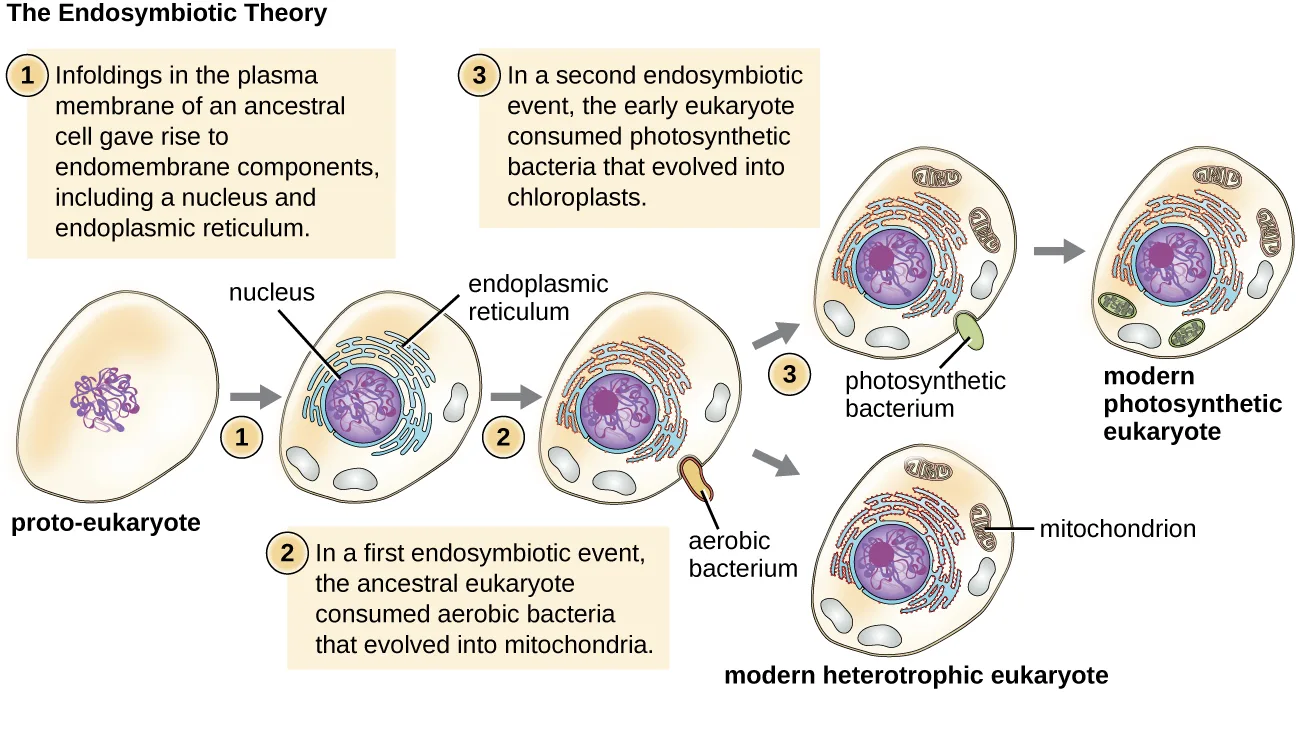
Again, this hypothesis was not initially popular, but mounting genetic evidence due to the advent of DNA sequencing supported the endosymbiotic theory, which is now defined as the theory that mitochondria and chloroplasts arose as a result of prokaryotic cells establishing a symbiotic relationship within a eukaryotic host (shown below). With Margulis’ initial endosymbiotic theory gaining wide acceptance, she expanded on the theory in her 1981 book Symbiosis in Cell Evolution. In it, she explains how endosymbiosis is a major driving factor in the evolution of organisms. More recent genetic sequencing and phylogenetic analysis show that mitochondrial DNA and chloroplast DNA are highly related to their bacterial counterparts, both in DNA sequence and chromosome structure. However, mitochondrial DNA and chloroplast DNA are reduced compared with nuclear DNA because many of the genes have moved from the organelles into the host cell’s nucleus. Additionally, mitochondrial and chloroplast ribosomes are structurally similar to bacterial ribosomes, rather than to the eukaryotic ribosomes of their hosts. Last, the binary fission of these organelles strongly resembles the binary fission of bacteria, as compared with mitosis performed by eukaryotic cells. Since Margulis’ original proposal, scientists have observed several examples of bacterial endosymbionts in modern-day eukaryotic cells. Examples include the endosymbiotic bacteria found within the guts of certain insects, such as cockroaches (A.E. Douglas, “The Microbial Dimension in Insect Nutritional Ecology,” Functional Ecology 23 (2009): 38–47), and photosynthetic bacteria-like organelles found in protists (J.M. Jaynes and L.P. Vernon, “The Cyanelle of Cyanophora paradoxa: Almost a Cyanobacterial Chloroplast,” Trends in Biochemical Sciences 7, no. 1 (1982): 22–24).
Extended description
Reading left to right: a plain oval proto-eukaryote cell containing only DNA. Step 1: infoldings in the plasma membrane of the ancestral cell give rise to endomembrane components, including a nucleus and endoplasmic reticulum — the cell now shows a membrane-bound nucleus and folds of endoplasmic reticulum. Step 2: in a first endosymbiotic event, the ancestral eukaryote consumes an aerobic bacterium, drawn entering the cell and then labeled a mitochondrion once inside; the resulting cell is labeled a modern heterotrophic eukaryote. Step 3: in a second endosymbiotic event, the early eukaryote consumes a photosynthetic bacterium, drawn entering the cell and then labeled a chloroplast once inside, alongside the mitochondrion already present; the resulting cell is labeled a modern photosynthetic eukaryote.
Check Your Understanding
What does the modern endosymbiotic theory state?
Think about which two organelles this theory concerns and what kind of relationship it says gave rise to them.What evidence supports the endosymbiotic theory?
Show model answer
Did your answer mention:
The Germ Theory of Disease
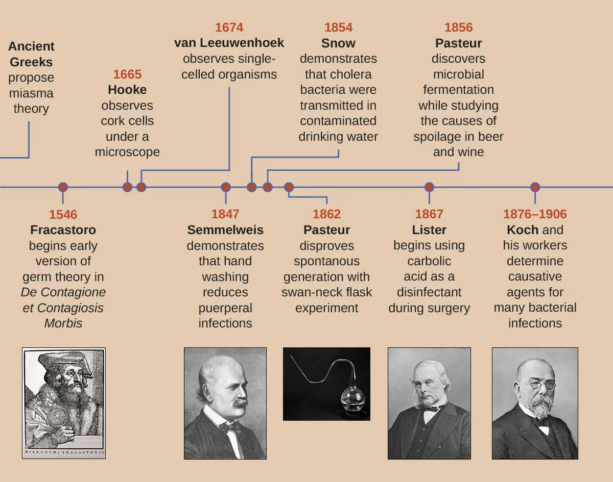
Prior to the discovery of microbes during the 17th century, other theories circulated about the origins of disease. For example, the ancient Greeks proposed the miasma theory, which held that disease originated from particles emanating from decomposing matter, such as that in sewage or cesspits. Such particles infected humans in close proximity to the rotting material. Diseases including the Black Death, which ravaged Europe’s population during the Middle Ages, were thought to have originated in this way. In the 11th Century, Persian physician Ibn Sina (sometimes referred to as Avicenna) proposed that tuberculosis was likely spread by people’s breath when in close proximity. Arab physician Ibn Zuhr, writing in about 1155, documented that the common skin condition scabies was caused by tiny mites that bored into the skin. Though scabies mites are about half a millimeter long (and therefore not technically microscopic) and their skin tunnels are often visible, Ibn Zuhr’s discovery gave more evidence that unseen substances or creatures caused diseases.
In 1546, Italian physician Girolamo Fracastoro proposed, in his essay De Contagione et Contagiosis Morbis, that seed-like spores may be transferred between individuals through direct contact, exposure to contaminated clothing, or through the air. We now recognize Fracastoro as an early proponent of the germ theory of disease, which states that diseases may result from microbial infection. However, in the 16th century, Fracastoro’s ideas were not widely accepted and would be largely forgotten until the 19th century.
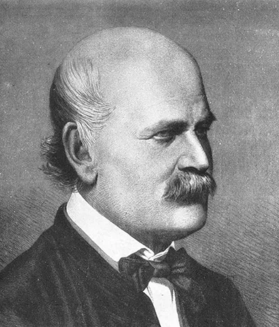
In 1847, Hungarian obstetrician Ignaz Semmelweis (shown below) observed that people who gave birth in hospital wards staffed by physicians and medical students were more likely to suffer and die from puerperal fever after childbirth (10%–20% mortality rate) than were people in wards staffed by midwives (1% mortality rate). Semmelweis observed medical students performing autopsies and then subsequently carrying out vaginal examinations on living patients without washing their hands in between. He suspected that the students carried disease from the autopsies to the patients they examined. His suspicions were supported by the untimely death of a friend, a physician who contracted a fatal wound infection after a postmortem examination of a woman who had died of a puerperal infection. The dead physician’s wound had been caused by a scalpel used during the examination, and his subsequent illness and death closely paralleled that of the dead patient.
Although Semmelweis did not know the true cause of puerperal fever, he proposed that physicians were somehow transferring the causative agent to their patients. He suggested that the number of puerperal fever cases could be reduced if physicians and medical students simply washed their hands with chlorinated lime water before and after examining every patient. When this practice was implemented, the maternal mortality rate in people cared for by physicians dropped to the same 1% mortality rate observed among people cared for by midwives. This demonstrated that handwashing was a very effective method for preventing disease transmission. Despite this great success, many discounted Semmelweis’s work at the time, and physicians were slow to adopt the simple procedure of handwashing to prevent infections in their patients because it contradicted established norms for that time period.
Around the same time Semmelweis was promoting handwashing, in 1854, British physician John Snow conducted studies to track the source of cholera outbreaks in London. (Source note: the source prints 1848 here, but its own timeline figure in this section dates Snow’s water-source study to 1854, the year of his Broad Street and water-company investigations; the page follows the timeline.) By tracing the outbreaks to two specific water sources, both of which were contaminated by sewage, Snow ultimately demonstrated that cholera bacteria were transmitted via drinking water. Snow’s work is influential in that it represents the first known epidemiological study, and it resulted in the first known public health response to an epidemic. The work of both Semmelweis and Snow clearly refuted the prevailing miasma theory of the day, showing that disease is not only transmitted through the air but also through contaminated items.
Although the work of Semmelweis and Snow successfully showed the role of sanitation in preventing infectious disease, the cause of disease was not fully understood. The subsequent work of Louis Pasteur, Robert Koch, and Joseph Lister would further substantiate the germ theory of disease.
While studying the causes of beer and wine spoilage in 1856, Pasteur discovered properties of fermentation by microorganisms. He had demonstrated with his swan-neck flask experiments (described in Spontaneous Generation) that airborne microbes, not spontaneous generation, were the cause of food spoilage, and he suggested that if microbes were responsible for food spoilage and fermentation, they could also be responsible for causing infection. This was the foundation for the germ theory of disease.
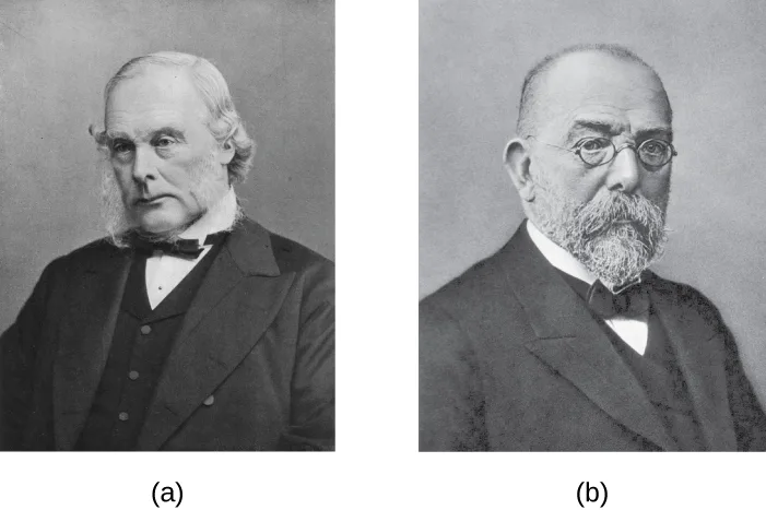
Meanwhile, British surgeon Joseph Lister (shown below) was trying to determine the causes of postsurgical infections. Many physicians did not give credence to the idea that microbes on their hands, on their clothes, or in the air could infect patients’ surgical wounds, despite the fact that 50% of surgical patients, on average, were dying of postsurgical infections (J. Wesley Alexander, “The Contributions of Infection Control to a Century of Progress,” Annals of Surgery 201 (1985): 423–428). Lister, however, was familiar with the work of Semmelweis and Pasteur; therefore, he insisted on handwashing and extreme cleanliness during surgery. In 1867, to further decrease the incidence of postsurgical wound infections, Lister began using carbolic acid (phenol) spray disinfectant/antiseptic during surgery. His extremely successful efforts to reduce postsurgical infection caused his techniques to become a standard medical practice.
A few years later, Robert Koch (shown below) proposed a series of postulates (Koch’s postulates) based on the idea that the cause of a specific disease could be attributed to a specific microbe. Using these postulates, Koch and his colleagues were able to definitively identify the causative pathogens of specific diseases, including anthrax, tuberculosis, and cholera. Koch’s “one microbe, one disease” concept was the culmination of the 19th century’s paradigm shift away from miasma theory and toward the germ theory of disease. Koch’s postulates are discussed more thoroughly in How Pathogens Cause Disease.
Check Your Understanding
Compare and contrast the miasma theory of disease with the germ theory of disease.
Miasma theory
Germ theory
How did Joseph Lister’s work contribute to the debate between the miasma theory and germ theory and how did this increase the success of medical procedures?
Show model answer
Did your answer mention:
Clinical Focus. Part 2
After suffering a fever, congestion, cough, and increasing aches and pains for several days, Barbara suspects that she has a case of the flu. She decides to visit the health center at her university. The PA tells Barbara that her symptoms could be due to a range of diseases, such as influenza, bronchitis, pneumonia, or tuberculosis.
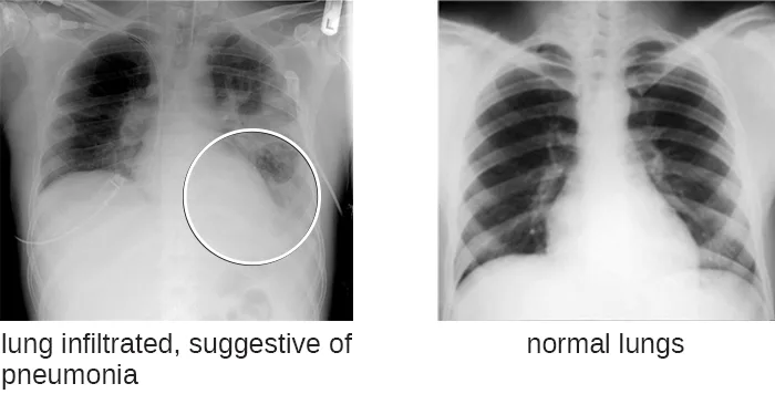
During her physical examination, the PA notes that Barbara’s heart rate is slightly elevated. Using a pulse oximeter, a small device that clips on her finger, he finds that Barbara has hypoxemia—a lower-than-normal level of oxygen in the blood. Using a stethoscope, the PA listens for abnormal sounds made by Barbara’s heart, lungs, and digestive system. As Barbara breathes, the PA hears a crackling sound and notes a slight shortness of breath. He collects a sputum sample, noting the greenish color of the mucus, and orders a chest radiograph, which shows a “shadow” in the left lung. All of these signs are suggestive of pneumonia, a condition in which the lungs fill with mucus (shown below).
- What kinds of infectious agents are known to cause pneumonia?
The case continues in Unique Characteristics of Prokaryotic Cells. The case began in Spontaneous Generation.
Extended description
Reading left to right along the timeline: Ancient Greeks propose the miasma theory. 1546, Fracastoro begins an early version of germ theory in De Contagione et Contagiosis Morbis, illustrated with a woodcut portrait. 1665, Hooke observes cork cells under a microscope. 1674, van Leeuwenhoek observes single-celled organisms. 1847, Semmelweis demonstrates that handwashing reduces puerperal infections, illustrated with his portrait. 1854, Snow demonstrates that cholera bacteria were transmitted in contaminated drinking water. 1856, Pasteur discovers microbial fermentation while studying the cause of spoilage in beer and wine. 1862, Pasteur disproves spontaneous generation with the swan-neck flask experiment, illustrated with a photograph of a swan-necked flask. 1867, Lister begins using carbolic acid as a disinfectant during surgery, illustrated with his portrait. 1876–1906, Koch and his workers determine the causative agents for many bacterial infections, illustrated with his portrait.
Summary
- Although cells were first observed in the 1660s by Robert Hooke, cell theory was not well accepted for another 200 years. The work of scientists such as Schleiden, Schwann, Remak, and Virchow contributed to its acceptance.
- Endosymbiotic theory states that mitochondria and chloroplasts, organelles found in many types of organisms, have their origins in bacteria. Significant structural and genetic information support this theory.
- The miasma theory of disease was widely accepted until the 19th century, when it was replaced by the germ theory of disease thanks to the work of Semmelweis, Snow, Pasteur, Lister, and Koch, and others.
Key terms
- endosymbiotic theory — the theory that mitochondria and chloroplasts arose as a result of prokaryotic cells establishing a symbiotic relationship within a eukaryotic host.
- germ theory of disease — the theory that many diseases are the result of microbial infection.
Practice
Explain the key points of cell theory and the individual contributions of Hooke, Schleiden, Schwann, Remak, and Virchow
Which of the following individuals did not contribute to the establishment of cell theory?
This individual is associated instead with an early version of the germ theory of disease, proposed nearly three centuries before the others on this list did their work.The scientist who first described cells was ________.
This English scientist coined the term after observing thin sections of cork under a microscope of his own design in 1665.How did the explanation of Virchow and Remak for the origin of cells differ from that of Schleiden and Schwann?
Think about what Schleiden believed caused new cells to form, and what Remak’s evidence showed instead.Explain the key points of endosymbiotic theory and cite the evidence that supports this concept
Whose proposal of the endosymbiotic theory of mitochondrial and chloroplast origin was ultimately accepted by the greater scientific community?
This American geneticist published her ideas in 1967, after the discovery of mitochondrial and chloroplast DNA revived a hypothesis that had been largely ignored for decades.________ states that mitochondria and chloroplasts, organelles found in many types of organisms, have their origins in bacteria.
This theory holds that these organelles began as free-living prokaryotes taken up by an ancestral eukaryotic host cell.Why are mitochondria and chloroplasts unable to multiply outside of a host cell?
Show model answer
Did your answer mention:
Explain the contributions of Semmelweis, Snow, Pasteur, Lister, and Koch to the development of germ theory
Which of the following developed a set of postulates for determining whether a particular disease is caused by a particular pathogen?
These postulates, associated with this scientist’s name, let him and his colleagues definitively identify the causative pathogens of diseases such as anthrax, tuberculosis, and cholera.John Snow is known as the Father of ________.
Snow’s tracing of London’s cholera outbreaks to two contaminated water sources represents the first known study of this kind.The ________ theory states that disease may originate from proximity to decomposing matter and is not due to person-to-person contact.
The ancient Greeks proposed this theory; it held that particles from sources such as sewage or cesspits caused disease in anyone nearby.What were the differences in mortality rates due to puerperal fever that Ignaz Semmelweis observed? How did he propose to reduce the occurrence of puerperal fever? Did it work?
Show model answer
Did your answer mention:
Why was the work of Snow so important in supporting the germ theory?
Show model answer
Did your answer mention:
This section is adapted from Microbiology, Section 3.2: Foundations of Modern Cell Theory by Nina Parker, Mark Schneegurt, Anh-Hue Thi Tu, Philip Lister, Brian M. Forster, and OpenStax, © OpenStax, licensed under CC BY-NC-SA 4.0. Access the original for free at openstax.org. Changes: figures re-encoded as WebP, with kind="diagram" set on the three drawn figures (Hooke’s cork illustration, the endosymbiotic-theory schematic, and the germ-theory timeline, the last because its dated milestone track is itself drawn even though five entries carry a small photo or drawing) and kind="photo" on the four portrait and radiograph figures, overriding the media manifest’s JPEG-based guess; alts rewritten for all seven figures to describe what is visibly drawn or photographed, since the source alt for the endosymbiotic-theory figure garbles “plasma membrane” as “plasm amembrane” and misprints “now” as “how” in its Step 2 sentence (both reported as source defects); longdesc added for the endosymbiotic-theory diagram and the germ-theory timeline, walking each in reading order; the eleven footnoted citations condensed into seven inline parenthetical citations placed after the sentences they support (grouped where several consecutive footnotes support one sentence boundary) (none of them carries an access URL or a DOI); the Eye on Ethics box rendered as a callout, with its figure kept inside the callout at its source position; three of the six Check Your Understanding bullets (one from each of the three boxes) rendered as body self-checks, with model answers and rubrics assembled from this section’s own preceding text; the Clinical Focus Part 2 box’s closing links replaced with a plain sentence noting that the case continues in Section 3.3 and began in Section 3.1, both of which exist; the cross-reference to Pasteur’s swan-neck flask figure in Section 3.1 rendered as a link to that section page, since it exists; the cross-reference to How Pathogens Cause Disease (m58867, a later, unauthored chapter) kept as plain italicized text naming the module; the end-of-section Multiple Choice and Fill in the Blank questions keep the source’s own keys, options, and order; model answers for one Short Answer and two Critical Thinking questions are written from this section’s own text, since the source prints no answer key for either set; one summary-derived cloze textin (“Endosymbiotic theory states that mitochondria and chloroplasts…”) fills out the second objective group after its own Short Answer question (“What evidence exists that supports the endosymbiotic theory?”) was dropped as a near-duplicate of the body Check Your Understanding bullet asking the same thing, and is named here rather than used in the Practice block; key terms compiled from the module’s two defined terms, both taken from the book’s Glossary appendix (none sentence-derived). One Short Answer question and three Check Your Understanding questions are graded from the module’s own sentences rather than answered in prose; the source prints no key for them: the “key points of cell theory” and “what does the endosymbiotic theory state” Check Your Understanding bullets became multiplechoice items keyed by the section’s own tenet and theory statements, the “compare and contrast the miasma theory … with the germ theory” Check Your Understanding bullet became a sort-into-bins item over the module’s own statements of each theory, and the Practice group’s “How did the explanation of Virchow and Remak … differ from that of Schleiden and Schwann?” Short Answer question (fs-id1172100616952) became a multiplechoice item keyed by the section’s own crystallization-versus-cell-division sentences. John Snow’s cholera study is dated 1854 rather than the source prose’s 1848, matching the module’s own timeline figure, with a visible Source note (erratum 379).