Unique Characteristics of Prokaryotic Cells
By the end of this section, you will be able to:
- Explain the distinguishing characteristics of prokaryotic cells
- Describe common cell morphologies and cellular arrangements typical of prokaryotic cells and explain how cells maintain their morphology
- Describe internal and external structures of prokaryotic cells in terms of their physical structure, chemical structure, and function
- Compare the distinguishing characteristics of bacterial and archaeal cells
Cell theory states that the cell is the fundamental unit of life. However, cells vary significantly in size, shape, structure, and function. At the simplest level of construction, all cells possess a few fundamental components. These include cytoplasm (a gel-like substance composed of water and dissolved chemicals needed for growth), which is contained within a plasma membrane (also called a cell membrane or cytoplasmic membrane); one or more chromosomes, which contain the genetic blueprints of the cell; and ribosomes, organelles used for the production of proteins.
Beyond these basic components, cells can vary greatly between organisms, and even within the same multicellular organism. The two largest categories of cells—prokaryotic cells and eukaryotic cells—are defined by major differences in several cell structures. Prokaryotic cells lack a nucleus surrounded by a complex nuclear membrane and generally have a single, circular chromosome located in a nucleoid. Eukaryotic cells have a nucleus surrounded by a complex nuclear membrane that contains multiple, rod-shaped chromosomes (Chan and Marshall, Organogenesis 6, no. 2 [2010]: 88–96).
All plant cells and animal cells are eukaryotic. Some microorganisms are composed of prokaryotic cells, whereas others are composed of eukaryotic cells. Prokaryotic microorganisms are classified within the domains Archaea and Bacteria, whereas eukaryotic organisms are classified within the domain Eukarya.
The structures inside a cell are analogous to the organs inside a human body, with unique structures suited to specific functions. Some of the structures found in prokaryotic cells are similar to those found in some eukaryotic cells; others are unique to prokaryotes. Although there are some exceptions, eukaryotic cells tend to be larger than prokaryotic cells. The comparatively larger size of eukaryotic cells dictates the need to compartmentalize various chemical processes within different areas of the cell, using complex membrane-bound organelles. In contrast, prokaryotic cells generally lack membrane-bound organelles; however, they often contain inclusions that compartmentalize their cytoplasm. The figure below illustrates structures typically associated with prokaryotic cells. These structures are described in more detail in the next section.
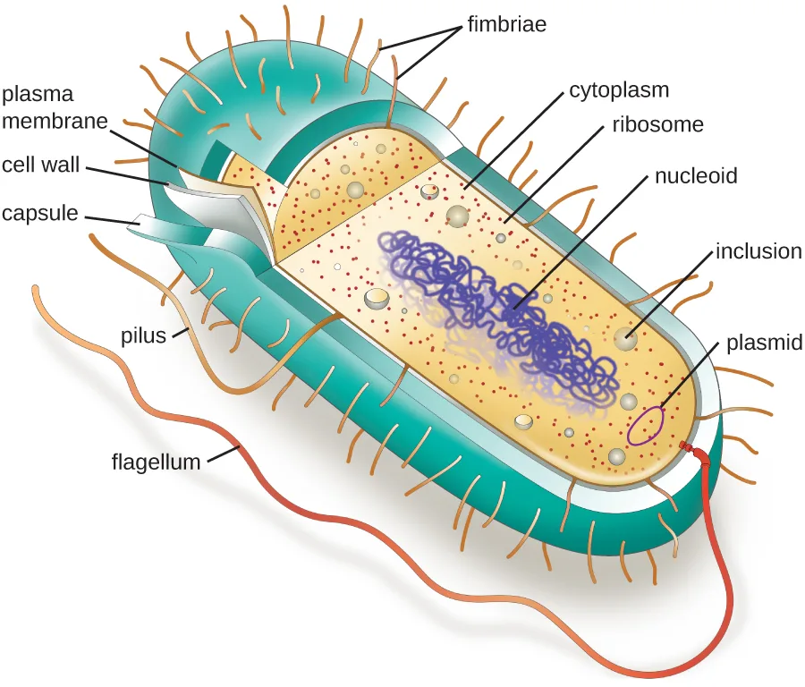
Extended description
Reading from outside in at the cell’s cut edge: a thick teal capsule, a thin cell wall, and a thinner plasma membrane enclose the cytoplasm. Inside, small dots mark ribosomes, larger pale spheres mark inclusions, a small looped strand marks a plasmid, and a long tangled loop marks the nucleoid. Short bristle-like fimbriae cover the cell surface by the hundreds, a single longer pilus extends from one side, and one long flagellum trails from one end of the cell.
Common Cell Morphologies and Arrangements
Individual cells of a particular prokaryotic organism are typically similar in shape, or cell morphology. Although thousands of prokaryotic organisms have been identified, only a handful of cell morphologies are commonly seen microscopically. The table below names and illustrates cell morphologies commonly found in prokaryotic cells. In addition to cellular shape, prokaryotic cells of the same species may group together in certain distinctive arrangements depending on the plane of cell division. Some common arrangements are shown in the figure after it.
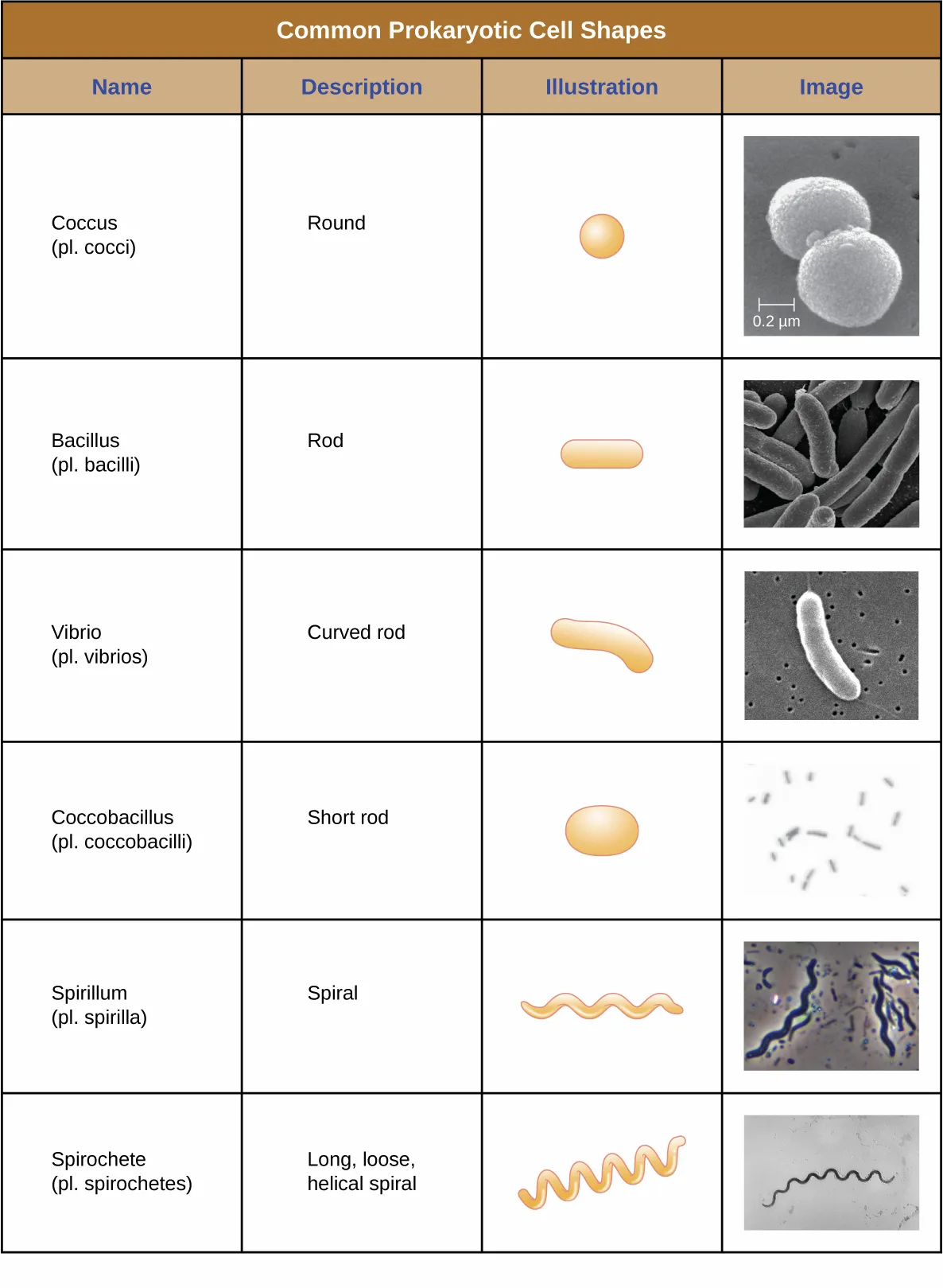
Extended description
The table has four columns — Name, Description, Illustration, Image — and six rows. Coccus (plural cocci): round; drawn as a sphere; micrograph shows two round cells. Bacillus (plural bacilli): rod; drawn as a rounded rectangle; micrograph shows several elongated rods. Vibrio (plural vibrios): curved rod; drawn as a comma shape; micrograph shows one curved rod. Coccobacillus (plural coccobacilli): short rod; drawn as an oval; micrograph shows small oval cells. Spirillum (plural spirilla): rigid spiral; drawn as a corkscrew; micrograph shows spiral chains. Spirochete (plural spirochetes): long, loose helical spiral; drawn as a looser corkscrew than the spirillum; micrograph shows a single loose spiral filament.
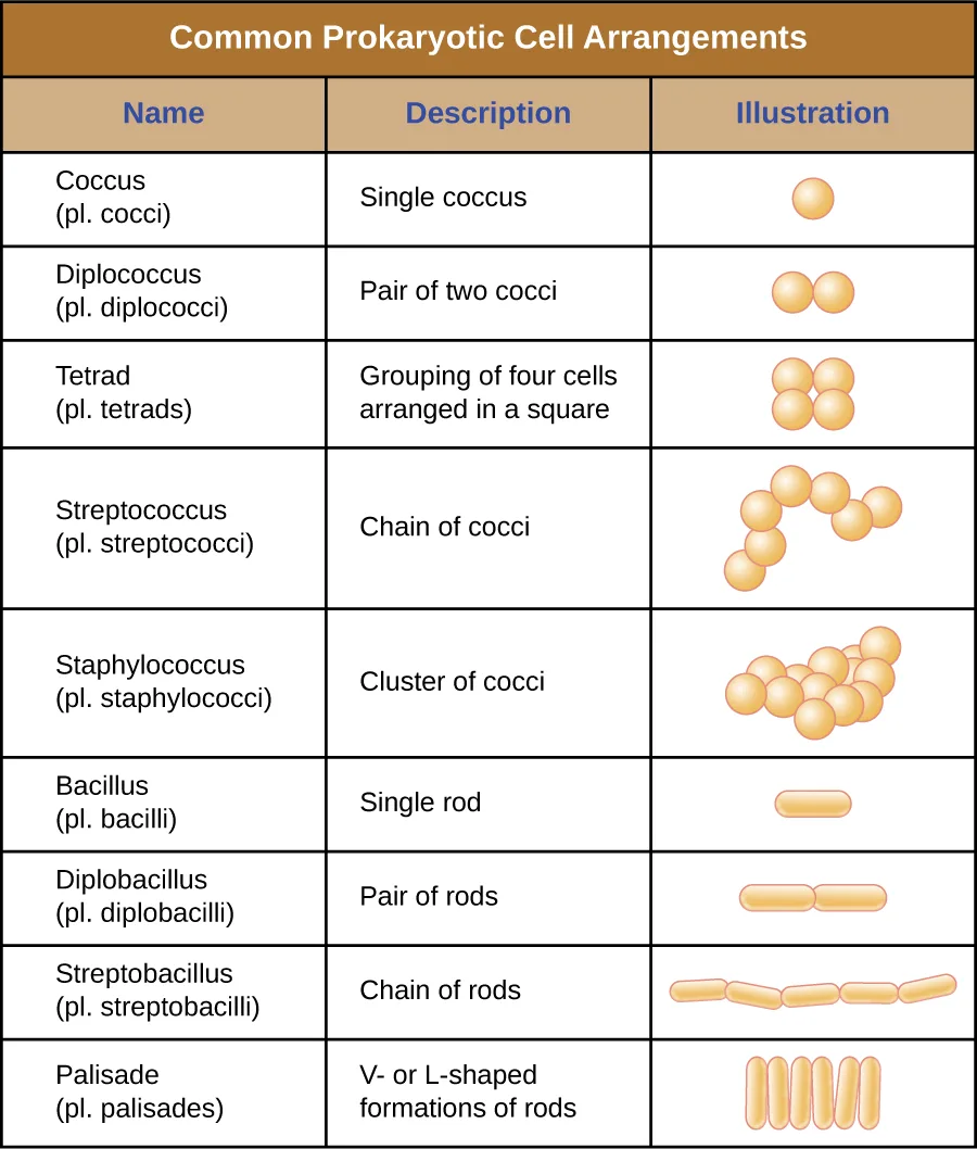
Extended description
Nine rows, each naming an arrangement and showing a drawn icon. Coccus (plural cocci): a single round cell. Diplococcus (plural diplococci): a pair of two cocci joined together. Tetrad (plural tetrads): four cocci arranged in a square. Streptococcus (plural streptococci): a chain of cocci. Staphylococcus (plural staphylococci): a cluster of cocci grouped in a bundle. Bacillus (plural bacilli): a single rod. Diplobacillus (plural diplobacilli): a pair of rods. Streptobacillus (plural streptobacilli): a chain of rods. Palisade (plural palisades): rods arranged in a V or L shape.
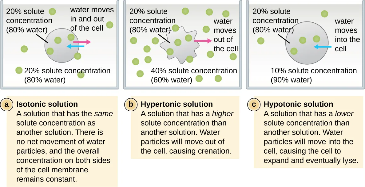
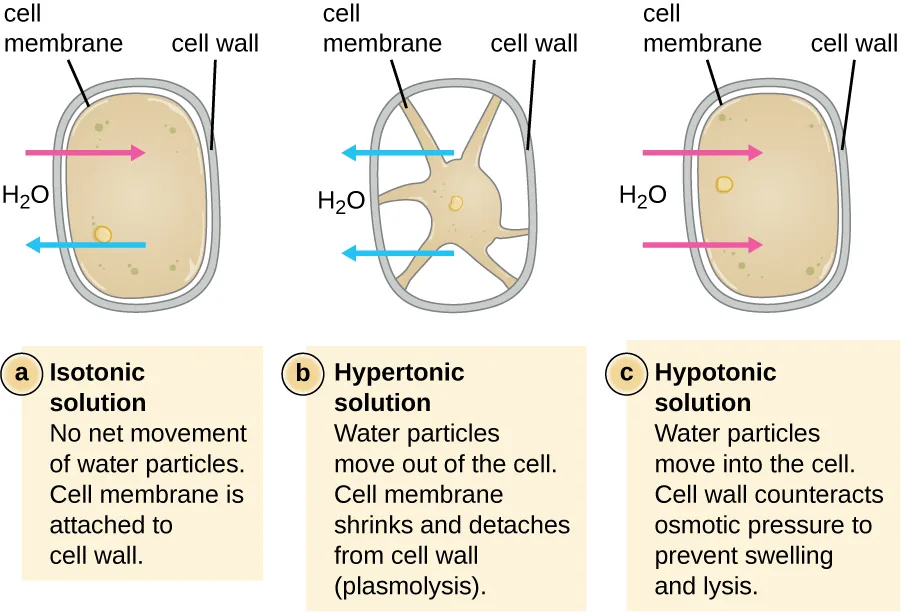
In most prokaryotic cells, morphology is maintained by the cell wall in combination with cytoskeletal elements. The cell wall is a structure found in most prokaryotes and some eukaryotes; it envelopes the cell membrane, protecting the cell from changes in osmotic pressure (shown below). Osmotic pressure occurs because of differences in the concentration of solutes on opposing sides of a semipermeable membrane. Water is able to pass through a semipermeable membrane, but solutes (dissolved molecules like salts, sugars, and other compounds) cannot. When the concentration of solutes is greater on one side of the membrane, water diffuses across the membrane from the side with the lower concentration (more water) to the side with the higher concentration (less water) until the concentrations on both sides become equal. This diffusion of water is called osmosis, and it can cause extreme osmotic pressure on a cell when its external environment changes.
The external environment of a cell can be described as an isotonic, hypertonic, or hypotonic medium. In an isotonic medium, the solute concentrations inside and outside the cell are approximately equal, so there is no net movement of water across the cell membrane. In a hypertonic medium, the solute concentration outside the cell exceeds that inside the cell, so water diffuses out of the cell and into the external medium. In a hypotonic medium, the solute concentration inside the cell exceeds that outside of the cell, so water will move by osmosis into the cell. This causes the cell to swell and potentially lyse, or burst.
The degree to which a particular cell is able to withstand changes in osmotic pressure is called tonicity. Cells that have a cell wall are better able to withstand subtle changes in osmotic pressure and maintain their shape. In hypertonic environments, cells that lack a cell wall can become dehydrated, causing crenation, or shriveling of the cell; the plasma membrane contracts and appears scalloped or notched (shown below). By contrast, cells that possess a cell wall undergo plasmolysis rather than crenation. In plasmolysis, the plasma membrane contracts and detaches from the cell wall, and there is a decrease in interior volume, but the cell wall remains intact, thus allowing the cell to maintain some shape and integrity for a period of time (shown below). Likewise, cells that lack a cell wall are more prone to lysis in hypotonic environments. The presence of a cell wall allows the cell to maintain its shape and integrity for a longer time before lysing.
Check Your Understanding
Explain the difference between cell morphology and arrangement.
Think about what each term describes: the shape of a single cell, or how multiple cells of a species group together.What advantages do cell walls provide prokaryotic cells?
Show model answer
Did your answer mention:
The Nucleoid
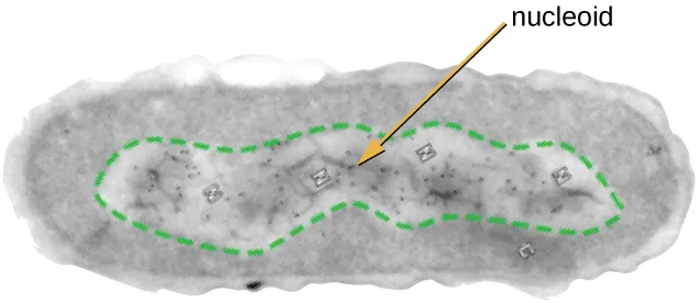
All cellular life has a DNA genome organized into one or more chromosomes. Prokaryotic chromosomes are typically circular, haploid (unpaired), and not bound by a complex nuclear membrane. Prokaryotic DNA and DNA-associated proteins are concentrated within the nucleoid region of the cell (shown below). In general, prokaryotic DNA interacts with nucleoid-associated proteins (NAPs) that assist in the organization and packaging of the chromosome. In bacteria, NAPs function similar to histones, which are the DNA-organizing proteins found in eukaryotic cells. In archaea, the nucleoid is organized by either NAPs or histone-like DNA organizing proteins.
Plasmids
Prokaryotic cells may also contain extrachromosomal DNA, or DNA that is not part of the chromosome. This extrachromosomal DNA is found in plasmids, which are small, circular, double-stranded DNA molecules. Cells that have plasmids often have hundreds of them within a single cell. Plasmids are more commonly found in bacteria; however, plasmids have been found in archaea and eukaryotic organisms. Plasmids often carry genes that confer advantageous traits such as antibiotic resistance; thus, they are important to the survival of the organism. We will discuss plasmids in more detail in the chapter introduction to Mechanisms of Microbial Genetics.
Ribosomes
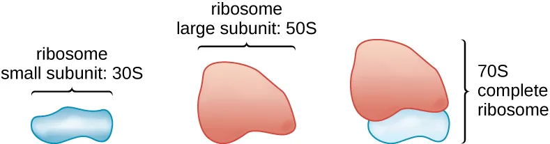
All cellular life synthesizes proteins, and organisms in all three domains of life possess ribosomes, structures responsible for protein synthesis. However, ribosomes in each of the three domains are structurally different. Ribosomes, themselves, are constructed from proteins, along with ribosomal RNA (rRNA). Prokaryotic ribosomes are found in the cytoplasm. They are called 70S ribosomes because they have a size of 70S (shown below), whereas eukaryotic cytoplasmic ribosomes have a size of 80S. (The S stands for Svedberg unit, a measure of sedimentation in an ultracentrifuge, which is based on size, shape, and surface qualities of the structure being analyzed). Although they are the same size, bacterial and archaeal ribosomes have different proteins and rRNA molecules, and the archaeal versions are more similar to their eukaryotic counterparts than to those found in bacteria.
Inclusions
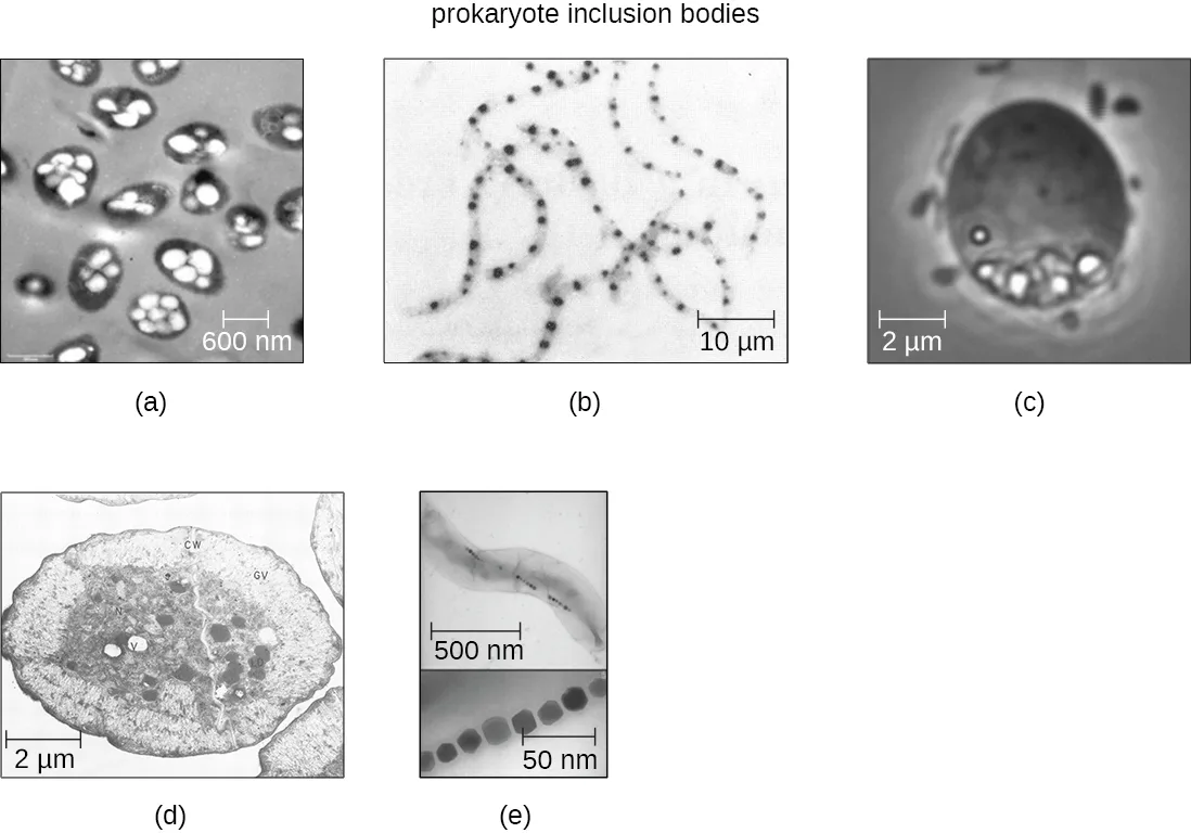
As single-celled organisms living in unstable environments, some prokaryotic cells have the ability to store excess nutrients within cytoplasmic structures called inclusions. Storing nutrients in a polymerized form is advantageous because it reduces the buildup of osmotic pressure that occurs as a cell accumulates solutes. Various types of inclusions store glycogen and starches, which contain carbon that cells can access for energy. Volutin granules, also called metachromatic granules because of their staining characteristics, are inclusions that store polymerized inorganic phosphate that can be used in metabolism and assist in the formation of biofilms. Microbes known to contain volutin granules include the archaea Methanosarcina, the bacterium Corynebacterium diphtheriae, and the unicellular eukaryotic alga Chlamydomonas. Sulfur granules, another type of inclusion, are found in sulfur bacteria of the genus Thiobacillus; these granules store elemental sulfur, which the bacteria use for metabolism.
Occasionally, certain types of inclusions are surrounded by a phospholipid monolayer embedded with protein. Polyhydroxybutyrate (PHB), which can be produced by species of Bacillus and Pseudomonas, is an example of an inclusion that displays this type of monolayer structure. Industrially, PHB has also been used as a source of biodegradable polymers for bioplastics. Several different types of inclusions are shown below.
Extended description
(a) A transmission electron micrograph of gray spheres, each containing several smaller white lipid droplets — polyhydroxybutyrate inclusions. (b) A light micrograph of thin, branching chains dotted with small dark granules — volutin granules. (c) A phase-contrast micrograph of one gray sphere with a cluster of smaller white spheres near its edge — sulfur granules. (d) A transmission electron micrograph of a large sphere packed with many smaller internal spheres — gas vacuoles. (e) A transmission electron micrograph of a long filament with a chain of small dark spheres along its length, magnified in an inset — magnetosomes.
Some prokaryotic cells have other types of inclusions that serve purposes other than nutrient storage. For example, some prokaryotic cells produce gas vacuoles, accumulations of small, protein-lined vesicles of gas. These gas vacuoles allow the prokaryotic cells that synthesize them to alter their buoyancy so that they can adjust their location in the water column. Magnetotactic bacteria, such as Magnetospirillum magnetotacticum, contain magnetosomes, which are inclusions of magnetic iron oxide or iron sulfide surrounded by a lipid layer. These allow cells to align along a magnetic field, aiding their movement. Cyanobacteria such as Anabaena cylindrica and bacteria such as Halothiobacillus neapolitanus produce carboxysome inclusions. Carboxysomes are composed of outer shells of thousands of protein subunits. Their interior is filled with ribulose-1,5-bisphosphate carboxylase/oxygenase (RuBisCO) and carbonic anhydrase. Both of these compounds are used for carbon metabolism. Some prokaryotic cells also possess carboxysomes that sequester functionally related enzymes in one location. These structures are considered proto-organelles because they compartmentalize important compounds or chemical reactions, much like many eukaryotic organelles.
Endospores
Bacterial cells are generally observed as vegetative cells, but some genera of bacteria have the ability to form endospores, structures that essentially protect the bacterial genome in a dormant state when environmental conditions are unfavorable. Endospores (not to be confused with the reproductive spores formed by fungi) allow some bacterial cells to survive long periods without food or water, as well as exposure to chemicals, extreme temperatures, and even radiation. The table below compares the characteristics of vegetative cells and endospores.
| Characteristics of Vegetative Cells versus Endospores | |
|---|---|
| Vegetative Cells | Endospores |
| Sensitive to extreme temperatures and radiation | Resistant to extreme temperatures and radiation |
| Nearly all endospore-producing bacteria gram-positive as vegetative cells | Do not absorb Gram stain, only special endospore stains (see Staining Microscopic Specimens) |
| Normal water content and enzymatic activity | Dehydrated; no metabolic activity |
| Capable of active growth and metabolism | Dormant; no growth or metabolic activity |
The process by which vegetative cells transform into endospores is called sporulation, and it generally begins when nutrients become depleted or environmental conditions become otherwise unfavorable (shown below). The process begins with the formation of a septum in the vegetative bacterial cell. The septum divides the cell asymmetrically, separating a DNA forespore from the mother cell. The forespore, which will form the core of the endospore, is essentially a copy of the cell’s chromosomes, and is separated from the mother cell by a second membrane. A cortex gradually forms around the forespore by laying down layers of calcium and dipicolinic acid between membranes. A protein coat then forms around the cortex while the DNA of the mother cell disintegrates. Further maturation of the endospore occurs with the formation of an outermost exosporium. The endospore is released upon disintegration of the mother cell, completing sporulation.
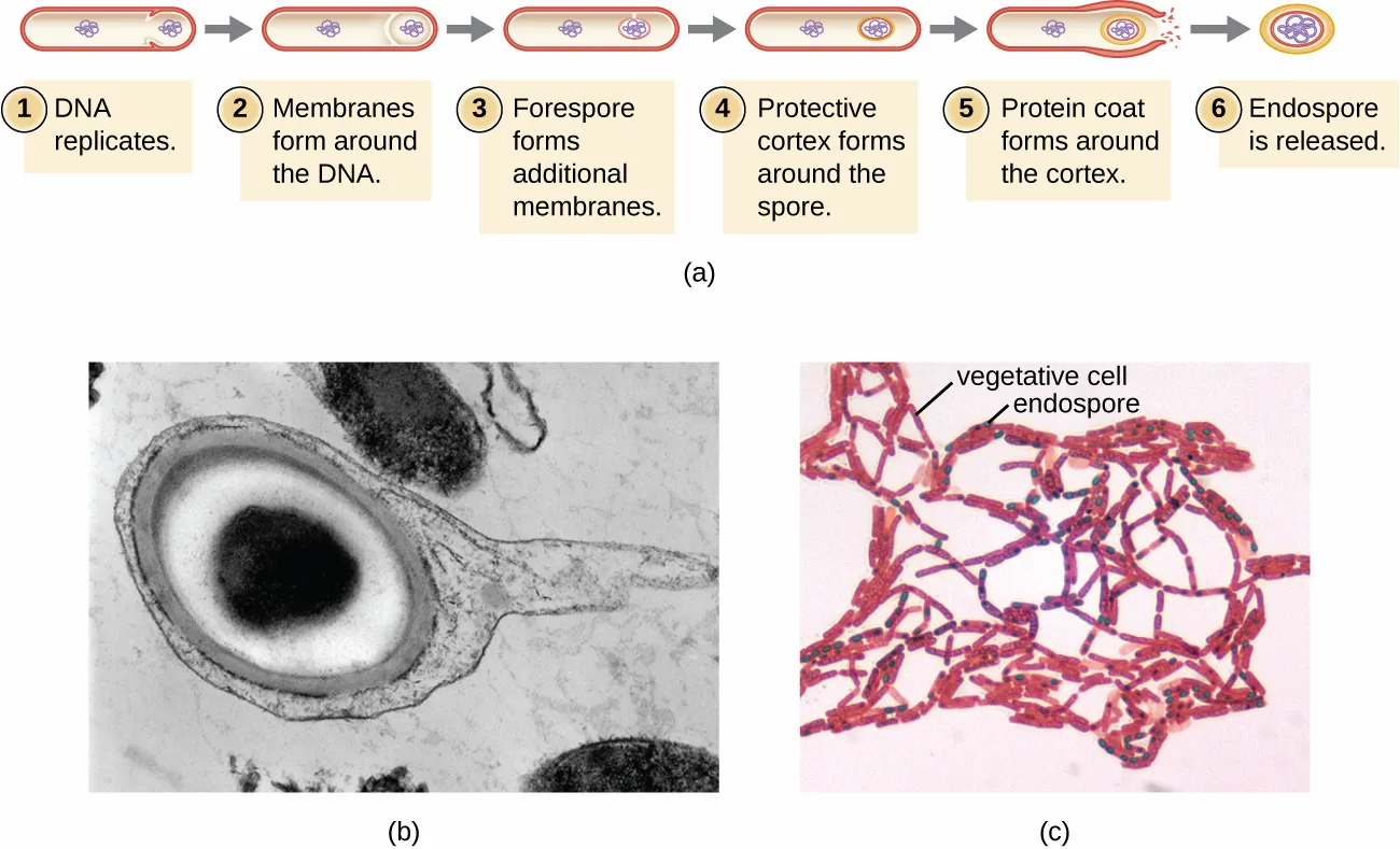
Extended description
Panel (a), six drawn steps: 1, a rod-shaped cell holds two loops of DNA, one central and one near an end, as the DNA replicates. 2, a membrane begins to encircle the DNA loop at the end of the cell. 3, the forespore forms an additional membrane layer, thickening the encircling lines. 4, a protective cortex forms, thickening the lines further. 5, a protein coat forms around the cortex, thickening the lines again, and the outer cell begins to lyse. 6, the mature endospore is released as a small layered spherical structure. Panel (b), an electron micrograph shows an oval endospore with a dark central core inside a lighter surrounding region, encased in thick outer layers. Panel (c), a micrograph shows red-stained rod-shaped cells in chains, several holding a small green-stained endospore near their center.
Endospores of certain species have been shown to persist in a dormant state for extended periods of time, up to thousands of years (Rothfuss, Bender, and Conrad, Microbial Ecology 33, no. 1 [1997]: 69–77). However, when living conditions improve, endospores undergo germination, reentering a vegetative state. After germination, the cell becomes metabolically active again and is able to carry out all of its normal functions, including growth and cell division.
Not all bacteria have the ability to form endospores; however, there are a number of clinically significant endospore-forming gram-positive bacteria of the genera Bacillus and Clostridium. These include B. anthracis, the causative agent of anthrax, which produces endospores capable of surviving for many decades (Sinclair et al., Applied and Environmental Microbiology 74, no. 3 [2008]: 555–563); C. tetani (causes tetanus); C. perfringens (causes gas gangrene); and C. botulinum (causes botulism). Clostridioides difficile is a distant relative causing pseudomembranous colitis. Pathogens such as these are particularly difficult to combat because their endospores are so hard to kill. Special sterilization methods for endospore-forming bacteria are discussed in the chapter introduction to Control of Microbial Growth.
Check Your Understanding
What is an inclusion?
Think about where in the cell this structure sits and what kind of substance it typically stores.What is the function of an endospore?
Think about what environmental conditions trigger endospore formation and what the structure protects the cell from.Plasma Membrane
Structures that enclose the cytoplasm and internal structures of the cell are known collectively as the cell envelope. In prokaryotic cells, the structures of the cell envelope vary depending on the type of cell and organism. Most (but not all) prokaryotic cells have a cell wall, but the makeup of this cell wall varies. All cells (prokaryotic and eukaryotic) have a plasma membrane (also called cytoplasmic membrane or cell membrane) that exhibits selective permeability, allowing some molecules to enter or leave the cell while restricting the passage of others.
The structure of the plasma membrane is often described in terms of the fluid mosaic model, which refers to the ability of membrane components to move fluidly within the plane of the membrane, as well as the mosaic-like composition of the components, which include a diverse array of lipid and protein components (shown below). The plasma membrane structure of most bacterial and eukaryotic cell types is a bilayer composed mainly of phospholipids formed with ester linkages and proteins. These phospholipids and proteins have the ability to move laterally within the plane of the membranes as well as between the two phospholipid layers.
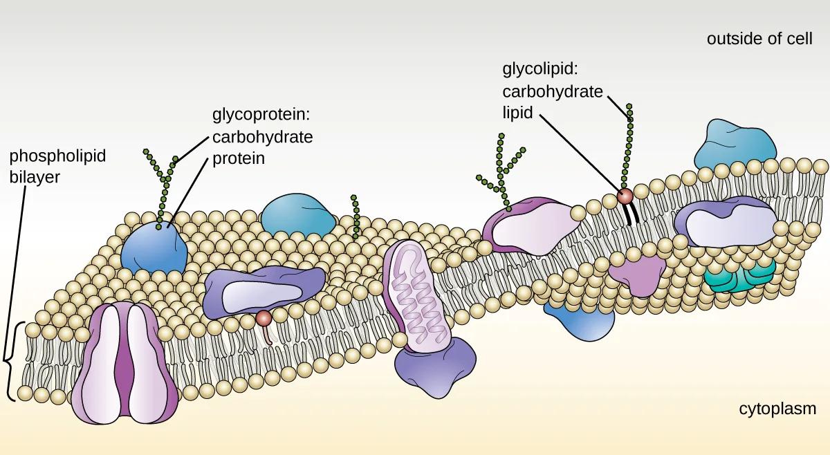
Extended description
Labeled top to bottom: outside of cell above the bilayer, cytoplasm below it. The phospholipid bilayer is two layers of sphere-and-two-tail phospholipids, tails facing inward. Embedded proteins include protein channels that span the bilayer with a central pore connecting outside to cytoplasm, peripheral proteins sitting on one face of the bilayer, and transmembrane proteins spanning its full width. Glycolipids (a carbohydrate chain on a single phospholipid) and glycoproteins (a carbohydrate chain on a protein) both carry their carbohydrate chains on the outside of the membrane only. A thin cytoskeleton layer runs just inside the inner face of the bilayer.
Archaeal membranes are fundamentally different from bacterial and eukaryotic membranes in a few significant ways. First, archaeal membrane phospholipids are formed with ether linkages, in contrast to the ester linkages found in bacterial or eukaryotic cell membranes. Second, archaeal phospholipids have branched chains, whereas those of bacterial and eukaryotic cells are straight chained. Finally, although some archaeal membranes can be formed of bilayers like those found in bacteria and eukaryotes, other archaeal plasma membranes are lipid monolayers.
Proteins on the cell’s surface are important for a variety of functions, including cell-to-cell communication, and sensing environmental conditions and pathogenic virulence factors. Membrane proteins and phospholipids may have carbohydrates (sugars) associated with them and are called glycoproteins or glycolipids, respectively. These glycoprotein and glycolipid complexes extend out from the surface of the cell, allowing the cell to interact with the external environment. Glycoproteins and glycolipids in the plasma membrane can vary considerably in chemical composition among archaea, bacteria, and eukaryotes, allowing scientists to use them to characterize unique species.
Plasma membranes from different cells types also contain unique phospholipids, which contain fatty acids. As described in the chapter Using Biochemistry to Identify Microorganisms, phospholipid-derived fatty acid analysis (PLFA) profiles can be used to identify unique types of cells based on differences in fatty acids. Archaea, bacteria, and eukaryotes each have a unique PFLA profile.
Membrane Transport Mechanisms
One of the most important functions of the plasma membrane is to control the transport of molecules into and out of the cell. Internal conditions must be maintained within a certain range despite any changes in the external environment. The transport of substances across the plasma membrane allows cells to do so.
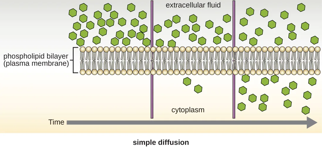
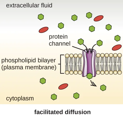
Cells use various modes of transport across the plasma membrane. For example, molecules moving from a higher concentration to a lower concentration with the concentration gradient are transported by simple diffusion, also known as passive transport (shown below). Some small molecules, like carbon dioxide, may cross the membrane bilayer directly by simple diffusion. However, charged molecules, as well as large molecules, need the help of carriers or channels in the membrane. These structures ferry molecules across the membrane, a process known as facilitated diffusion (shown below).
Active transport occurs when cells move molecules across their membrane against concentration gradients (shown below). A major difference between passive and active transport is that active transport requires adenosine triphosphate (ATP) or other forms of energy to move molecules “uphill.” Therefore, active transport structures are often called “pumps.”
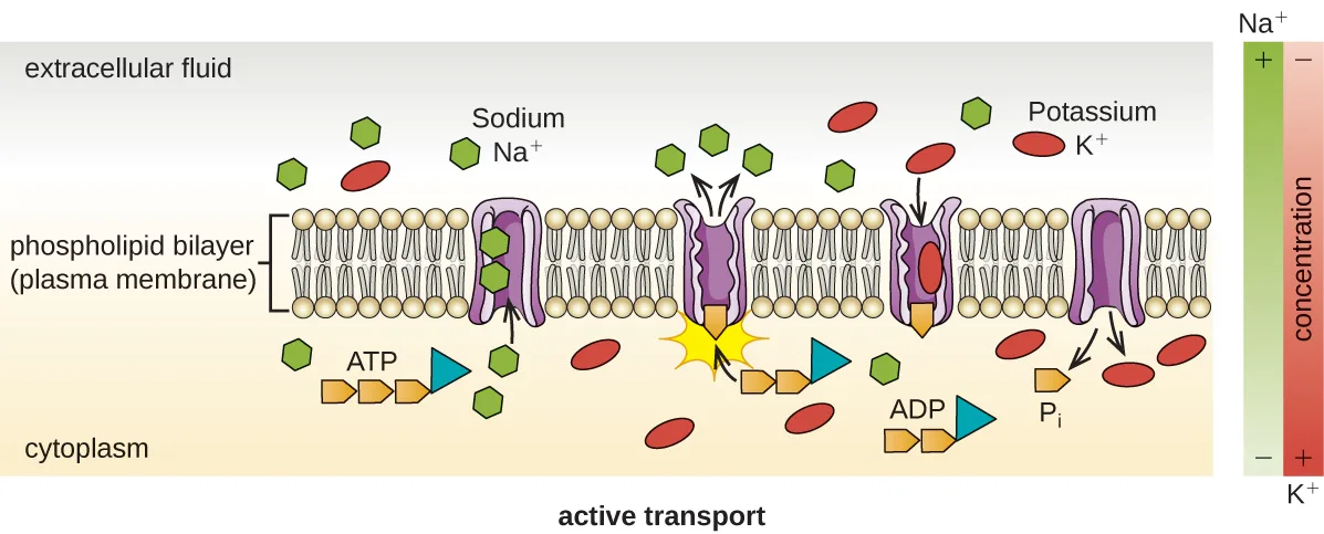
Group translocation also transports substances into bacterial cells. In this case, as a molecule moves into a cell against its concentration gradient, it is chemically modified so that it does not require transport against an unfavorable concentration gradient. A common example of this is the bacterial phosphotransferase system, a series of carriers that phosphorylates (i.e., adds phosphate ions to) glucose or other sugars upon entry into cells. Since the phosphorylation of sugars is required during the early stages of sugar metabolism, the phosphotransferase system is considered to be an energy neutral system.
Photosynthetic Membrane Structures
Some prokaryotic cells, namely cyanobacteria and photosynthetic bacteria, have membrane structures that enable them to perform photosynthesis. These structures consist of an infolding of the plasma membrane that encloses photosynthetic pigments such as green chlorophylls and bacteriochlorophylls. In cyanobacteria, these membrane structures are called thylakoids; in photosynthetic bacteria, they are called chromatophores, lamellae, or chlorosomes.
Cell Wall
The primary function of the cell wall is to protect the cell from harsh conditions in the outside environment. When present, there are notable similarities and differences among the cell walls of archaea, bacteria, and eukaryotes.
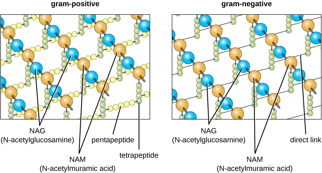
The major component of bacterial cell walls is called peptidoglycan (or murein); it is only found in bacteria. Structurally, peptidoglycan resembles a layer of meshwork or fabric (shown below). Each layer is composed of long chains of alternating molecules of N-acetylglucosamine (NAG) and N-acetylmuramic acid (NAM). The structure of the long chains has significant two-dimensional tensile strength due to the formation of peptide bridges that connect NAG and NAM within each peptidoglycan layer. In gram-negative bacteria, tetrapeptide chains extending from each NAM unit are directly cross-linked, whereas in gram-positive bacteria, these tetrapeptide chains are linked by pentaglycine cross-bridges. Peptidoglycan subunits are made inside of the bacterial cell and then exported and assembled in layers, giving the cell its shape.
Since peptidoglycan is unique to bacteria, many antibiotic drugs are designed to interfere with peptidoglycan synthesis, weakening the cell wall and making bacterial cells more susceptible to the effects of osmotic pressure (see the chapter Mechanisms of Antibacterial Drugs). In addition, certain cells of the human immune system are able to “recognize” bacterial pathogens by detecting peptidoglycan on the surface of a bacterial cell; these cells then engulf and destroy the bacterial cell, using enzymes such as lysozyme, which breaks down and digests the peptidoglycan in their cell walls (see the chapter Pathogen Recognition and Phagocytosis).
Extended description
Left panel, gram-positive: rows of alternating orange (NAG) and blue (NAM) spheres form parallel chains; each NAM in one chain connects to the NAM of a neighboring chain by two bridges, one labeled pentapeptide (a chain of small yellow spheres) and one labeled tetrapeptide (a chain of small green spheres). Right panel, gram-negative: the same alternating NAG and NAM chains appear, but neighboring chains are linked directly to each other by a single line, labeled direct link, rather than by the two peptide bridges.
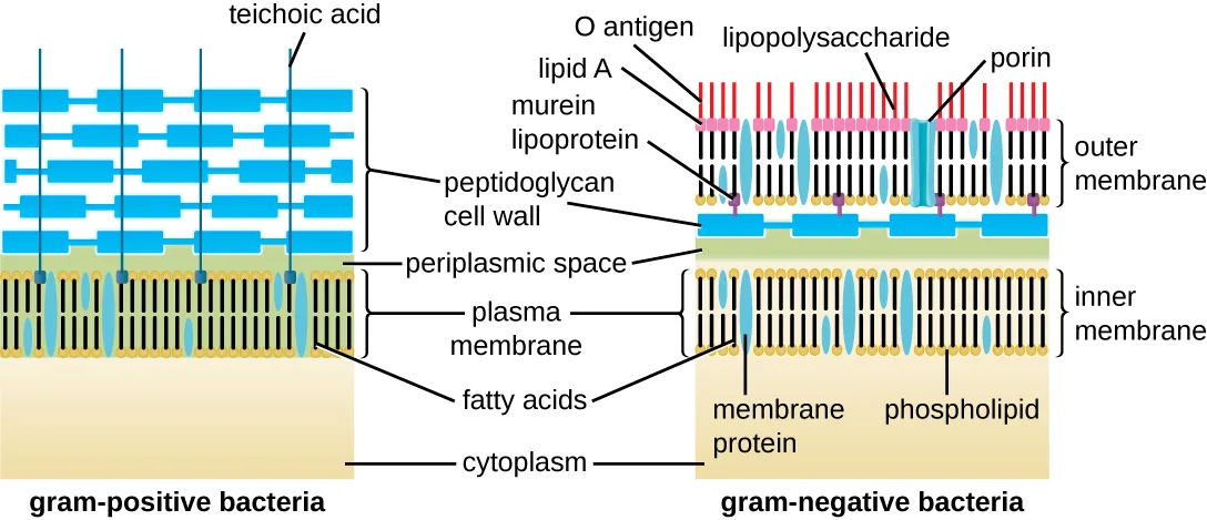
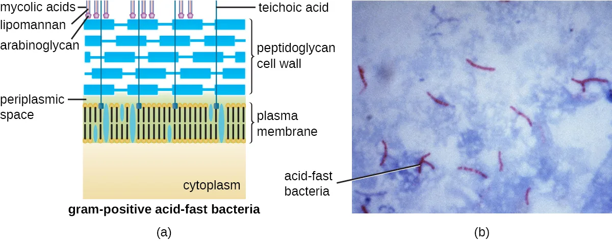
The Gram staining protocol (see Staining Microscopic Specimens) is used to differentiate two common types of cell wall structures (shown below). Gram-positive cells have a cell wall consisting of many layers of peptidoglycan totaling 30–100 nm in thickness. These peptidoglycan layers are commonly embedded with teichoic acids (TAs), carbohydrate chains that extend through and beyond the peptidoglycan layer (Silhavy, Kahne, and Walker, Cold Spring Harbor Perspectives in Biology 2, no. 5 [2010]: a000414). TA is thought to stabilize peptidoglycan by increasing its rigidity. TA also plays a role in the ability of pathogenic gram-positive bacteria such as Streptococcus to bind to certain proteins on the surface of host cells, enhancing their ability to cause infection. In addition to peptidoglycan and TAs, bacteria of the family Mycobacteriaceae have an external layer of waxy mycolic acids in their cell wall; as described in Staining Microscopic Specimens, these bacteria are referred to as acid-fast, since acid-fast stains must be used to penetrate the mycolic acid layer for purposes of microscopy (shown below).
Extended description
Left, gram-positive bacteria: cytoplasm at the base, then the plasma membrane, then a thick peptidoglycan cell wall built of stacked rectangular units threaded vertically by teichoic acid strands. Right, gram-negative bacteria: cytoplasm, inner (plasma) membrane with embedded membrane proteins and phospholipids, a thin periplasmic space, a thin peptidoglycan cell wall, and an outer membrane; murein lipoprotein anchors the outer membrane to the peptidoglycan, and the outer membrane’s outer face carries lipid A, a core, and O-antigen chains (lipopolysaccharide) alongside porin channels.
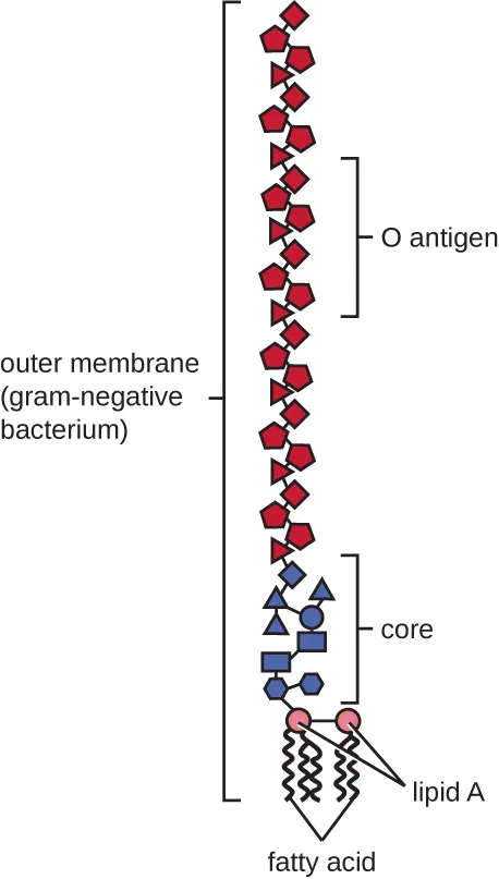
Gram-negative cells have a much thinner layer of peptidoglycan (no more than about 4 nm thick; Gan, Chen, and Jensen, Proceedings of the National Academy of Sciences of the United States of America 105, no. 48 [2008]: 18953–18957) than gram-positive cells, and the overall structure of their cell envelope is more complex. In gram-negative cells, a gel-like matrix occupies the periplasmic space between the cell wall and the plasma membrane, and there is a second lipid bilayer called the outer membrane, which is external to the peptidoglycan layer (shown below). This outer membrane is attached to the peptidoglycan by murein lipoprotein. The outer leaflet of the outer membrane contains the molecule lipopolysaccharide (LPS), which functions as an endotoxin in infections involving gram-negative bacteria, contributing to symptoms such as fever, hemorrhaging, and septic shock. Each LPS molecule is composed of Lipid A, a core polysaccharide, and an O side chain that is composed of sugar-like molecules that comprise the external face of the LPS (shown below). The composition of the O side chain varies between different species and strains of bacteria. Parts of the O side chain called antigens can be detected using serological or immunological tests to identify specific pathogenic strains like Escherichia coli O157:H7, a deadly strain of bacteria that causes bloody diarrhea and kidney failure.
Archaeal cell wall structure differs from that of bacteria in several significant ways. First, archaeal cell walls do not contain peptidoglycan; instead, they contain a similar polymer called pseudopeptidoglycan (pseudomurein) in which NAM is replaced with a different subunit. Other archaea may have a layer of glycoproteins or polysaccharides that serves as the cell wall instead of pseudopeptidoglycan. Last, as is the case with some bacterial species, there are a few archaea that appear to lack cell walls entirely.
Glycocalyces and S-Layers
Although most prokaryotic cells have cell walls, some may have additional cell envelope structures exterior to the cell wall, such as glycocalyces and S-layers. A glycocalyx is a sugar coat, of which there are two important types: capsules and slime layers. A capsule is an organized layer located outside of the cell wall and usually composed of polysaccharides or proteins (shown below). A slime layer is a less tightly organized layer that is only loosely attached to the cell wall and can be more easily washed off. Slime layers may be composed of polysaccharides, glycoproteins, or glycolipids.
Glycocalyces allows cells to adhere to surfaces, aiding in the formation of biofilms (colonies of microbes that form in layers on surfaces). In nature, most microbes live in mixed communities within biofilms, partly because the biofilm affords them some level of protection. Biofilms generally hold water like a sponge, preventing desiccation. They also protect cells from predation and hinder the action of antibiotics and disinfectants. All of these properties are advantageous to the microbes living in a biofilm, but they present challenges in a clinical setting, where the goal is often to eliminate microbes.
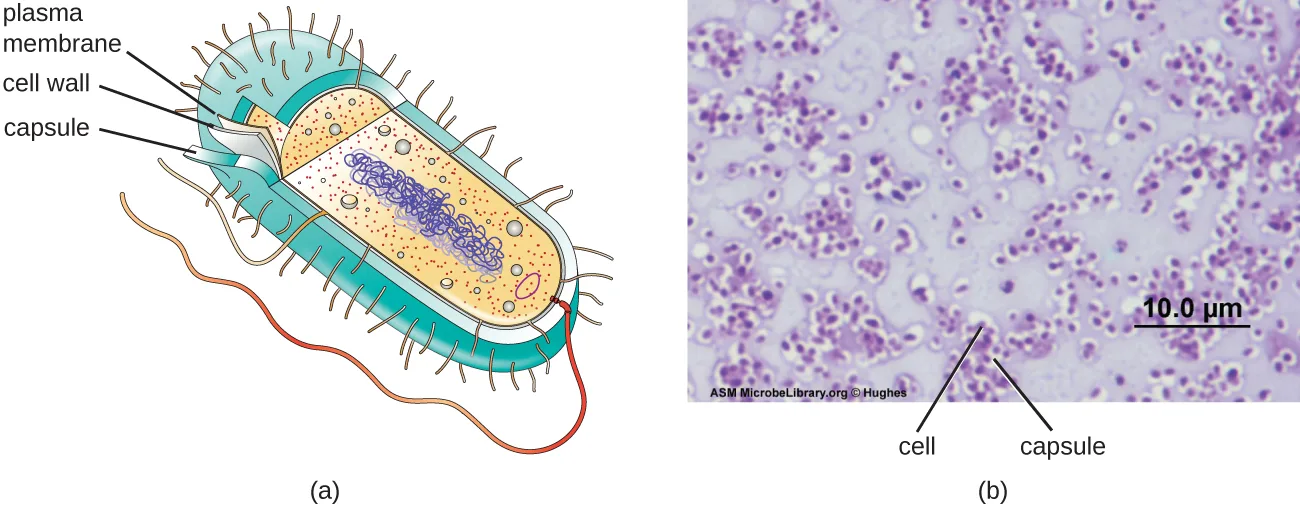
The ability to produce a capsule can contribute to a microbe’s pathogenicity (ability to cause disease) because the capsule can make it more difficult for phagocytic cells (such as white blood cells) to engulf and kill the microorganism. Streptococcus pneumoniae, for example, produces a capsule that is well known to aid in this bacterium’s pathogenicity. As explained in Staining Microscopic Specimens, capsules are difficult to stain for microscopy; negative staining techniques are typically used.
An S-layer is another type of cell envelope structure; it is composed of a mixture of structural proteins and glycoproteins. In bacteria, S-layers are found outside the cell wall, but in some archaea, the S-layer serves as the cell wall. The exact function of S-layers is not entirely understood, and they are difficult to study; but available evidence suggests that they may play a variety of functions in different prokaryotic cells, such as helping the cell withstand osmotic pressure and, for certain pathogens, interacting with the host immune system.
Clinical Focus. Part 3
After diagnosing Barbara with pneumonia, the PA writes her a prescription for amoxicillin, a commonly-prescribed type of penicillin derivative. More than a week later, despite taking the full course as directed, Barbara still feels weak and is not fully recovered, although she is still able to get through her daily activities. She returns to the health center for a follow-up visit.
Many types of bacteria, fungi, and viruses can cause pneumonia. Amoxicillin targets the peptidoglycan of bacterial cell walls. Since the amoxicillin has not resolved Barbara’s symptoms, the PA concludes that the causative agent probably lacks peptidoglycan, meaning that the pathogen could be a virus, a fungus, or a bacterium that lacks peptidoglycan. Another possibility is that the pathogen is a bacterium containing peptidoglycan but has developed resistance to amoxicillin.
- How can the PA definitively identify the cause of Barbara’s pneumonia?
- What form of treatment should the PA prescribe, given that the amoxicillin was ineffective?
The case continues in Unique Characteristics of Eukaryotic Cells. The case began in Spontaneous Generation.
Filamentous Appendages
Many bacterial cells have protein appendages embedded within their cell envelopes that extend outward, allowing interaction with the environment. These appendages can attach to other surfaces, transfer DNA, or provide movement. Filamentous appendages include fimbriae, pili, and flagella.
Fimbriae and Pili
Fimbriae and pili are structurally similar and, because differentiation between the two is problematic, these terms are often used interchangeably (Garnett et al., Proceedings of the National Academy of Sciences of the United States of America 109, no. 10 [2012]: 3950–3955; Proft and Baker, Cellular and Molecular Life Sciences 66 [2009]: 613). The term fimbriae commonly refers to short bristle-like proteins projecting from the cell surface by the hundreds. Fimbriae enable a cell to attach to surfaces and to other cells. For pathogenic bacteria, adherence to host cells is important for colonization, infectivity, and virulence. Adherence to surfaces is also important in biofilm formation.
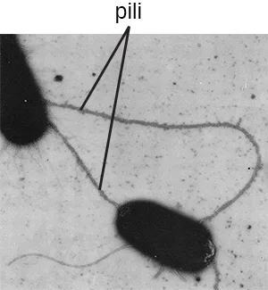
The term pili (singular: pilus) commonly refers to longer, less numerous protein appendages that aid in attachment to surfaces (shown below). A specific type of pilus, called the F pilus or sex pilus, is important in the transfer of DNA between bacterial cells, which occurs between members of the same generation when two cells physically transfer or exchange parts of their respective genomes (see the chapter How Asexual Prokaryotes Achieve Genetic Diversity).
Micro Connection. Group A Strep
Before the structure and function of the various components of the bacterial cell envelope were well understood, scientists were already using cell envelope characteristics to classify bacteria. In 1933, Rebecca Lancefield proposed a method for serotyping various β-hemolytic strains of Streptococcus species using an agglutination assay, a technique using the clumping of bacteria to detect specific cell-surface antigens. In doing so, Lancefield discovered that one group of S. pyogenes, found in Group A, was associated with a variety of human diseases. She determined that various strains of Group A strep could be distinguished from each other based on variations in specific cell surface proteins that she named M proteins.
Today, more than 80 different strains of Group A strep have been identified based on M proteins. Various strains of Group A strep are associated with a wide variety of human infections, including streptococcal pharyngitis (strep throat), impetigo, toxic shock syndrome, scarlet fever, rheumatic fever, and necrotizing fasciitis. The M protein is an important virulence factor for Group A strep, helping these strains evade the immune system. Changes in M proteins appear to alter the infectivity of a particular strain of Group A strep.
Flagella
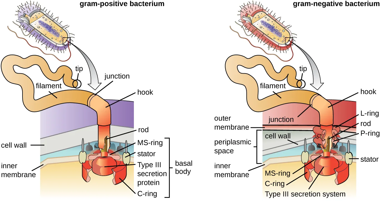
Flagella are structures used by cells to move in aqueous environments. Bacterial flagella act like propellers. They are stiff spiral filaments composed of flagellin protein subunits that extend outward from the cell and spin in solution. The basal body is the motor for the flagellum and is embedded in the plasma membrane (shown below). A hook region connects the basal body to the filament. Gram-positive and gram-negative bacteria have different basal body configurations due to differences in cell wall structure.
Different types of motile bacteria exhibit different arrangements of flagella (shown below). A bacterium with a singular flagellum, typically located at one end of the cell (polar), is said to have a monotrichous flagellum. An example of a monotrichously flagellated bacterial pathogen is Vibrio cholerae, the gram-negative bacterium that causes cholera. Cells with amphitrichous flagella have a flagellum or tufts of flagella at each end. An example is Spirillum minor, the cause of spirillary (Asian) rat-bite fever or sodoku. Cells with lophotrichous flagella have a tuft at one end of the cell. The gram-negative bacillus Pseudomonas aeruginosa, an opportunistic pathogen known for causing many infections, including “swimmer’s ear” and burn wound infections, has lophotrichous flagella. Flagella that cover the entire surface of a bacterial cell are called peritrichous flagella. The gram-negative bacterium E. coli shows a peritrichous arrangement of flagella.
Extended description
Both panels show, from a cutaway corner of the cell, a wavy filament ending in a tip, connected through a junction and a bent hook to a rod. In the gram-positive panel, the rod passes through the cell wall directly to a basal body in the plasma membrane, made of a C-ring at the base, a central type III secretion protein, surrounding stator proteins, and an MS-ring on top. In the gram-negative panel, the rod passes through an L-ring seated in the outer membrane, then a P-ring seated in the cell wall, then the periplasmic space, before reaching the same C-ring, type III secretion system, MS-ring, and stator arrangement in the inner membrane.

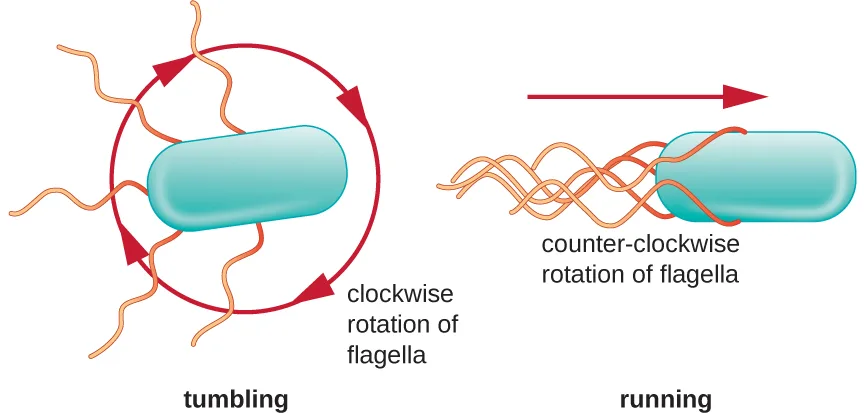
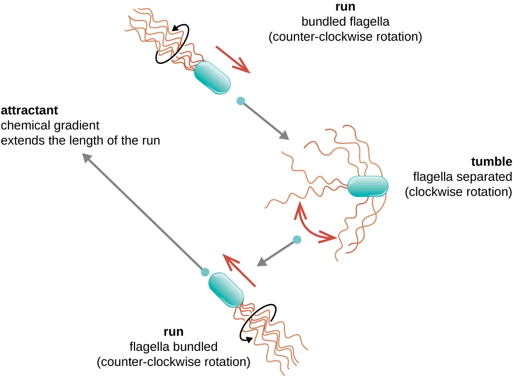
Directional movement depends on the configuration of the flagella. Bacteria can move in response to a variety of environmental signals, including light (phototaxis), magnetic fields (magnetotaxis) using magnetosomes, and, most commonly, chemical gradients (chemotaxis). Purposeful movement toward a chemical attractant, like a food source, or away from a repellent, like a poisonous chemical, is achieved by increasing the length of runs and decreasing the length of tumbles. When running, flagella rotate in a counterclockwise direction, allowing the bacterial cell to move forward. In a peritrichous bacterium, the flagella are all bundled together in a very streamlined way (shown below), allowing for efficient movement. When tumbling, flagella are splayed out while rotating in a clockwise direction, creating a looping motion and preventing meaningful forward movement but reorienting the cell toward the direction of the attractant. When an attractant exists, runs and tumbles still occur; however, the length of runs is longer, while the length of the tumbles is reduced, allowing overall movement toward the higher concentration of the attractant. When no chemical gradient exists, the lengths of runs and tumbles are more equal, and overall movement is more random (shown below).
Extended description
The diagram cycles through three positions of a cell joined by arrows: a run with flagella bundled, rotating counter-clockwise, moving in a straight line; a tumble with flagella separated, rotating clockwise, reorienting the cell; and a second run in a new direction, heading toward the upper left. A label at upper left reads ‘attractant: chemical gradient extends the length of the run,’ next to the longest of the drawn runs, showing that a run oriented toward the attractant persists longer than the others.
Check Your Understanding
What is the peptidoglycan layer and how does it differ between gram-positive and gram-negative bacteria?
Gram-positive
Gram-negative
Compare and contrast monotrichous, amphitrichous, lophotrichous, and peritrichous flagella.
Monotrichous
Amphitrichous
Lophotrichous
Peritrichous
Summary
- Prokaryotic cells differ from eukaryotic cells in that their genetic material is contained in a nucleoid rather than a membrane-bound nucleus. In addition, prokaryotic cells generally lack membrane-bound organelles.
- Prokaryotic cells of the same species typically share a similar cell morphology and cellular arrangement.
- Most prokaryotic cells have a cell wall that helps the organism maintain cellular morphology and protects it against changes in osmotic pressure.
- Outside of the nucleoid, prokaryotic cells may contain extrachromosomal DNA in plasmids.
- Prokaryotic ribosomes that are found in the cytoplasm have a size of 70S.
- Some prokaryotic cells have inclusions that store nutrients or chemicals for other uses.
- Some prokaryotic cells are able to form endospores through sporulation to survive in a dormant state when conditions are unfavorable. Endospores can germinate, transforming back into vegetative cells when conditions improve.
- In prokaryotic cells, the cell envelope includes a plasma membrane and usually a cell wall.
- Bacterial membranes are composed of phospholipids with integral or peripheral proteins. The fatty acid components of these phospholipids are ester-linked and are often used to identify specific types of bacteria. The proteins serve a variety of functions, including transport, cell-to-cell communication, and sensing environmental conditions. Archaeal membranes are distinct in that they are composed of fatty acids that are ether-linked to phospholipids.
- Some molecules can move across the bacterial membrane by simple diffusion, but most large molecules must be actively transported through membrane structures using cellular energy.
- Prokaryotic cell walls may be composed of peptidoglycan (bacteria) or pseudopeptidoglycan (archaea).
- Gram-positive bacterial cells are characterized by a thick peptidoglycan layer, whereas gram-negative bacterial cells are characterized by a thin peptidoglycan layer surrounded by an outer membrane.
- Some prokaryotic cells produce glycocalyx coatings, such as capsules and slime layers, that aid in attachment to surfaces and/or evasion of the host immune system.
- Some prokaryotic cells have fimbriae or pili, filamentous appendages that aid in attachment to surfaces. Pili are also used in the transfer of genetic material between cells.
- Some prokaryotic cells use one or more flagella to move through water. Peritrichous bacteria, which have numerous flagella, use runs and tumbles to move purposefully in the direction of a chemical attractant.
Key terms
- cytoplasm — the gel-like material composed of water and dissolved or suspended chemicals contained within the plasma membrane of a cell.
- ribosomes — a complex intracellular structure that synthesizes proteins.
- prokaryotic cells — a cell lacking a nucleus bound by a complex nuclear membrane.
- eukaryotic cells — has a nucleus surrounded by a complex nuclear membrane that contains multiple, rod-shaped chromosomes.
- morphology — cell shape, structure, and arrangement, as viewed microscopically.
- cell wall — a structure in the cell envelope of some cells that helps the cell maintain its shape and withstand changes in osmotic pressure.
- osmotic pressure — the force or pressure generated by water diffusing across a semipermeable membrane, driven by differences in solute concentration across the membrane.
- osmosis — diffusion of water across a semipermeable membrane.
- isotonic medium — a solution in which the solute concentrations inside and outside the cell are approximately equal, thereby creating no net movement of water molecules across the cell membrane.
- hypertonic medium — an environment in which the solute concentration outside a cell exceeds that inside the cell, causing water molecules to move out of the cell, resulting in crenation (shriveling) or plasmolysis.
- hypotonic medium — an environment in which the solute concentration inside a cell exceeds that outside the cell, causing water molecules to move into the cell, possibly leading to swelling and possibly lysis.
- crenation — shriveling of a cell.
- plasmolysis — the separation of the plasma membrane away from the cell wall when a cell is exposed to a hypertonic environment.
- nucleoid — concentrated area of DNA genome and associated proteins found in a prokaryotic cell that is not surrounded by a membrane.
- nucleoid-associated proteins (NAPs) — protein that assists in the organization and packaging of the chromosome in prokaryotic cells.
- plasmid — small, circular, double-stranded DNA molecule that is typically independent from the bacterial chromosome.
- 70S ribosome — a ribosome composed of 50S and 30S subunits.
- inclusions — prokaryotic cell cytoplasmic structures for storing specific nutrients and other resources needed by cells.
- volutin — inclusions of polymerized inorganic phosphate; also called metachromatic granules.
- metachromatic granules — a type of inclusion containing volutin, a polymerized inorganic phosphate that appears red when stained with methylene blue.
- polyhydroxybutyrate (PHB) — a type of cellular inclusion surrounded by a phospholipid monolayer embedded with protein.
- magnetosomes — inclusions in certain bacterial cells containing magnetic iron oxide or iron sulfide, which allows bacteria to align along a magnetic field by magnetotaxis.
- carboxysome — an inclusion composed of an outer shell of thousands of protein subunits. Its interior is filled with ribulose-1,5-bisphosphate carboxylase/oxygenase (RuBisCO) and carbonic anhydrase, which are both used for carbon metabolism.
- vegetative cells — a cell that is actively growing and dividing, and does not contain an endospore.
- endospores — a cellular structure formed by some bacteria in response to adverse conditions; preserves DNA of the cell in a dormant state until conditions are favorable again.
- sporulation — the process by which a vegetative cell produces a dormant endospore.
- germination — process of an endospore returning to the vegetative state.
- cell envelope — the combination of external cellular structures (e.g., plasma membrane, cell wall, outer membrane, glycocalyces) that collectively contain the cytoplasm and internal structures of a cell.
- plasma membrane — lipid bilayer with embedded proteins that defines the boundary of the cell (also called the cell membrane or cytoplasmic membrane).
- cytoplasmic membrane — see cell membrane.
- cell membrane — lipid bilayer with embedded proteins and carbohydrates that defines the boundary of the cell (also called the cytoplasmic membrane or plasma membrane).
- fluid mosaic model — refers to the ability of membrane components to move fluidly within the plane of the membrane, as well as the mosaic-like composition of the components.
- chlorophylls — a type of photosynthetic pigment found in some prokaryotic and eukaryotic cells.
- peptidoglycan — the polymer of alternating N-acetylmuramic acid (NAM) and N-acetylglucosamine (NAG) subunits linked together by peptide chains; a major constituent of bacterial cell walls.
- mycolic acids — waxy molecules associated with peptidoglycan in some gram-positive, acid-fast bacteria, chiefly mycobacteria.
- periplasmic space — the space between the cell wall and the plasma membrane, primarily in gram-negative bacteria.
- outer membrane — a phospholipid bilayer external to the peptidoglycan layer found in gram-negative cell walls.
- lipopolysaccharide (LPS) — lipid molecules with attached sugars that are found as components of gram-negative outer membranes.
- glycocalyx — cell envelope structure (either capsules or slime layer) outside the cell wall in some bacteria; allows bacteria to adhere to surfaces, aids in biofilm formation, and provides protection from predation.
- capsule — type of glycocalyx with organized layers of polysaccharides that aid in bacterial adherence to surfaces and in evading destruction by immune cells.
- slime layer — a type of glycocalyx with unorganized layers of polysaccharides that aid bacterial adherence to surfaces.
- S-layer — cell envelope layer composed of protein covering the cell walls of some bacteria and archaea; in some archaea, may function as the cell wall.
- fimbriae — filamentous appendages found by the hundreds on some bacterial cells; they aid adherence to host cells.
- pili — long protein extensions on the surface of some bacterial cells; specialized F or sex pilus aids in DNA transfer between cells.
- F pilus — specialized type of pilus that aids in DNA transfer between cells; conjugation pilus of E. coli.
- sex pilus — specialized type of pilus that aids in DNA transfer between some prokaryotic cells.
- flagella — long, rigid, spiral structures used by prokaryotic cells for motility in aqueous environments; composed of a filament made of flagellin, a hook, and a motor (basal body) that are attached to the cell envelope.
- basal body — the motor for a bacterial flagellum, embedded in the plasma membrane and connected to the filament by a hook.
- monotrichous — having one flagellum, typically located on one end of the bacterial cell.
- amphitrichous — having two flagella or tufts of multiple flagella, with one flagellum or tuft located at each end of the bacterial cell.
- lophotrichous — having a single tuft of flagella located at one end of a bacterial cell.
- peritrichous — having numerous flagella covering the entire surface of a bacterial cell.
- phototaxis — directional movement using flagella in response to light.
- magnetotaxis — directional movement of bacterial cells using flagella in response to a magnetic field.
- chemotaxis — directional movement of a cell in response to a chemical attractant.
- runs — purposeful, directional movement of a prokaryotic cell propelled by counterclockwise flagellar rotation.
- tumbles — random, circuitous movement of a bacterial cell, propelled by clockwise flagellar rotation.
Practice
Explain the distinguishing characteristics of prokaryotic cells
Which of the following do prokaryotic cells generally lack, in contrast to eukaryotic cells?
This section’s introduction explains what prokaryotic cells generally lack, while noting that they often contain inclusions instead.Prokaryotic microorganisms are classified within the domains Bacteria and ________.
This domain is the other prokaryotic one; its members are not bacteria, and their cell walls, when present, are built from a different polymer.Bacteria have 80S ribosomes each composed of a 60S large subunit and a 40S small subunit.
Recall the S values given earlier in this section for prokaryotic ribosomes and their subunits, and compare them with the values in this statement.Describe common cell morphologies and cellular arrangements typical of prokaryotic cells and explain how cells maintain their morphology
Which of the following terms refers to a prokaryotic cell that is comma shaped?
This shape is named in the cell-shapes table above, described as a curved rod.Prokaryotic cells that are rod-shaped are called ________.
This is the plural form of the shape named in the cell-shapes table above, alongside coccus, vibrio, and spirillum.What is the direction of water flow for a bacterial cell living in a hypotonic environment? How do cell walls help bacteria living in such environments?
Think about which way water moves when the solute concentration is higher inside the cell than outside, and what protects the cell from swelling too far.
Which of the micrographs above is a good example of staphylococci? Explain your answer.
Recall that a staphylococcus arrangement is a cluster of cocci, and compare that with the shapes and arrangements shown in the four micrographs.Describe internal and external structures of prokaryotic cells in terms of their physical structure, chemical structure, and function
Which of the following cell wall components is unique to gram-negative cells?
Recall which structure sits in the outer membrane found only in gram-negative cell walls, described in the Cell Wall section above.Which of the following terms refers to a bacterial cell having a single tuft of flagella at one end?
This differs from a single flagellum at one end and from a flagellum at both ends.Bacterial cell walls are primarily composed of which of the following?
This polymer of alternating NAG and NAM subunits was named earlier in the Cell Wall section.Sort each structure by whether it is one the section names as important for a bacterial cell’s adherence to surfaces.
Aids adherence to surfaces
Does not aid adherence to surfaces
The type of inclusion containing polymerized inorganic phosphate is called ________.
This inclusion is also known by a name describing its staining characteristics.How do bacterial flagella respond to a chemical gradient of an attractant to move toward a higher concentration of the chemical?
Think about how the balance between straight-line movement and reorienting movement changes when an attractant is present.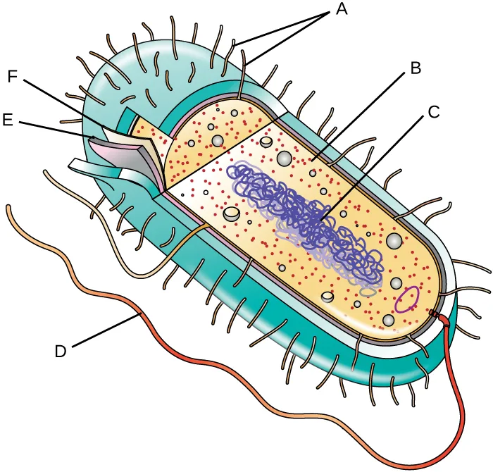
Extended description
Reading the cutaway from outside in: the thick outer layer is unlabeled. The next layer in, thinner, is marked E. An even thinner layer inside that is marked F. Inside the cell, small dots are marked B, and a long tangled loop is marked C. On the outside of the cell, short projections are marked A, and one long projection is marked D.
Using the letters in the figure above, identify each of the six labeled structures of the prokaryotic cell.
Show model answer
Did your answer mention:
Provide some examples of bacterial structures that might be used as antibiotic targets and explain why.
Show model answer
Did your answer mention:
The causative agent of botulism, a deadly form of food poisoning, is an endospore-forming bacterium called Clostridium botulinum. Why might it be difficult to kill this bacterium in contaminated food?
Show model answer
Did your answer mention:
Sort each characteristic of the Vegetative Cells versus Endospores table to the cell type it describes.
Vegetative cells
Endospores
Compare the distinguishing characteristics of bacterial and archaeal cells
Which of the following is true of archaeal membrane phospholipids, unlike those of bacteria?
Recall the linkage chemistry that distinguishes archaeal membrane phospholipids from bacterial and eukaryotic ones, described in the Plasma Membrane section above.Unlike bacterial cell walls, most archaeal cell walls contain a polymer called ________ instead of peptidoglycan.
This polymer differs from peptidoglycan in its subunit chemistry, even though its name signals that it resembles peptidoglycan without truly being the same molecule.How do the ribosomes of archaea compare with those of bacteria and eukaryotes?
Think about which measurement — size or molecular composition — archaeal ribosomes share with bacteria, and which they share with eukaryotes.This section is adapted from Microbiology, Section 3.3: Unique Characteristics of Prokaryotic Cells by Nina Parker, Mark Schneegurt, Anh-Hue Thi Tu, Philip Lister, Brian M. Forster, and OpenStax, © OpenStax, licensed under CC BY-NC-SA 4.0. Access the original for free at openstax.org. Changes: figures re-encoded as WebP, with kind="diagram" or kind="photo" set explicitly on all twenty-five figures (twenty-three ordinary figures plus the two Art Connection images) after inspecting each image, overriding the media manifest’s JPEG-based guess — four of the twenty-five are photographic or micrograph-only (Nucleoid, IncBodies, FimbrPili, ExStaph_img) and the rest are diagrams, including five composite figures (ProkTable, ProkArrang, Sporulation, Capsules, acidfast) whose drawn half is what the caption teaches; alts rewritten for all twenty-five figures to describe what is visibly drawn or photographed rather than reused verbatim, because this chapter’s source alts contain one-word typos (“loo” for “loop” in the ProkCell and Art Connection cell diagrams, “mving” for “moving” and “partices” for “particles” in the Tonicity and Plasmolysis alts, “arrangments” for “arrangements” in the ProkArrang alt, “forma” for “forms” in the Sporulation alt, “techoic” for “teichoic” and “Proins” for “Proteins” in the CellWalls alt, and “Amphitrichouls” for “Amphitrichous” in the FlagellaAr alt) and one content defect (the Plasmolysis alt mislabels its third, hypotonic panel as “hypertonic,” repeating the second panel’s label — reported as a source defect); two one-word typos in source captions corrected in place with no inline note (“carbolfuschin” to carbolfuchsin in the acidfast caption, “polyhydroxybutryrate” to polyhydroxybutyrate in the IncBodies caption; two footnoted author lists print a stray affiliation letter after each surname in the source (“Garnetta et al.” and “Gana, S. Chena, G.J. Jensena”) and are cited here as Garnett et al. and Gan, Chen, and Jensen); longdesc added for eleven labeled diagrams, walking each in reading order; the peptidoglycan key-term definition adds the parentheses around “NAM” that the Glossary appendix omits; two Art Connection figures (PartsProk_img, in the module’s Short Answer set rather than its Critical Thinking set as originally scoped, and ExStaph_img, in the Critical Thinking set) rendered as mediafigures immediately followed by their items inside the Practice group of the objective each serves, with author-written captions describing what each image shows and alts that describe the visible shapes without naming which one answers its paired item; the vegetative-cells-versus-endospores table transcribed as a Markdown table from the table cells, with its link to Section 2.4 kept as an absolute link; feature boxes rendered as callouts; one of the six Check Your Understanding bullets (the cell-wall-advantages bullet of the first box) rendered as a body self-check, with its model answer and rubric assembled from this section’s own preceding text; the Clinical Focus box’s “Jump to the next / Go back to the previous Clinical Focus box” links replaced with two plain sentences linking to Section 3.4 and Section 3.1 (not Section 3.2, where the case’s second part ran); eight footnotes (two of them supporting the same sentence, about fimbriae and pili being used interchangeably, folded into one combined citation) rendered as seven inline parenthetical citations, author, title or article title, publication, and pages, with no bare access URLs or DOIs to drop or keep; cross-references to Section 2.4 rendered as absolute links (four occurrences); the cross-references to the not-yet-authored Using Biochemistry to Identify Microorganisms, How Asexual Prokaryotes Achieve Genetic Diversity, and Mechanisms of Antibacterial Drugs and Pathogen Recognition and Phagocytosis sections, and to the Mechanisms of Microbial Genetics and Control of Microbial Growth chapter introductions, left as plain text naming the section or chapter; the end-of-section Multiple Choice, True/False, Fill in the Blank, and unkeyed Short Answer and Critical Thinking questions adapted into the closing interactive Practice block, sorted under the objective each supports; the “select all that apply” Multiple Choice item on structures important for surface adherence (keyed C, D) rebuilt as a sort-into-bins item, because the site’s multiple-choice component grades a single answer and cannot represent a two-answer key; a model answer for one Short Answer question (the Art Connection labeling item, fs-id1172101988000) and two Critical Thinking questions (antibiotic targets, botulism) is written from this section’s own text, because the source prints no answer key for either set; three filler items for the first objective (a multiple choice and a text-recall item built from this section’s own introductory sentences, plus the section’s own True/False item moved here from a general ribosome-size objective) and three filler items for the fourth objective (two multiple choice items and a text-recall item, all built from this section’s own sentences on archaeal membranes, cell walls, and ribosomes — the ribosome-comparison item was a self-check and is now graded from the module’s own sentence, since one sentence fixes its answer) fill out the two objectives the source’s own exercise set does not reach; key terms compiled from the module’s fifty-seven defined terms and the book’s Glossary appendix (fifty-five taken directly from the glossary; two taken from the module’s own defining sentence — morphology, whose only glossary entry is under the nearest headword cell morphology, used because its sense matches the module’s, and basal body, whose only glossary entry defines the eukaryotic flagellum’s basal body — nine microtubule triplets — a different structure from the bacterial motor this module describes, so the module’s own sentence is used instead). Three of the source’s unkeyed Short Answer and Critical Thinking questions, five Check Your Understanding questions, and one author-written filler question are graded from the module’s own sentences, tables, or figures rather than answered in prose; the sort-into-bins item built from the Vegetative Cells versus Endospores table leaves out the table’s Gram-stain row, whose vegetative-cell entry describes only endospore-producing bacteria; the source prints no key for them: the “morphology versus arrangement,” “what is an inclusion,” “function of an endospore,” “peptidoglycan gram-positive versus gram-negative” (sort-into-bins), and “compare and contrast the four flagellar arrangements” (sort-into-bins, four bins) Check Your Understanding bullets; the Practice group’s hypotonic-environment and chemotaxis Short Answer questions (fs-id1172099614303, fs-id1172099597455), both multiplechoice; and the staphylococci-micrograph Critical Thinking question (fs-id1172101798592), a figure-keyed multiplechoice over the four lettered slides, keyed from the ExStaph_img micrograph itself (panel D: round cells grouped in a dense cluster). A sort-into-bins item built from the Vegetative Cells versus Endospores table is added to the third objective’s Practice group.