Unique Characteristics of Eukaryotic Cells
By the end of this section, you will be able to:
- Explain the distinguishing characteristics of eukaryotic cells
- Describe internal and external structures of eukaryotic cells in terms of their physical structure, chemical structure, and function
- Identify and describe structures and organelles unique to eukaryotic cells
- Compare and contrast similar structures found in prokaryotic and eukaryotic cells
- Describe the processes of eukaryotic mitosis and meiosis, and compare to prokaryotic binary fission
Eukaryotic organisms include protozoans, algae, fungi, plants, and animals. Some eukaryotic cells are independent, single-celled microorganisms, whereas others are part of multicellular organisms. The cells of eukaryotic organisms have several distinguishing characteristics. Above all, eukaryotic cells are defined by the presence of a nucleus surrounded by a complex nuclear membrane. Also, eukaryotic cells are characterized by the presence of membrane-bound organelles in the cytoplasm. Organelles such as mitochondria, the endoplasmic reticulum (ER), Golgi apparatus, lysosomes, and peroxisomes are held in place by the cytoskeleton, an internal network that supports transport of intracellular components and helps maintain cell shape (shown below). The genome of eukaryotic cells is packaged in multiple, rod-shaped chromosomes as opposed to the single, circular-shaped chromosome that characterizes most prokaryotic cells. The table below compares the characteristics of eukaryotic cell structures with those of bacteria and archaea.
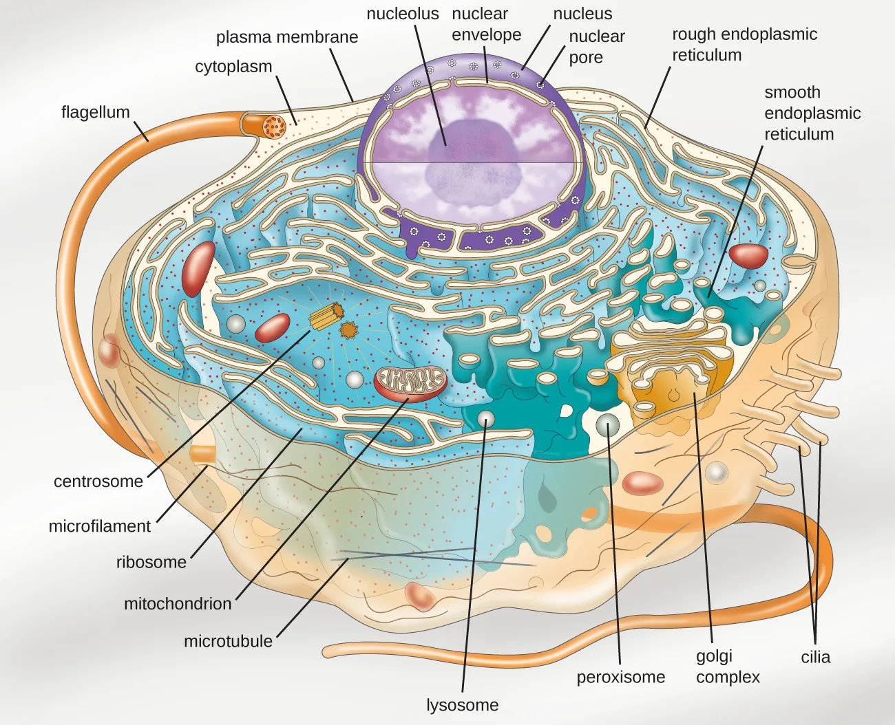
Extended description
Reading the diagram outward from the nucleus: a large central sphere is labeled nucleus, its boundary labeled nuclear envelope with openings labeled nuclear pores, and a smaller sphere inside it labeled nucleolus. Outside the nucleus, folded membrane studded with dots is labeled rough endoplasmic reticulum and ribosomes; the same membrane where it is bare is labeled smooth endoplasmic reticulum. An oval organelle with internal folds is labeled mitochondrion; round organelles are labeled peroxisome and lysosome; a stack of curved membranes is labeled Golgi complex; two short paired tubes are labeled centrosome; a single dot in the cytoplasm, apart from the endoplasmic reticulum, is labeled ribosome. Thin lines running through the cytoplasm are labeled microfilament and microtubule, and the ground substance is labeled cytoplasm. At the cell’s outer boundary, labeled plasma membrane, a long tail-like projection is labeled flagellum and several shorter projections are labeled cilia.
| Cell Structure | Prokaryotes (Bacteria) | Prokaryotes (Archaea) | Eukaryotes |
|---|---|---|---|
| Size | ~0.5–1 µm | ~0.5–1 µm | ~5–20 µm |
| Surface area-to-volume ratio | High | High | Low |
| Nucleus | No | No | Yes |
| Genome characteristics | Single chromosome; circular; haploid; lacks histones | Single chromosome; circular; haploid; contains histones | Multiple chromosomes; linear; haploid or diploid; contains histones |
| Cell division | Binary fission | Binary fission | Mitosis, meiosis |
| Membrane lipid composition | Ester-linked; straight-chain fatty acids; bilayer | Ether-linked; branched isoprenoids; bilayer or monolayer | Ester-linked; straight-chain fatty acids; sterols; bilayer |
| Cell wall composition | Peptidoglycan, or none | Pseudopeptidoglycan, or glycopeptide, or polysaccharide, or protein (S-layer), or none | Cellulose (plants, some algae); chitin (fungi); silica (some algae); most others lack cell walls |
| Motility structures | Rigid spiral flagella composed of flagellin | Rigid spiral flagella composed of archaeal flagellins | Flexible flagella and cilia composed of microtubules |
| Membrane-bound organelles | No | No | Yes |
| Endomembrane system | No | No | Yes (ER, Golgi, lysosomes) |
| Ribosomes | 70S | 70S | 80S in cytoplasm and rough ER; 70S in mitochondria, chloroplasts |
Cell Morphologies
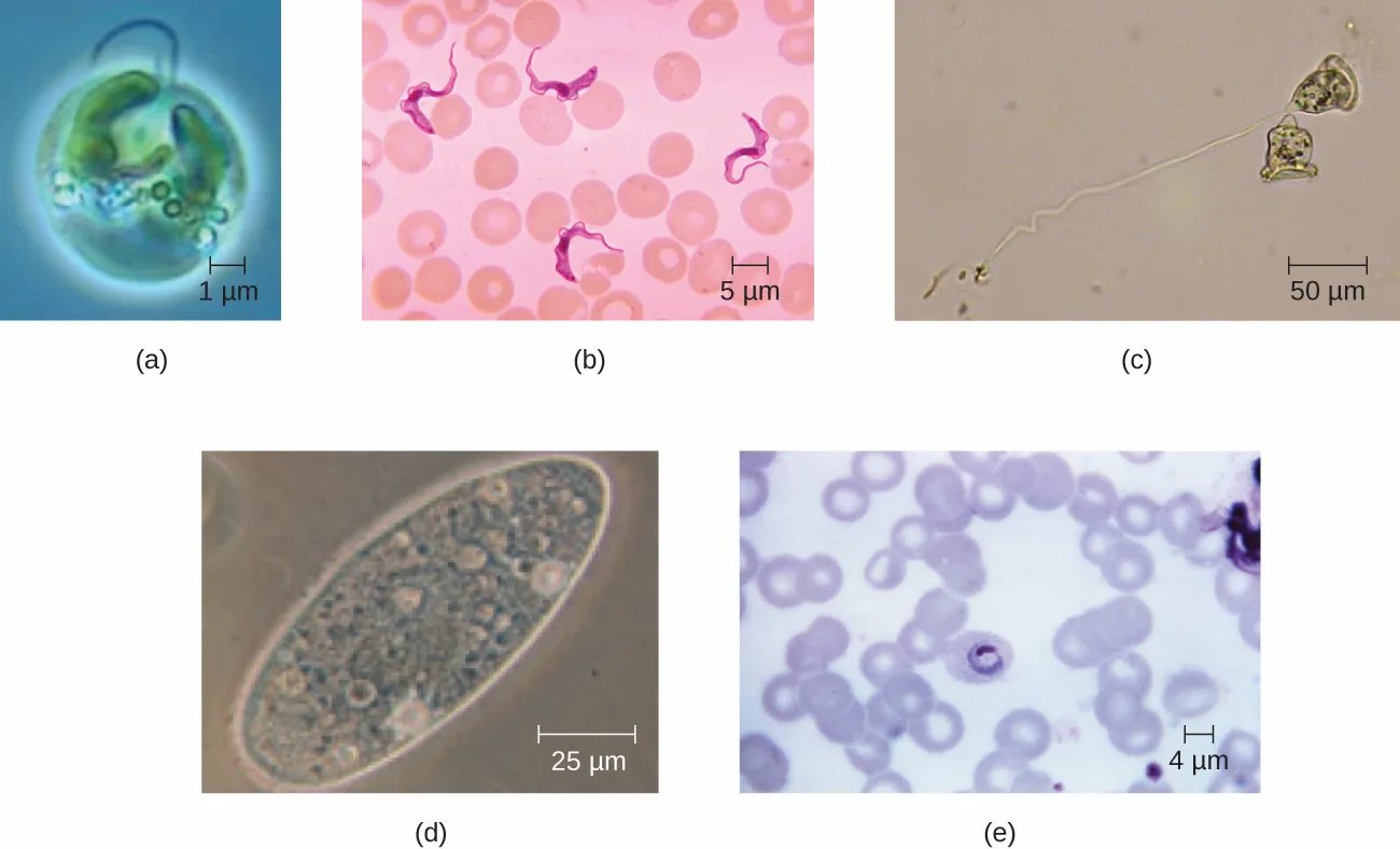
Eukaryotic cells display a wide variety of different cell morphologies. Possible shapes include spheroid, ovoid, cuboidal, cylindrical, flat, lenticular, fusiform, discoidal, crescent, ring stellate, and polygonal (shown below). Some eukaryotic cells are irregular in shape, and some are capable of changing shape. The shape of a particular type of eukaryotic cell may be influenced by factors such as its primary function, the organization of its cytoskeleton, the viscosity of its cytoplasm, the rigidity of its cell membrane or cell wall (if it has one), and the physical pressure exerted on it by the surrounding environment and/or adjoining cells.
Check Your Understanding
Identify two differences between eukaryotic and prokaryotic cells.
Show model answer
Did your answer mention:
Nucleus
Unlike prokaryotic cells, in which DNA is loosely contained in the nucleoid region, eukaryotic cells possess a nucleus, which is surrounded by a complex nuclear membrane that houses the DNA genome (shown below). By containing the cell’s DNA, the nucleus ultimately controls all activities of the cell and also serves an essential role in reproduction and heredity. Eukaryotic cells typically have their DNA organized into multiple linear chromosomes. The DNA within the nucleus is highly organized and condensed to fit inside the nucleus, which is accomplished by wrapping the DNA around proteins called histones.
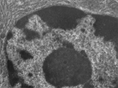
Although most eukaryotic cells have only one nucleus, exceptions exist. For example, protozoans of the genus Paramecium typically have two complete nuclei: a small nucleus that is used for reproduction (micronucleus) and a large nucleus that directs cellular metabolism (macronucleus). Additionally, some fungi transiently form cells with two nuclei, called heterokaryotic cells, during sexual reproduction. Cells whose nuclei divide, but whose cytoplasm does not, are called coenocytes.
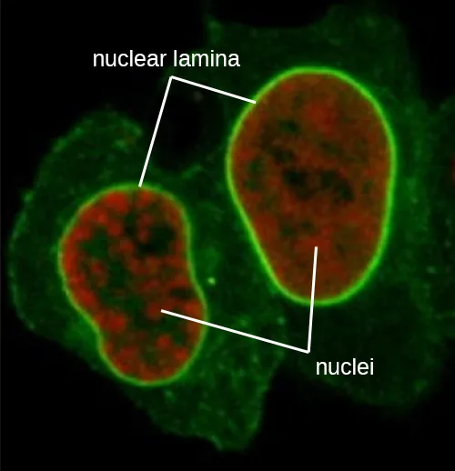
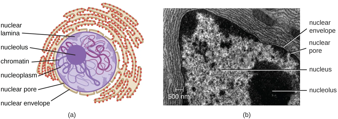
The nucleus is bound by a complex nuclear membrane, often called the nuclear envelope, that consists of two distinct lipid bilayers that are contiguous with each other (shown below). Despite these connections between the inner and outer membranes, each membrane contains unique lipids and proteins on its inner and outer surfaces. The nuclear envelope contains nuclear pores, which are large, rosette-shaped protein complexes that control the movement of materials into and out of the nucleus. The overall shape of the nucleus is determined by the nuclear lamina, a meshwork of intermediate filaments found just inside the nuclear envelope membranes. Outside the nucleus, additional intermediate filaments form a looser mesh and serve to anchor the nucleus in position within the cell.
Eukaryotes are able to multiply through asexual reproduction, during which a single parent cell becomes two identical daughter cells. This process of clonal reproduction is called mitosis. Although mitosis may sound similar to asexual binary fission in prokaryotes, the processes are very different. In contrast to the single chromosome in most prokaryotes, eukaryotic cells possess multiple chromosomes that must be replicated and strategically divided between daughter cells. Therefore, mitosis is a much more complex cellular process than binary fission.
The eukaryotic cell cycle is an ordered and carefully regulated series of events involving cell growth, DNA replication, and cell division to produce two clonal daughter cells. One “turn” or cycle of the cell cycle consist of two general phases: interphase and the mitotic phase (shown below). During interphase, the cell is not dividing, but rather is undergoing normal growth processes and DNA is replicated preparing for cell division. The three stages of interphase are called G1, S, and G2.
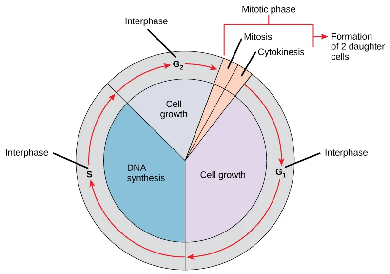
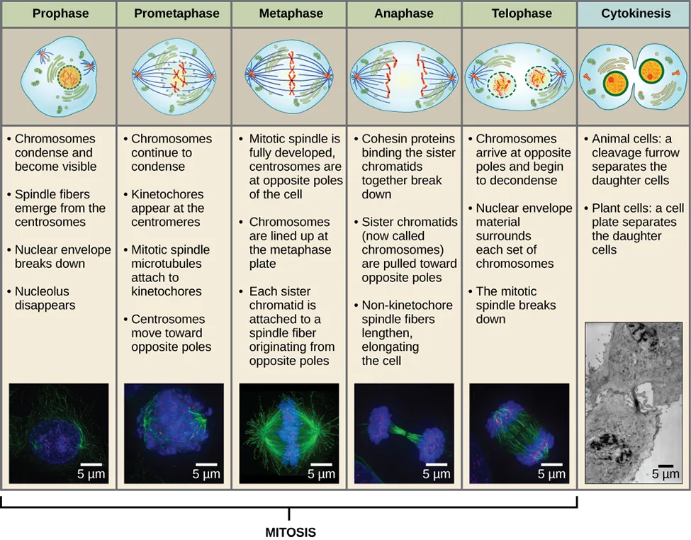
The mitotic phase is a multistep process during which the duplicated chromosomes are aligned, separated, move to opposite poles of the cell, and then are divided into two identical daughter cells. The first portion of the mitotic phase is called karyokinesis, or nuclear division. Karyokinesis is divided into a series of phases—prophase, prometaphase, metaphase, anaphase, and telophase—that result in the division of the cell nucleus (shown below). The second portion of the mitotic phase, called cytokinesis, is the physical separation of the cytoplasmic components into the two daughter cells.
Link to Learning
Go to this University of Arizona website about the stages of mitosis to learn more.
In addition to the mitotic asexual reproduction described above, most eukaryotic microorganisms also have the option of sexual reproduction involving meiosis. Although mitosis and meiosis both require DNA replication, nuclear division, and share procedural similarities, there are important differences between the process and outcomes (shown below).
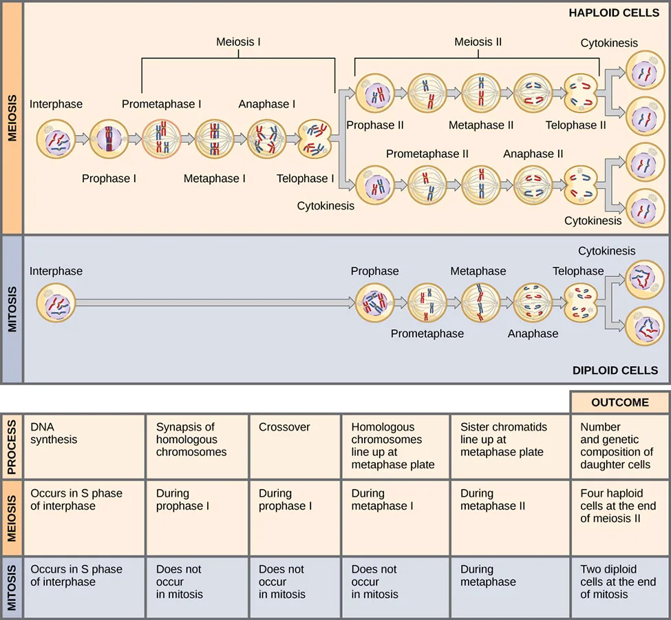
In contrast to the single nuclear division that completes mitosis, meiosis involves two separate nuclear divisions. Rather than creating two clonal daughter cells, the goal of meiosis is to create four genetically-distinct gametes, with each gamete possessing half the number of chromosomes found in the original cell. This strategic chromosome reduction is essential for the fertilization that occurs during sexual reproduction to produce in a zygote with a full complement of chromosomes.
Nucleolus
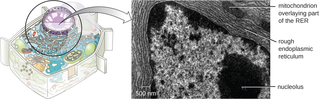
The nucleolus is a dense region within the nucleus where ribosomal RNA (rRNA) biosynthesis occurs. In addition, the nucleolus is also the site where assembly of ribosomes begins. Preribosomal complexes are assembled from rRNA and proteins in the nucleolus; they are then transported out to the cytoplasm, where ribosome assembly is completed (shown below).
Ribosomes
Ribosomes found in eukaryotic organelles such as mitochondria or chloroplasts have 70S ribosomes—the same size as prokaryotic ribosomes. However, nonorganelle-associated ribosomes in eukaryotic cells are 80S ribosomes, composed of a 40S small subunit and a 60S large subunit. In terms of size and composition, this makes them distinct from the ribosomes of prokaryotic cells.
The two types of nonorganelle-associated eukaryotic ribosomes are defined by their location in the cell: free ribosomes and membrane-bound ribosomes. Free ribosomes are found in the cytoplasm and serve to synthesize water-soluble proteins; membrane-bound ribosomes are found attached to the rough endoplasmic reticulum and make proteins for insertion into the cell membrane or proteins destined for export from the cell.
The differences between eukaryotic and prokaryotic ribosomes are clinically relevant because certain antibiotic drugs are designed to target one or the other. For example, cycloheximide targets eukaryotic action, whereas chloramphenicol targets prokaryotic ribosomes (A.E. Barnhill, M.T. Brewer, S.A. Carlson, “Adverse Effects of Antimicrobials via Predictable or Idiosyncratic Inhibition of Host Mitochondrial Components,” Antimicrobial Agents and Chemotherapy 56 no. 8 [2012]: 4046–4051). Since human cells are eukaryotic, they generally are not harmed by antibiotics that destroy the prokaryotic ribosomes in bacteria. However, sometimes negative side effects may occur because mitochondria in human cells contain prokaryotic ribosomes.
Endomembrane System
The endomembrane system, unique to eukaryotic cells, is a series of membranous tubules, sacs, and flattened disks that synthesize many cell components and move materials around within the cell (shown below). Because of their larger cell size, eukaryotic cells require this system to transport materials that cannot be dispersed by diffusion alone. The endomembrane system comprises several organelles and connections between them, including the endoplasmic reticulum, Golgi apparatus, lysosomes, and vesicles.
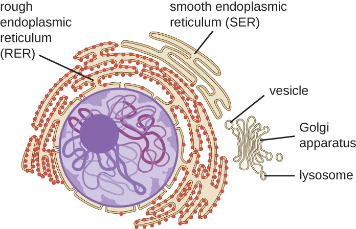
Extended description
Reading outward from the center: the nucleus is drawn as a purple sphere holding a darker sphere and threadlike loops, none of them labeled in this figure. Its boundary is continuous with a folded membrane network outside the nucleus — the part studded with dots is the rough endoplasmic reticulum (RER) and the bare part is the smooth endoplasmic reticulum (SER), and the two are continuous with each other. To one side, a small vesicle sits near a stack of curved membranes labeled the Golgi apparatus, next to a separate sac labeled lysosome.
Endoplasmic Reticulum
The endoplasmic reticulum (ER) is an interconnected array of tubules and cisternae (flattened sacs) with a single lipid bilayer (shown below). The spaces inside of the cisternae are called lumen of the ER. There are two types of ER, rough endoplasmic reticulum (RER) and smooth endoplasmic reticulum (SER). These two different types of ER are sites for the synthesis of distinctly different types of molecules. RER is studded with ribosomes bound on the cytoplasmic side of the membrane. These ribosomes make proteins destined for the plasma membrane. Following synthesis, these proteins are inserted into the membrane of the RER. Small sacs of the RER containing these newly synthesized proteins then bud off as transport vesicles and move either to the Golgi apparatus for further processing, directly to the plasma membrane, to the membrane of another organelle, or out of the cell. Transport vesicles are single-lipid, bilayer, membranous spheres with hollow interiors that carry molecules. SER does not have ribosomes and, therefore, appears “smooth.” It is involved in biosynthesis of lipids, carbohydrate metabolism, and detoxification of toxic compounds within the cell.
Golgi Apparatus
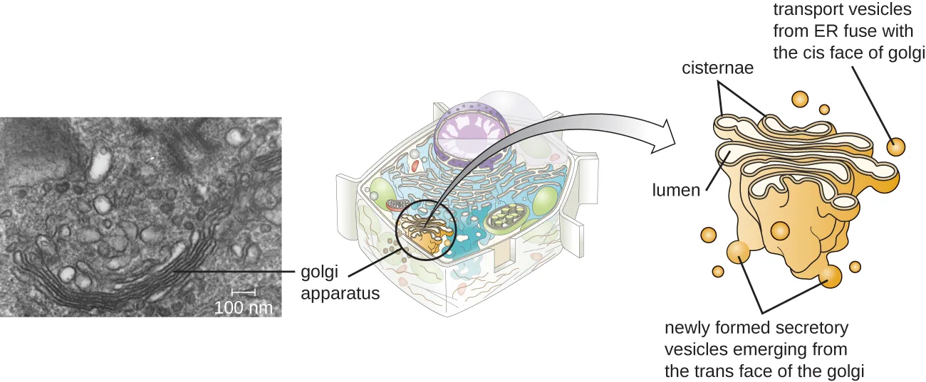
The Golgi apparatus was discovered within the endomembrane system in 1898 by Italian scientist Camillo Golgi (1843–1926), who developed a novel staining technique that showed stacked membrane structures within nerve cells. (Source note: the source says Golgi saw these structures in the cells of Plasmodium, the malaria parasite. Golgi’s 1898 report described the “internal reticular apparatus” in neurons of the cerebellum and spinal ganglia; his malaria work was separate and earlier.) The Golgi apparatus is composed of a series of membranous disks called dictyosomes, each having a single lipid bilayer, that are stacked together (shown below).
Enzymes in the Golgi apparatus modify lipids and proteins transported from the ER to the Golgi, often adding carbohydrate components to them, producing glycolipids, glycoproteins, or proteoglycans. Glycolipids and glycoproteins are often inserted into the plasma membrane and are important for signal recognition by other cells or infectious particles. Different types of cells can be distinguished from one another by the structure and arrangement of the glycolipids and glycoproteins contained in their plasma membranes. These glycolipids and glycoproteins commonly also serve as cell surface receptors.
Transport vesicles leaving the ER fuse with a Golgi apparatus on its receiving, or cis, face. The proteins are processed within the Golgi apparatus, and then additional transport vesicles containing the modified proteins and lipids pinch off from the Golgi apparatus on its outgoing, or trans, face. These outgoing vesicles move to and fuse with the plasma membrane or the membrane of other organelles.
Exocytosis is the process by which secretory vesicles (spherical membranous sacs) release their contents to the cell’s exterior (shown below). All cells have constitutive secretory pathways in which secretory vesicles transport soluble proteins that are released from the cell continually (constitutively). Certain specialized cells also have regulated secretory pathways, which are used to store soluble proteins in secretory vesicles. Regulated secretion involves substances that are only released in response to certain events or signals. For example, certain cells of the human immune system (e.g., mast cells) secrete histamine in response to the presence of foreign objects or pathogens in the body. Histamine is a compound that triggers various mechanisms used by the immune system to eliminate pathogens.
Lysosomes
In 1955, Belgian scientist Christian de Duve (1917–2013) discovered lysosomes, (Source note: the source says “in the 1960s”; de Duve’s group proposed the lysosome in 1955, and it was peroxisomes that his laboratory characterized in the 1960s.) membrane-bound organelles of the endomembrane system that contain digestive enzymes. Certain types of eukaryotic cells use lysosomes to break down various particles, such as food, damaged organelles or cellular debris, microorganisms, or immune complexes. Compartmentalization of the digestive enzymes within the lysosome allows the cell to efficiently digest matter without harming the cytoplasmic components of the cell.
Check Your Understanding
Name the components of the endomembrane system and describe the function of each component.
Endoplasmic reticulum
Golgi apparatus
Lysosomes
Peroxisomes
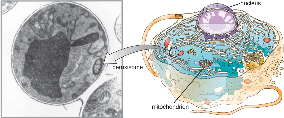
Christian de Duve is also credited with the discovery of peroxisomes, membrane-bound organelles that are not part of the endomembrane system (shown below). Peroxisomes form independently in the cytoplasm from the synthesis of peroxin proteins by free ribosomes and the incorporation of these peroxin proteins into existing peroxisomes. Growing peroxisomes then divide by a process similar to binary fission.
Peroxisomes were first named for their ability to produce hydrogen peroxide, a highly reactive molecule that helps to break down molecules such as uric acid, amino acids, and fatty acids. Peroxisomes also possess the enzyme catalase, which can degrade hydrogen peroxide. Along with the SER, peroxisomes also play a role in lipid biosynthesis. Like lysosomes, the compartmentalization of these degradative molecules within an organelle helps protect the cytoplasmic contents from unwanted damage.
The peroxisomes of certain organisms are specialized to meet their particular functional needs. For example, glyoxysomes are modified peroxisomes of yeasts and plant cells that perform several metabolic functions, including the production of sugar molecules. Similarly, glycosomes are modified peroxisomes made by certain trypanosomes, the pathogenic protozoans that cause Chagas disease and African sleeping sickness.
Cytoskeleton
Eukaryotic cells have an internal cytoskeleton made of microfilaments, intermediate filaments, and microtubules. This matrix of fibers and tubes provides structural support as well as a network over which materials can be transported within the cell and on which organelles can be anchored (shown below). For example, the process of exocytosis involves the movement of a vesicle via the cytoskeletal network to the plasma membrane, where it can release its contents.
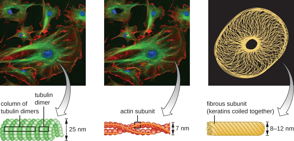
Microfilaments are composed of two intertwined strands of actin, each composed of actin monomers forming filamentous cables 6 nm in diameter (Fuchs E, Cleveland DW, “A Structural Scaffolding of Intermediate Filaments in Health and Disease,” Science 279 no. 5350 [1998]: 514–519) (shown below). The actin filaments work together with motor proteins, like myosin, to effect muscle contraction in animals or the amoeboid movement of some eukaryotic microbes. In ameboid organisms, actin can be found in two forms: a stiffer, polymerized, gel form and a more fluid, unpolymerized soluble form. Actin in the gel form creates stability in the ectoplasm, the gel-like area of cytoplasm just inside the plasma membrane of ameboid protozoans.
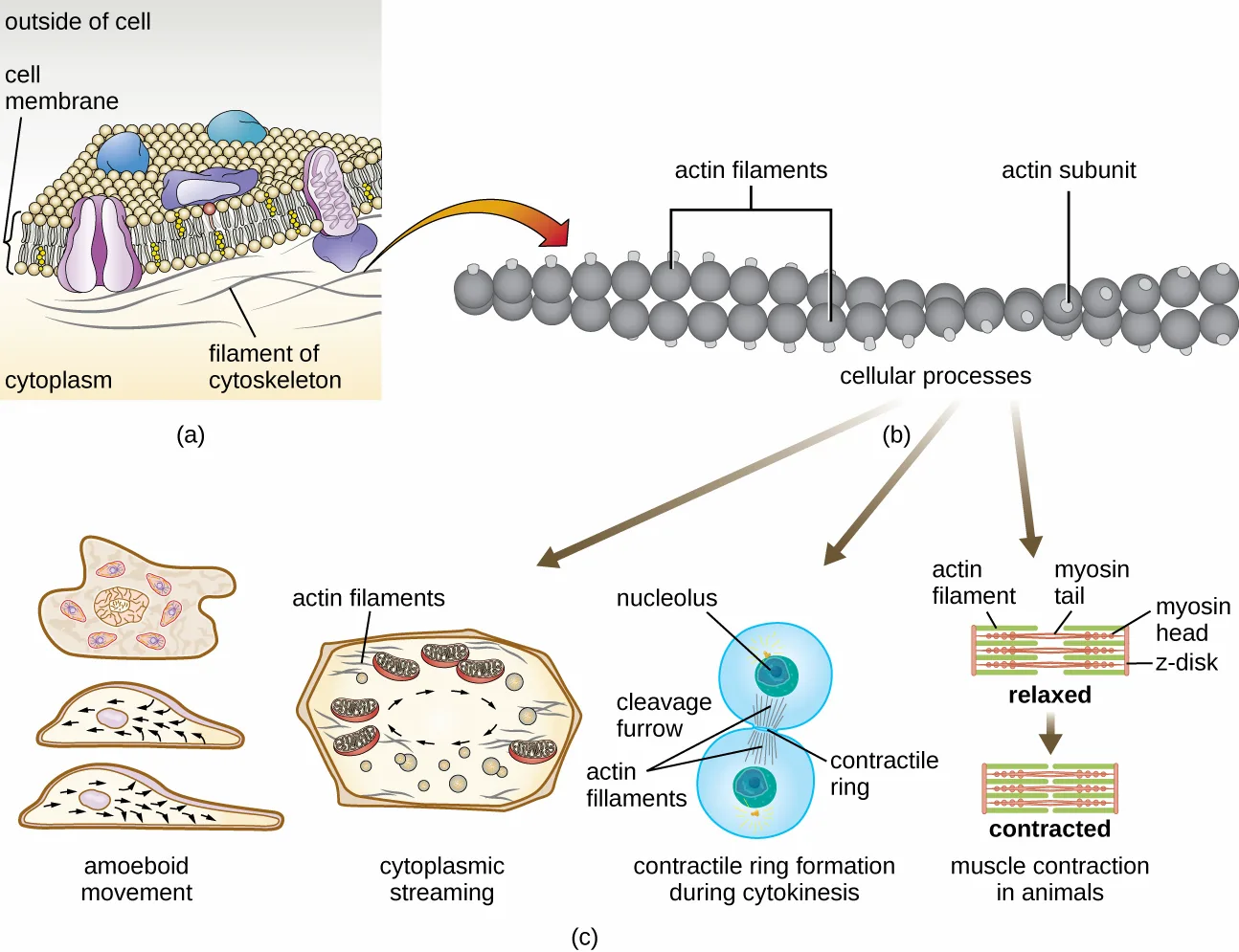
Temporary extensions of the cytoplasmic membrane called pseudopodia (meaning “false feet”) are produced through the forward flow of soluble actin filaments into the pseudopodia, followed by the gel-sol cycling of the actin filaments, resulting in cell motility. Once the cytoplasm extends outward, forming a pseudopodium, the remaining cytoplasm flows up to join the leading edge, thereby creating forward locomotion. Beyond amoeboid movement, microfilaments are also involved in a variety of other processes in eukaryotic cells, including cytoplasmic streaming (the movement or circulation of cytoplasm within the cell), cleavage furrow formation during cell division, and muscle movement in animals (shown below). These functions are the result of the dynamic nature of microfilaments, which can polymerize and depolymerize relatively easily in response to cellular signals, and their interactions with molecular motors in different types of eukaryotic cells.
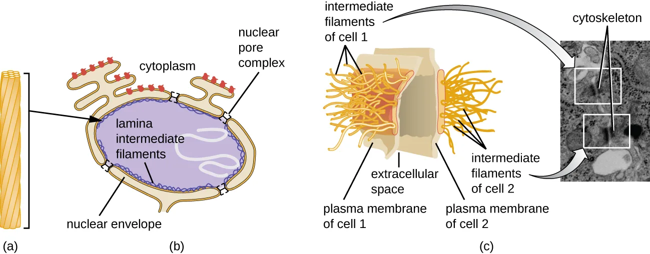
Intermediate filaments (shown below) are a diverse group of cytoskeletal filaments that act as cables within the cell. They are termed “intermediate” because their 10-nm diameter is thicker than that of actin but thinner than that of microtubules (E. Fuchs, D.W. Cleveland, “A Structural Scaffolding of Intermediate Filaments in Health and Disease,” Science 279 no. 5350 [1998]: 514–519). They are composed of several strands of polymerized subunits that, in turn, are made up of a wide variety of monomers. Intermediate filaments tend to be more permanent in the cell and maintain the position of the nucleus. They also form the nuclear lamina (lining or layer) just inside the nuclear envelope. Additionally, intermediate filaments play a role in anchoring cells together in animal tissues. The intermediate filament protein desmin is found in desmosomes, the protein structures that join muscle cells together and help them resist external physical forces. The intermediate filament protein keratin is a structural protein found in hair, skin, and nails.
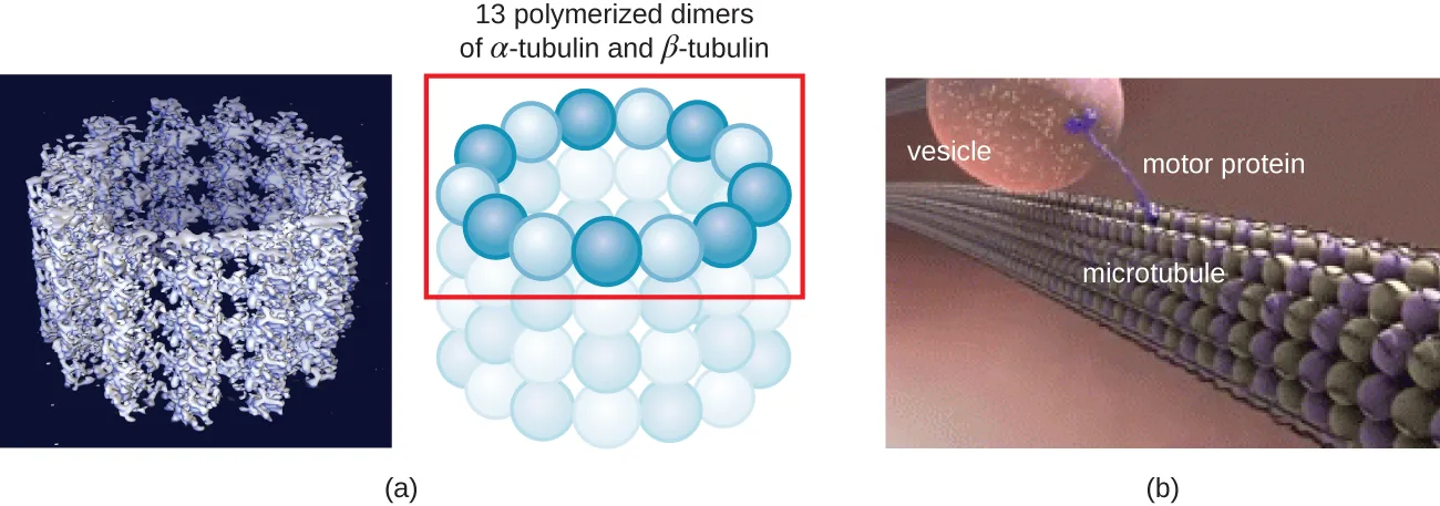
Microtubules (shown below) are a third type of cytoskeletal fiber composed of tubulin dimers (α tubulin and β tubulin). These form hollow tubes 23 nm in diameter that are used as girders within the cytoskeleton (E. Fuchs, D.W. Cleveland, “A Structural Scaffolding of Intermediate Filaments in Health and Disease,” Science 279 no. 5350 [1998]: 514–519). Like microfilaments, microtubules are dynamic and have the ability to rapidly assemble and disassemble. Microtubules also work with motor proteins (such as dynein and kinesin) to move organelles and vesicles around within the cytoplasm. Additionally, microtubules are the main components of eukaryotic flagella and cilia, composing both the filament and the basal body components (shown below).
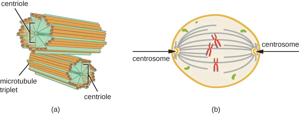
In addition, microtubules are involved in cell division, forming the mitotic spindle that serves to separate chromosomes during mitosis and meiosis. The mitotic spindle is produced by two centrosomes, which are essentially microtubule-organizing centers, at opposite ends of the cell. Each centrosome is composed of a pair of centrioles positioned at right angles to each other, and each centriole is an array of twenty-seven parallel microtubules arranged in triplets (shown below).
Check Your Understanding
Compare and contrast the three types of cytoskeletal structures described in this section.
Microfilaments
Intermediate filaments
Microtubules
Mitochondria
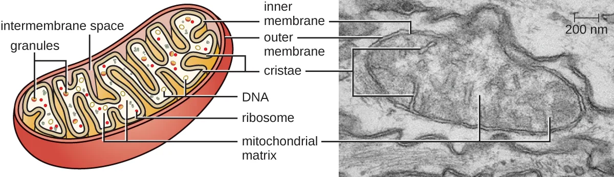
The large, complex organelles in which aerobic cellular respiration occurs in eukaryotic cells are called mitochondria (shown below). The term “mitochondrion” was first coined by German microbiologist Carl Benda in 1898 and was later connected with the process of respiration by Otto Warburg in 1913. Scientists during the 1960s discovered that mitochondria have their own genome and 70S ribosomes. The mitochondrial genome was found to be bacterial, when it was sequenced in 1976. These findings ultimately supported the endosymbiotic theory proposed by Lynn Margulis, which states that mitochondria originally arose through an endosymbiotic event in which a bacterium capable of aerobic cellular respiration was taken up by phagocytosis into a host cell and remained as a viable intracellular component.
Each mitochondrion has two lipid membranes. The outer membrane is a remnant of the original host cell’s membrane structures. The inner membrane was derived from the bacterial plasma membrane. The electron transport chain for aerobic respiration uses integral proteins embedded in the inner membrane. The mitochondrial matrix, corresponding to the location of the original bacterium’s cytoplasm, is the current location of many metabolic enzymes. It also contains mitochondrial DNA and 70S ribosomes. Invaginations of the inner membrane, called cristae, evolved to increase surface area for the location of biochemical reactions. The folding patterns of the cristae differ among various types of eukaryotic cells and are used to distinguish different eukaryotic organisms from each other.
Chloroplasts
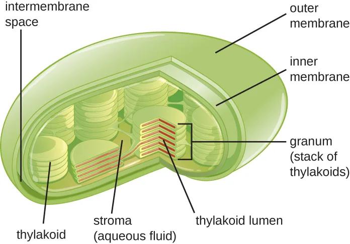
Plant cells and algal cells contain chloroplasts, the organelles in which photosynthesis occurs (shown below). All chloroplasts have at least three membrane systems: the outer membrane, the inner membrane, and the thylakoid membrane system. Inside the outer and inner membranes is the chloroplast stroma, a gel-like fluid that makes up much of a chloroplast’s volume, and in which the thylakoid system floats. The thylakoid system is a highly dynamic collection of folded membrane sacs. It is where the green photosynthetic pigment chlorophyll is found and the light reactions of photosynthesis occur. In most plant chloroplasts, the thylakoids are arranged in stacks called grana (singular: granum), whereas in some algal chloroplasts, the thylakoids are free floating.
Other organelles similar to mitochondria have arisen in other types of eukaryotes, but their roles differ. Hydrogenosomes are found in some anaerobic eukaryotes and serve as the location of anaerobic hydrogen production. Hydrogenosomes typically lack their own DNA and ribosomes. Kinetoplasts are a variation of the mitochondria found in some eukaryotic pathogens. In these organisms, each cell has a single, long, branched mitochondrion in which kinetoplast DNA, organized as multiple circular pieces of DNA, is found concentrated at one pole of the cell.
Micro Connection. Mitochondria-Related Organelles in Protozoan Parasites
Many protozoans, including several protozoan parasites that cause infections in humans, can be identified by their unusual appearance. Distinguishing features may include complex cell morphologies, the presence of unique organelles, or the absence of common organelles. The protozoan parasites Giardia lamblia and Trichomonas vaginalis are two examples.
G. lamblia, a frequent cause of diarrhea in humans and many other animals, is an anaerobic parasite that possesses two nuclei and several flagella. Its Golgi apparatus and endoplasmic reticulum are greatly reduced, and it lacks mitochondria completely. However, it does have organelles known as mitosomes, double-membrane-bound organelles that appear to be severely reduced mitochondria. This has led scientists to believe that G. lamblia’s ancestors once possessed mitochondria that evolved to become mitosomes. T. vaginalis, which causes the sexually transmitted infection vaginitis, is another protozoan parasite that lacks conventional mitochondria. Instead, it possesses hydrogenosomes, mitochondrial-related, double-membrane-bound organelles that produce molecular hydrogen used in cellular metabolism. Scientists believe that hydrogenosomes, like mitosomes, also evolved from mitochondria (N. Yarlett, J.H.P. Hackstein, “Hydrogenosomes: One Organelle, Multiple Origins,” BioScience 55 no. 8 [2005]: 657–658).
Plasma Membrane
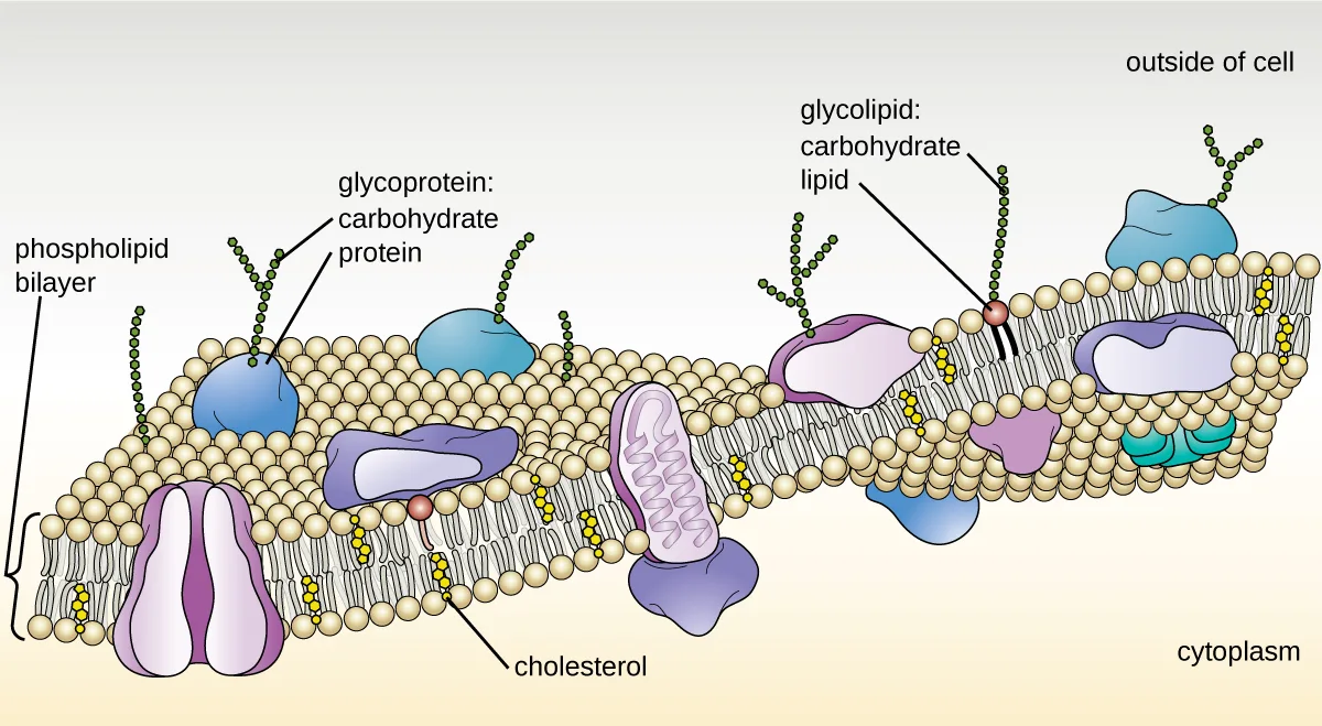
The plasma membrane of eukaryotic cells is similar in structure to the prokaryotic plasma membrane in that it is composed mainly of phospholipids forming a bilayer with embedded peripheral and integral proteins (shown below). These membrane components move within the plane of the membrane according to the fluid mosaic model. However, unlike the prokaryotic membrane, eukaryotic membranes contain sterols, including cholesterol, that alter membrane fluidity. Additionally, many eukaryotic cells contain some specialized lipids, including sphingolipids, which are thought to play a role in maintaining membrane stability as well as being involved in signal transduction pathways and cell-to-cell communication.
Membrane Transport Mechanisms
The processes of simple diffusion, facilitated diffusion, and active transport are used in both eukaryotic and prokaryotic cells. However, eukaryotic cells also have the unique ability to perform various types of endocytosis, the uptake of matter through plasma membrane invagination and vacuole/vesicle formation (shown below). A type of endocytosis involving the engulfment of large particles through membrane invagination is called phagocytosis, which means “cell eating.” In phagocytosis, particles (or other cells) are enclosed in a pocket within the membrane, which then pinches off from the membrane to form a vacuole that completely surrounds the particle. Another type of endocytosis is called pinocytosis, which means “cell drinking.” In pinocytosis, small, dissolved materials and liquids are taken into the cell through small vesicles. Saprophytic fungi, for example, obtain their nutrients from dead and decaying matter largely through pinocytosis.
Receptor-mediated endocytosis is a type of endocytosis that is initiated by specific molecules called ligands when they bind to cell surface receptors on the membrane. Receptor-mediated endocytosis is the mechanism that peptide and amine-derived hormones use to enter cells and is also used by various viruses and bacteria for entry into host cells.
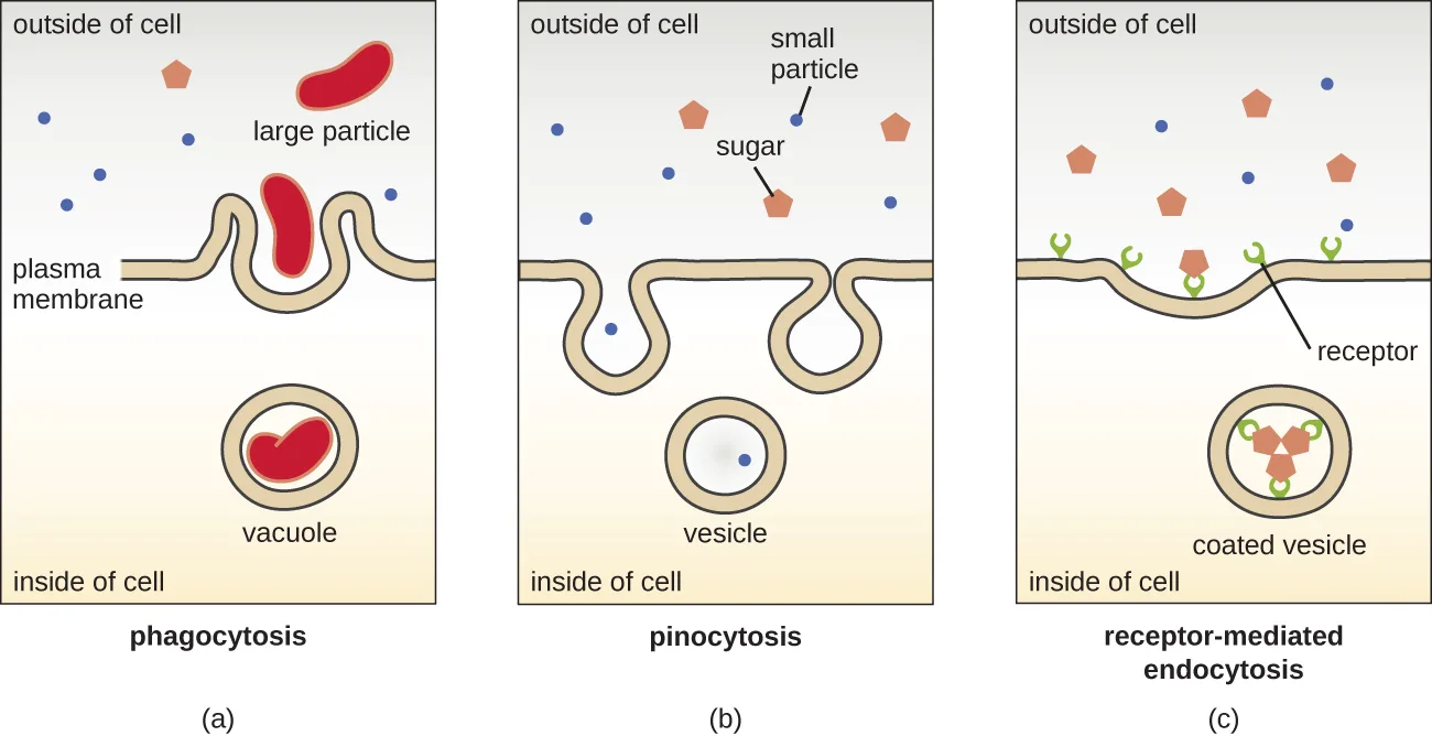
The process by which secretory vesicles release their contents to the cell’s exterior is called exocytosis. Vesicles move toward the plasma membrane and then meld with the membrane, ejecting their contents out of the cell. Exocytosis is used by cells to remove waste products and may also be used to release chemical signals that can be taken up by other cells.
Cell Wall
In addition to a plasma membrane, some eukaryotic cells have a cell wall. Cells of fungi, algae, plants, and even some protists have cell walls. Depending upon the type of eukaryotic cell, cell walls can be made of a wide range of materials, including cellulose (plants); biogenic silica, calcium carbonate, agar, and carrageenan (protists and algae); or chitin (fungi). In general, all cell walls provide structural stability for the cell and protection from environmental stresses such as desiccation, changes in osmotic pressure, and traumatic injury (M. Dudzick, “Protists,” OpenStax CNX, November 27, 2013).
Extracellular Matrix
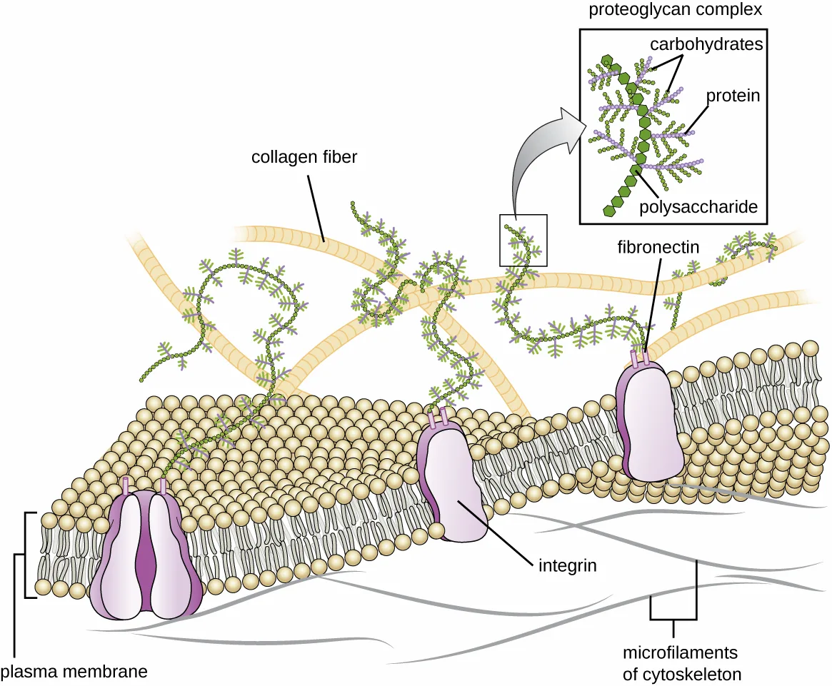
Cells of animals and some protozoans do not have cell walls to help maintain shape and provide structural stability. Instead, these types of eukaryotic cells produce an extracellular matrix for this purpose. They secrete a sticky mass of carbohydrates and proteins into the spaces between adjacent cells (shown below). Some protein components assemble into a basement membrane to which the remaining extracellular matrix components adhere. Proteoglycans typically form the bulky mass of the extracellular matrix while fibrous proteins, like collagen, provide strength. Both proteoglycans and collagen are attached to fibronectin proteins, which, in turn, are attached to integrin proteins. These integrin proteins interact with transmembrane proteins in the plasma membranes of eukaryotic cells that lack cell walls.
In animal cells, the extracellular matrix allows cells within tissues to withstand external stresses and transmits signals from the outside of the cell to the inside. The amount of extracellular matrix is quite extensive in various types of connective tissues, and variations in the extracellular matrix can give different types of tissues their distinct properties. In addition, a host cell’s extracellular matrix is often the site where microbial pathogens attach themselves to establish infection. For example, Streptococcus pyogenes, the bacterium that causes strep throat and various other infections, binds to fibronectin in the extracellular matrix of the cells lining the oropharynx (upper region of the throat).
Flagella and Cilia
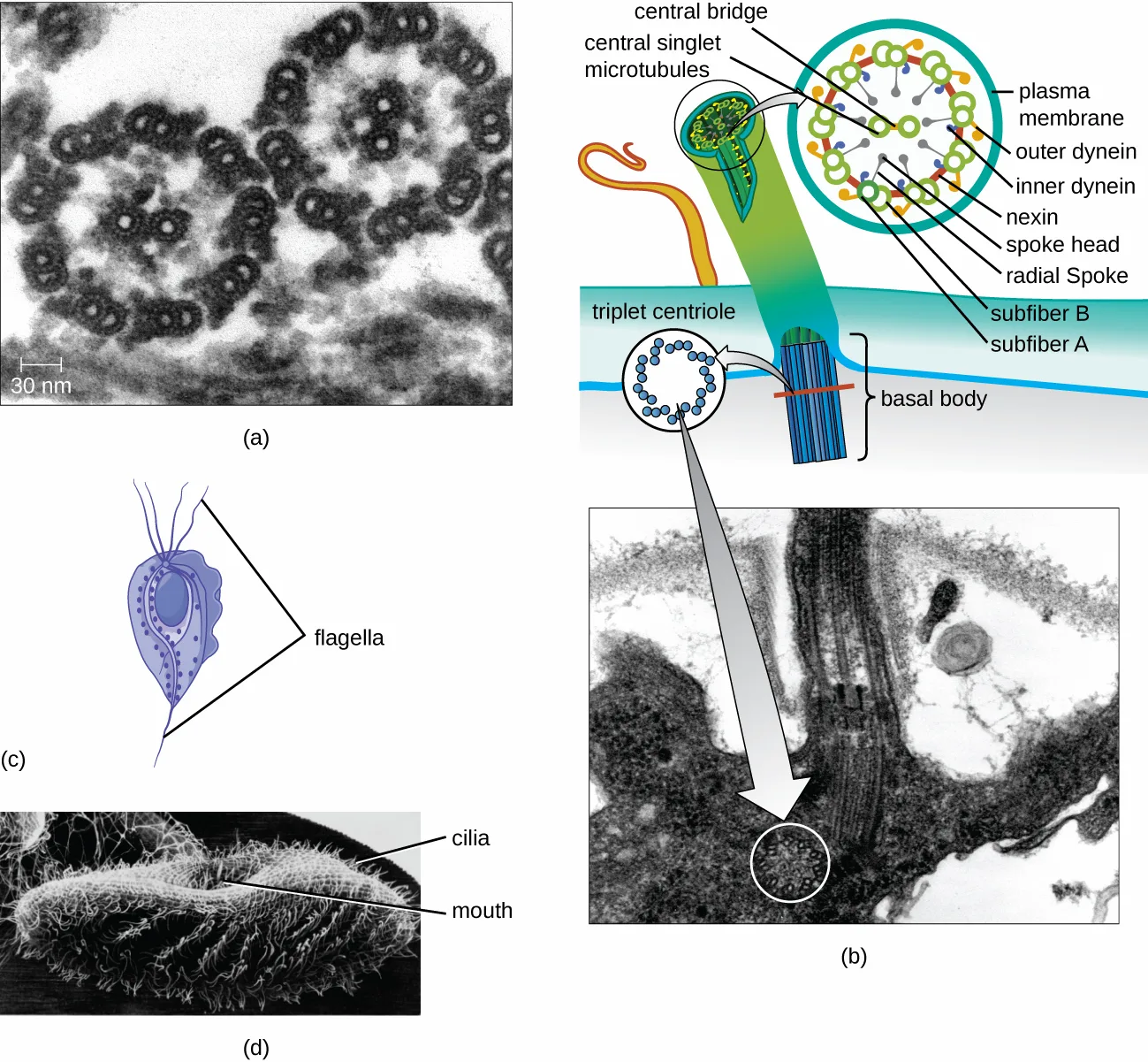
Some eukaryotic cells use flagella for locomotion; however, eukaryotic flagella are structurally distinct from those found in prokaryotic cells. Whereas the prokaryotic flagellum is a stiff, rotating structure, a eukaryotic flagellum is more like a flexible whip composed of nine parallel pairs of microtubules surrounding a central pair of microtubules. This arrangement is referred to as a 9+2 array (shown below). The parallel microtubules use dynein motor proteins to move relative to each other, causing the flagellum to bend.
Cilia (singular: cilium) are a similar external structure found in some eukaryotic cells. Unique to eukaryotes, cilia are shorter than flagella and often cover the entire surface of a cell; however, they are structurally similar to flagella (a 9+2 array of microtubules) and use the same mechanism for movement. A structure called a basal body is found at the base of each cilium and flagellum. The basal body, which attaches the cilium or flagellum to the cell, is composed of an array of triplet microtubules similar to that of a centriole but embedded in the plasma membrane. Because of their shorter length, cilia use a rapid, flexible, waving motion. In addition to motility, cilia may have other functions such as sweeping particles past or into cells. For example, ciliated protozoans use the sweeping of cilia to move food particles into their mouthparts, and ciliated cells in the mammalian respiratory tract beat in synchrony to sweep mucus and debris up and out of the lungs (shown below).
Extended description
Panel (b), reading from the top: a cross-section circle of the flagellum shows nine outer doublets of microtubules, each doublet labeled subfiber A and subfiber B, joined to its neighbors by nexin and carrying outer dynein and inner dynein arms; radial spokes with spoke heads run inward to two central singlet microtubules joined by a central bridge; the whole is enclosed by the plasma membrane. Below, the flagellum enters the cell at a basal body, whose own cross-section circle is labeled triplet centriole. A matching electron micrograph beneath the drawing shows the basal body in longitudinal section, with a circled cross-section of the triplet array.
Check Your Understanding
Explain how the cellular envelope of eukaryotic cells compares to that of prokaryotic cells.
Show model answer
Did your answer mention:
Explain the difference between eukaryotic and prokaryotic flagella.
Eukaryotic
Prokaryotic
Clinical Focus. Resolution
Since amoxicillin has not resolved Barbara’s case of pneumonia, the PA prescribes another antibiotic, azithromycin, which targets bacterial ribosomes rather than peptidoglycan. After taking the azithromycin as directed, Barbara’s symptoms resolve and she finally begins to feel like herself again. Presuming no drug resistance to amoxicillin was involved, and given the effectiveness of azithromycin, the causative agent of Barbara’s pneumonia is most likely Mycoplasma pneumoniae. Even though this bacterium is a prokaryotic cell, it is not inhibited by amoxicillin because it does not have a cell wall and, therefore, does not make peptidoglycan.
This resolves the case that began in Spontaneous Generation.
Summary
- Eukaryotic cells are defined by the presence of a nucleus containing the DNA genome and bound by a nuclear membrane (or nuclear envelope) composed of two lipid bilayers that regulate transport of materials into and out of the nucleus through nuclear pores.
- Eukaryotic cell morphologies vary greatly and may be maintained by various structures, including the cytoskeleton, the cell membrane, and/or the cell wall
- The nucleolus, located in the nucleus of eukaryotic cells, is the site of ribosomal synthesis and the first stages of ribosome assembly.
- Eukaryotic cells contain 80S ribosomes in the rough endoplasmic reticulum (membrane bound-ribosomes) and cytoplasm (free ribosomes). They contain 70s ribosomes in mitochondria and chloroplasts.
- Eukaryotic cells have evolved an endomembrane system, containing membrane-bound organelles involved in transport. These include vesicles, the endoplasmic reticulum, and the Golgi apparatus.
- The smooth endoplasmic reticulum plays a role in lipid biosynthesis, carbohydrate metabolism, and detoxification of toxic compounds. The rough endoplasmic reticulum contains membrane-bound 80S ribosomes that synthesize proteins destined for the cell membrane
- The Golgi apparatus processes proteins and lipids, typically through the addition of sugar molecules, producing glycoproteins or glycolipids, components of the plasma membrane that are used in cell-to-cell communication.
- Lysosomes contain digestive enzymes that break down small particles ingested by endocytosis, large particles or cells ingested by phagocytosis, and damaged intracellular components.
- The cytoskeleton, composed of microfilaments, intermediate filaments, and microtubules, provides structural support in eukaryotic cells and serves as a network for transport of intracellular materials.
- Centrosomes are microtubule-organizing centers important in the formation of the mitotic spindle in mitosis.
- Mitochondria are the site of cellular respiration. They have two membranes: an outer membrane and an inner membrane with cristae. The mitochondrial matrix, within the inner membrane, contains the mitochondrial DNA, 70S ribosomes, and metabolic enzymes.
- The plasma membrane of eukaryotic cells is structurally similar to that found in prokaryotic cells, and membrane components move according to the fluid mosaic model. However, eukaryotic membranes contain sterols, which alter membrane fluidity, as well as glycoproteins and glycolipids, which help the cell recognize other cells and infectious particles.
- In addition to active transport and passive transport, eukaryotic cell membranes can take material into the cell via endocytosis, or expel matter from the cell via exocytosis.
- Cells of fungi, algae, plants, and some protists have a cell wall, whereas cells of animals and some protozoans have a sticky extracellular matrix that provides structural support and mediates cellular signaling.
- Eukaryotic flagella are structurally distinct from prokaryotic flagella but serve a similar purpose (locomotion). Cilia are structurally similar to eukaryotic flagella, but shorter; they may be used for locomotion, feeding, or movement of extracellular particles.
Key terms
- cytoskeleton — a network of filaments or tubules in the eukaryotic cell that provides shape and structural support for cells; aids movement of materials throughout the cell.
- nucleus — a membrane-bound structure of eukaryotic cells that houses the DNA genome.
- coenocytes — multinucleated eukaryotic cell that forms as a result of multiple rounds of nuclear division without the accompanying division of the plasma membrane.
- nuclear membrane — a structure defining the boundary of the nucleus; composed of two distinct lipid bilayers that are contiguous with each other and with the endoplasmic reticulum.
- nuclear envelope — (also called the nuclear membrane) a structure defining the boundary of the nucleus; composed of two distinct lipid bilayers that are contiguous with each other and with the endoplasmic reticulum.
- nuclear lamina — a meshwork of intermediate filaments (mainly lamins) found just inside the nuclear envelope; provides structural support to the nucleus.
- mitosis — (also, karyokinesis) period of the cell cycle during which the duplicated chromosomes are separated into identical nuclei; includes prophase, prometaphase, metaphase, anaphase, and telophase.
- interphase — period of the cell cycle leading up to mitosis; includes G1, S, and G2 phases (the interim period between two consecutive cell divisions).
- mitotic phase — period of the cell cycle during which duplicated chromosomes are distributed into two nuclei and cytoplasmic contents are divided; includes karyokinesis (mitosis) and cytokinesis.
- karyokinesis — mitotic nuclear division.
- cytokinesis — division of the cytoplasm following mitosis that forms two daughter cells.
- meiosis — a two-stage nuclear division process that results in four genetically distinct gametes.
- nucleolus — a dense region within the nucleus where ribosomal RNA biosynthesis occurs and preribosomal complexes are made.
- 80S ribosomes — cytoplasmic eukaryotic ribosome composed of 60S and 40S subunits.
- free ribosomes — eukaryotic 80S ribosome found in the cytoplasm; synthesizes water-soluble proteins.
- membrane-bound ribosomes — 80S eukaryotic ribosome attached to rough endoplasmic reticulum.
- endomembrane system — a series of organelles (endoplasmic reticulum, Golgi apparatuses, lysosomes, and transport vesicles) arranged as membranous tubules, sacs, and disks that synthesize many cell components.
- endoplasmic reticulum (ER) — part of the endomembrane system that is an interconnected array of tubules and flattened sacs with a single lipid bilayer that may be either rough or smooth; important in synthesizing proteins and lipids.
- cisternae — the sacs of the endoplasmic reticulum.
- lumen — space inside the cisternae of the endoplasmic reticulum in eukaryotic cells.
- rough endoplasmic reticulum (RER) — a type of endoplasmic reticulum containing bound 80S ribosomes for the synthesis of proteins destined for the plasma membrane.
- smooth endoplasmic reticulum (SER) — a type of endoplasmic reticulum that lacks ribosomes, is involved in the biosynthesis of lipids and in carbohydrate metabolism, and serves as the site of detoxification of toxic compounds within the cell.
- transport vesicles — membranous sac that carries molecules between various components of the endomembrane system.
- Golgi apparatus — an organelle of the endomembrane system that is composed of a series of flattened membranous disks, called dictyosomes, each having a single lipid bilayer, that are stacked together; important in the processing of lipids and proteins.
- secretory vesicles — membranous sac that carries molecules through the plasma membrane to be released (secreted) from the cell.
- lysosomes — an organelle of the endomembrane system that contains digestive enzymes that break down engulfed material such as foodstuffs, infectious particles, or damaged cellular components.
- peroxisomes — in eukaryotic cells, a membrane-bound organelle (not part of the endomembrane system) that produces hydrogen peroxide to break down various types of molecules; also plays a role in lipid biosynthesis.
- microfilaments — cytoskeletal fiber composed of actin filaments.
- intermediate filaments — one of a diverse group of cytoskeletal fibers that act as cables within the cell and anchor the nucleus, comprise the nuclear lamina, or contribute to the formation of desmosomes.
- microtubules — hollow tube composed of tubulin dimers (α and β tubulin); the structural component of the cytoskeleton, centrioles, flagella, and cilia.
- actin — a protein that polymerizes to form microfilaments.
- pseudopodia — temporary projections involved in ameboid movement; these “false feet” form by gel-sol cycling of actin polymerization/depolymerization.
- centrosomes — a microtubule-organizing center for the mitotic spindle found in animal cells; it separates chromosomes during cell division and is composed of a pair of centrioles positioned at right angles to each other.
- centrioles — a component of a centrosome with the structural array of nine parallel microtubules arranged in triplets; involved in eukaryotic cell division.
- mitochondria — large, complex organelle that is the site of cellular respiration in eukaryotic cells.
- mitochondrial matrix — the innermost space of the mitochondrion enclosed by two membranes; the location of many metabolic enzymes as well as the mitochondrial DNA and 70S ribosomes.
- chloroplasts — organelle found in plant and algal cells in which photosynthesis occurs.
- stroma — a gel-like fluid that makes up much of a chloroplast’s volume, and in which the thylakoids floats.
- thylakoid — a highly dynamic collection of membranous sacs found in the stroma of chloroplasts; site of photosynthesis.
- endocytosis — the uptake of molecules through plasma membrane invagination and vacuole/vesicle formation.
- phagocytosis — a type of endocytosis in which large particles are engulfed by membrane invagination, after which the particles are enclosed in a pocket, which is pinched off from the membrane to form a vacuole.
- pinocytosis — a type of endocytosis in which small dissolved materials are endocytosed into smaller vesicles.
- receptor-mediated endocytosis — a type of endocytosis in which extracellular ligands are targeted to specific cells through their binding to specific cell surface receptors.
- exocytosis — the release of the contents of transport vesicles to the cell’s exterior by fusion of the transport vesicle’s membrane with the plasma membrane.
- extracellular matrix — material composed of proteoglycans and fibrous proteins secreted by some eukaryotic cells that lack cell walls; helps multicellular structures withstand physical stresses and coordinates signaling from the external surface of the cell to the interior of the cell.
- flagella — a flexible, whip-like structure that some eukaryotic cells use for locomotion, composed of nine parallel pairs of microtubules surrounding a central pair (a 9+2 array), structurally distinct from the rigid, rotating prokaryotic flagellum.
- dynein — motor proteins that interact with microtubules in eukaryotic flagella and cilia.
- basal body — component of eukaryotic flagellum or cilium composed of nine microtubule triplets and attaches the flagellum or cilium to the cell.
Practice
Explain the distinguishing characteristics of eukaryotic cells
Above all, eukaryotic cells are defined by the presence of which structure, surrounded by a complex nuclear membrane?
This structure houses the DNA genome and, unlike a prokaryotic cell’s loosely contained nucleoid region, is bound by a membrane.The internal network of filaments and tubules that holds organelles such as mitochondria and the endoplasmic reticulum in place and helps maintain cell shape is called the ________.
This structure also provides a network over which materials can be transported within the cell.Why do eukaryotic cells require an endomembrane system?
Think about what limits the distances that diffusion alone can move materials, and how eukaryotic cell size compares to a prokaryotic cell’s.Describe internal and external structures of eukaryotic cells in terms of their physical structure, chemical structure, and function
Sugar groups may be added to proteins in which of the following?
This organelle receives proteins transported from the ER and modifies them, often adding carbohydrate components to produce glycoproteins.Which type of nutrient uptake involves the engulfment of small dissolved molecules into vesicles?
This process’s name means ‘cell drinking,’ as opposed to the type of endocytosis that engulfs large particles.Why must the functions of both lysosomes and peroxisomes be compartmentalized?
Show model answer
Did your answer mention:
Identify and describe structures and organelles unique to eukaryotic cells
Which of the following organelles is not part of the endomembrane system?
The other three organelles listed are all connected, directly or by transport vesicles, in the pathway that begins at the endoplasmic reticulum.Which type of cytoskeletal fiber is important in the formation of the nuclear lamina?
This type of filament tends to be more permanent in the cell than the other cytoskeletal fibers, and also anchors the nucleus in position.Which of the following is not composed of microtubules?
This structure, which joins muscle cells together, is built from a different type of cytoskeletal fiber than the one shared by the other three options.Peroxisomes typically produce ________, a harsh chemical that helps break down molecules.
Peroxisomes were first named for their ability to produce this reactive molecule, which the enzyme catalase can also degrade.Microfilaments are composed of ________ monomers.
This protein forms two intertwined strands that make up each microfilament, and also produces the temporary pseudopodia used in amoeboid movement.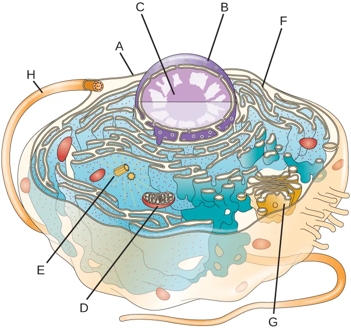
Label the lettered parts of this eukaryotic cell: A, the cell’s outer boundary; B, the outer boundary of the large central sphere; C, the smaller sphere inside it; D, the oval structure with internal lines; E, the two short paired tubes; F, the folded, dot-studded membranes outside the large sphere; G, the separate stack of folded membranes with small spheres budding from it; H, the long projection outside the cell.
Show model answer
Did your answer mention:
Compare and contrast similar structures found in prokaryotic and eukaryotic cells
Which of the following structures of a eukaryotic cell is not likely derived from endosymbiotic bacterium?
This mitochondrial membrane is a remnant of the original host cell’s own membrane, unlike the other three options, which trace back to the engulfed bacterium.Mitochondria in eukaryotic cells contain ribosomes that are structurally similar to those found in prokaryotic cells.
Recall the size class of ribosome (70S or 80S) found in mitochondria, and compare it with the size class of a prokaryotic ribosome.What existing evidence supports the theory that mitochondria are of prokaryotic origin?
Show model answer
Did your answer mention:
How are peroxisomes more like mitochondria than like the membrane-bound organelles of the endomembrane system? How do they differ from mitochondria?
Show model answer
Did your answer mention:
Sort each characteristic of the Summary of Cell Structures table to whether it describes prokaryotes (bacteria and archaea) or eukaryotes.
Prokaryotes
Eukaryotes
Describe the processes of eukaryotic mitosis and meiosis, and compare to prokaryotic binary fission
Eukaryotes are able to multiply through asexual reproduction, during which a single parent cell becomes two identical daughter cells; this process of clonal reproduction is called ________.
This process is much more complex than prokaryotic binary fission, because eukaryotic cells possess multiple chromosomes that must be replicated and divided between daughter cells.Which nuclear division process produces four genetically distinct gametes, each with half the number of chromosomes found in the original cell?
This process involves two separate nuclear divisions, rather than the single division that completes the other option.The portion of the mitotic phase in which the cytoplasmic components are physically separated into the two daughter cells is called ________.
This step follows karyokinesis, the nuclear-division portion of the mitotic phase.How does eukaryotic mitosis differ from prokaryotic binary fission?
Think about how many chromosomes a typical prokaryotic cell has to divide, compared to a eukaryotic cell.This section is adapted from Microbiology, Section 3.4: Unique Characteristics of Eukaryotic Cells by Nina Parker, Mark Schneegurt, Anh-Hue Thi Tu, Philip Lister, Brian M. Forster, and OpenStax, © OpenStax, licensed under CC BY-NC-SA 4.0. Access the original for free at openstax.org. Changes: figures re-encoded as WebP, with kind="diagram" set on twenty-one of the twenty-four figures (including the cell-cycle clock and the meiosis/mitosis comparison figure, both borrowed from Biology 2e and mis-guessed “photo” by the manifest’s file-extension heuristic) and kind="photo" on the three true micrograph figures (the cell-shapes panel, the nucleus TEM, and the nuclear-lamina fluorescence image); alts rewritten for all twenty-four figures to describe only what is visibly drawn or photographed, since several of this module’s source alts are incomplete or wrong rather than reused verbatim — the endomembrane-system diagram’s alt describes only the nucleus and ER and omits the vesicle, Golgi apparatus, and lysosome the image itself labels; the plasma-membrane diagram’s alt never mentions the cholesterol the image labels; the cytoskeleton figure’s alt states the microtubule column is “25 µm” in diameter where the image itself is labeled “25 nm” (all three reported as source defects); the nuclear-lamina micrograph’s alt describes “a large oval nucleus” (singular) where the image shows and labels two nuclei (“nuclei,” plural) — also reported; longdescs added for the opening labeled cell diagram, the endomembrane-system diagram, and the flagellum figure (reading order); the Summary of Cell Structures table (four columns, eleven rows) is transcribed as a Markdown table from the CNXML’s own table cells, not from the summary attribute, which independently misspells “chromosome’s” for “chromosomes,” “fagellins” for “flagellins,” labels the eukaryote membrane-lipid-composition cell “bacteria” a second time instead of “eukaryotes,” and gives the eukaryote cell-wall row as “chitin (molluscs, insects, crustaceans, and fungi)” where the table cell prints “Chitin (fungi)” (all four reported as source defects, uncorrected on the page because the real table cells the page transcribes are unaffected); that table also seeds one Practice sortbins item (twelve prokaryote/eukaryote characteristics, from the six binary-contrast rows the two prokaryote columns agree on) under the fourth objective; feature boxes rendered as callouts; the Link to Learning kept its source sentence and URL; the Micro Connection box’s title follows the bold feature name; the four Check Your Understanding boxes (five bullets total, none repeated or split); two of the five bullets (the two-differences bullet and the eukaryotic-versus-prokaryotic-envelope bullet) are rendered as body self-checks with model answers and rubrics assembled from this section’s own preceding text, and the other three (the endomembrane-components, cytoskeletal-structures, and eukaryotic-versus-prokaryotic-flagella bullets) are rendered as sort-into-bins items graded from this section’s own distinguishing phrases; the Art Connection exercise (OSC_Microbio_03_04_AniCell_img, a relettered version of the section’s own opening figure) is rendered as a mediafigure immediately followed by its selfcheck inside the third objective’s Practice group, with an author-written caption describing only what is drawn (never naming the labeled structures) and a model answer that maps each letter to the structure at the same position in the fully labeled figure shown earlier in this section; the Clinical Focus box, printed here as the Resolution, keeps the source’s own text and replaces its “go back to the previous Clinical Focus box” link — which pointed at Section 3.3, not where the case began — with a plain sentence naming Section 3.1, Spontaneous Generation, where the case actually began; six footnoted citations rendered as inline parenthetical citations after the sentences they support (bare access URLs dropped, DOIs kept — the Cell Wall section’s citation has no DOI, so its access URL is dropped and its author, title, publisher, and date are kept); model answers for one of the three Short Answer questions (the mitochondria-evidence question) and the three Critical Thinking questions (one of the three is the Art Connection labelling item) are written from this section’s own text, since the source prints no answer key for any of them (four, plus the two Check Your Understanding self-checks above — six author-written model answers on this page in total); the second Short Answer question, “Why do eukaryotic cells require an endomembrane system?” (fs-id1172100625770), is instead graded as a multiplechoice item keyed by this section’s own cell-size-and-diffusion sentence, with three wrong-reason distractors built from this section’s own claims about the nucleus, ribosome size, and cell division; the third Short Answer question, “Name at least two ways that prokaryotic flagella are different from eukaryotic flagella,” is not used because the Flagella and Cilia subsection’s Check Your Understanding self-check already asks it (and that self-check is itself now one of the sort-into-bins conversions above); the six Multiple Choice, one True/False, and two Fill in the Blank items keep the source’s own keys, options, and order and were sorted into the Practice block by objective; seven filler items round out the first, fourth, and fifth objective groups, which the section’s own exercise set does not reach on its own by itself: a select-the-term multiple choice and a text-recall for the first objective (nucleus, cytoskeleton), a sortbins built from the body’s own Summary of Cell Structures table for the fourth objective, and a select-the-term multiple choice, two cloze text-recalls, and one multiplechoice for the fifth objective (mitosis, meiosis, cytokinesis, and mitosis-versus-binary-fission, this last one graded by the module’s own chromosome-count contrast rather than answered in prose) — all built from this section’s own body sentences; key terms compiled from the module’s forty-eight defined terms and the book’s Glossary appendix, with two adaptations: the nuclear membrane/nuclear envelope pair share one appendix entry, so the nuclear membrane bullet drops that entry’s “(also called the nuclear membrane)” parenthetical to avoid printing a term inside its own definition, and the flagella bullet is written from this section’s own defining sentences rather than the appendix, because the appendix’s only flagella entry defines the prokaryotic structure (composed of flagellin, a hook, and a basal-body motor) while this section’s term names the structurally distinct eukaryotic flagellum (one definition taken from the defining sentence; the other forty-seven from the glossary); cross-reference to Section 3.3 (m58792) omitted from prose since the module’s own reference to it is the table link, already rendered as “the table below”; the module’s own inconsistency in how it cites the same Fuchs/Cleveland 1998 Science article across three footnotes (author order and initials differ each time) is reproduced as printed rather than normalized. One of the source’s unkeyed Short Answer questions and three Check Your Understanding questions are graded from the module’s own sentences rather than answered in prose; the source prints no key for them: the endomembrane-system Short Answer question above, and the endomembrane-components, cytoskeletal-structures, and eukaryotic-versus-prokaryotic-flagella Check Your Understanding bullets, all rendered as sort-into-bins items; the fifth objective’s filler multiplechoice on mitosis versus binary fission is likewise graded from this section’s own chromosome-count sentence rather than answered in prose. Two history-of-discovery claims are corrected with visible Source notes: the Golgi apparatus was first described in nerve cells, not Plasmodium, and lysosomes were discovered in 1955, not the 1960s (errata 380–381).