Nonproteobacteria Gram-Negative Bacteria and Phototrophic Bacteria
By the end of this section, you will be able to:
- Describe the unique features of nonproteobacteria gram-negative bacteria
- Give an example of a nonproteobacteria bacterium in each category
- Describe the unique features of phototrophic bacteria
- Identify phototrophic bacteria
The majority of the gram-negative bacteria belong to the phylum Proteobacteria, discussed in the previous section. Those that do not are called the nonproteobacteria. In this section, we will describe four classes of gram-negative nonproteobacteria: Chlamydia, the spirochetes, the CFB group, and the Planctomycetes. A diverse group of phototrophic bacteria that includes Proteobacteria and nonproteobacteria will be discussed at the end of this section.
Chlamydia
C. trachomatis is a human pathogen that causes trachoma, a disease of the eyes, often leading to blindness. C. trachomatis also causes the sexually transmitted disease lymphogranuloma venereum (LGV). This disease is often mildly symptomatic, manifesting as regional lymph node swelling, or it may be asymptomatic, but it is extremely contagious and is common on college campuses.
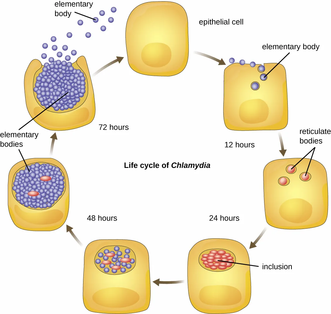
Members of the genus Chlamydia are gram-negative, obligate intracellular pathogens that are extremely resistant to the cellular defenses, giving them the ability to spread from host to host rapidly via elementary bodies. The metabolically and reproductively inactive elementary bodies are the endospore-like form of intracellular bacteria that enter an epithelial cell, where they become active. The diagram below illustrates the life cycle of Chlamydia.
Extended description
Following the arrows clockwise: free purple elementary bodies approach an epithelial cell and enter it. At 12 hours, the cell contains red reticulate bodies. At 24 hours, many red reticulate bodies fill a labeled inclusion. At 48 hours, the inclusion contains both red reticulate bodies and purple elementary bodies. At 72 hours, the cell ruptures and releases many purple elementary bodies, which can enter another epithelial cell.
Spirochetes
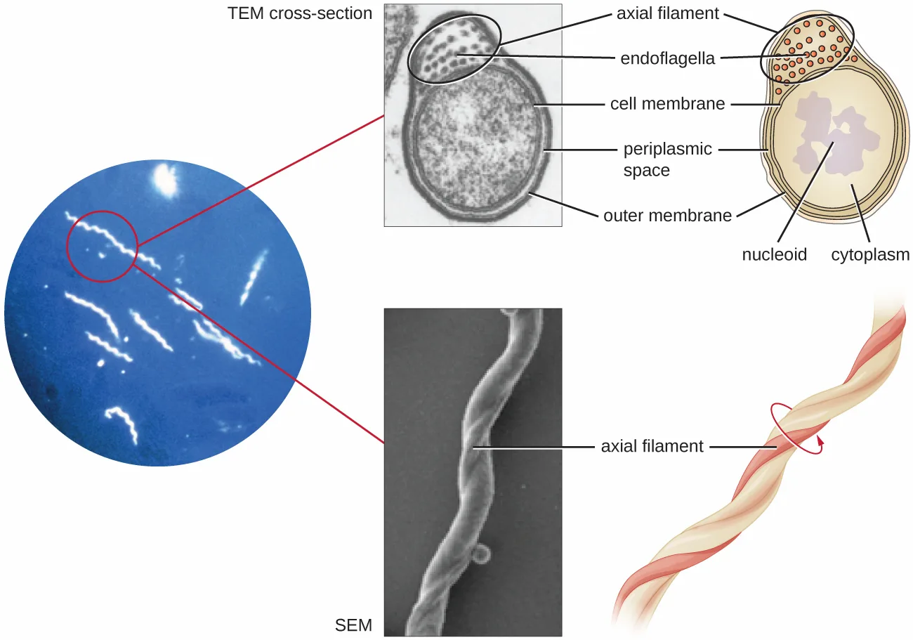
Spirochetes are characterized by their long (up to 250 µm), spiral-shaped bodies. Most spirochetes are also very thin, which makes it difficult to examine gram-stained preparations under a conventional brightfield microscope. Darkfield fluorescent microscopy is typically used instead. Spirochetes are also difficult or even impossible to culture. They are highly motile, using their axial filament to propel themselves. The axial filament is similar to a flagellum, but it wraps around the cell and runs inside the cell body of a spirochete in the periplasmic space between the outer membrane and the plasma membrane.
Extended description
At left, a circular darkfield micrograph shows several thin, bright white spiral spirochetes on blue. Red lines connect one cell to a TEM cross-section at top center and an SEM at bottom center. At top right, a cutaway cross-section labels axial filament and endoflagella in the periplasmic space between the cell membrane and outer membrane; it also labels the nucleoid and cytoplasm. At lower right, a schematic shows an axial filament wrapping around the long spiral body, with an arrow indicating the twisting motion.
Several genera of spirochetes include human pathogens. For example, the genus Treponema includes a species T. pallidum, which is further classified into four subspecies: T. pallidum pallidum, T. pallidum pertenue, T. pallidum carateum, and T. pallidum endemicum. The subspecies T. pallidum pallidum causes the sexually transmitted infection known as syphilis, the third most prevalent sexually transmitted bacterial infection in the United States, after chlamydia and gonorrhea. The other subspecies of T. pallidum cause tropical infectious diseases of the skin, bones, and joints.
Another genus of spirochete, Borrelia, contains a number of pathogenic species. B. burgdorferi causes Lyme disease, which is transmitted by several genera of ticks (notably Ixodes and Amblyomma) and often produces a “bull’s eye” rash, fever, fatigue, and, sometimes, debilitating arthritis. B. recurrens causes a condition known as relapsing fever. Appendix D lists the genera, species, and related diseases for spirochetes.
Check Your Understanding
Why do scientists typically use darkfield fluorescent microscopy to visualize spirochetes?
Compare the body shape described for these organisms with what a conventional brightfield preparation can show.Cytophaga, Fusobacterium, and Bacteroides
The gram-negative nonproteobacteria of the genera Cytophaga, Fusobacterium, and Bacteroides are classified together as a phylum and called the CFB group. Although they are phylogenetically diverse, bacteria of the CFB group share some similarities in the sequence of nucleotides in their DNA. They are rod-shaped bacteria adapted to anaerobic environments, such as the tissue of the gums, gut, and rumen of ruminating animals. CFB bacteria are avid fermenters, able to process cellulose in rumen, thus enabling ruminant animals to obtain carbon and energy from grazing.
Cytophaga are motile aquatic bacteria that glide. Fusobacteria inhabit the human mouth and may cause severe infectious diseases. The largest genus of the CFB group is Bacteroides, which includes dozens of species that are prevalent inhabitants of the human large intestine, making up about 30% of the entire gut microbiome. One gram of human feces contains up to 100 billion Bacteroides cells. Most Bacteroides are mutualistic. They benefit from nutrients they find in the gut, and humans benefit from their ability to prevent pathogens from colonizing the large intestine. Indeed, when populations of Bacteroides are reduced in the gut—as often occurs when a patient takes antibiotics—the gut becomes a more favorable environment for pathogenic bacteria and fungi, which can cause secondary infections.
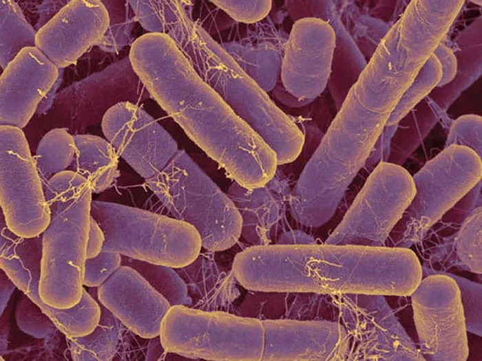
Only a few species of Bacteroides are pathogenic. B. melaninogenicus, for example, can cause wound infections in patients with weakened immune systems.
Check Your Understanding
Why are Cytophaga, Fusobacterium, and Bacteroides classified together as the CFB group?
Look for the stated shared basis of classification, rather than a habitat or a disease association.Planctomycetes
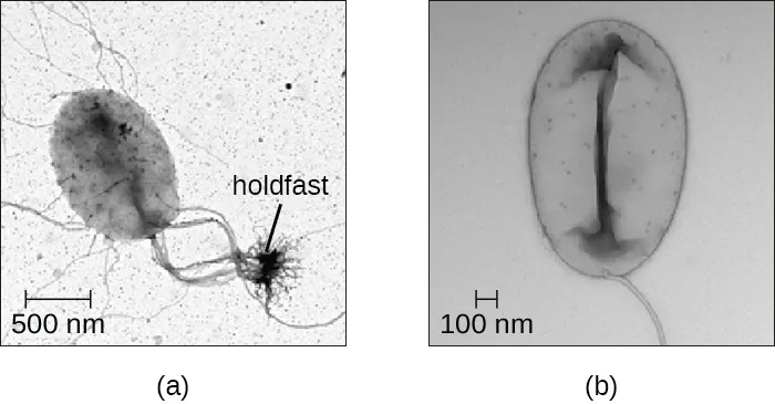
The Planctomycetes are found in aquatic environments, inhabiting freshwater, saltwater, and brackish water. Planctomycetes are unusual in that they reproduce by budding, meaning that instead of one maternal cell splitting into two equal daughter cells in the process of binary fission, the mother cell forms a bud that detaches from the mother cell and lives as an independent cell. These so-called swarmer cells are motile and not attached to a surface. However, they will soon differentiate into sessile (immobile) cells with an appendage called a holdfast that allows them to attach to surfaces in the water. Only the sessile cells are able to reproduce.
The table below summarizes the characteristics of some of the most clinically relevant genera of nonproteobacteria.
| Example Genus | Microscopic Morphology | Unique Characteristics |
|---|---|---|
| Chlamydia | Gram-negative, coccoid or ovoid bacterium | Obligatory intracellular bacteria; some cause chlamydia, trachoma, and pneumonia |
| Bacteroides | Gram-negative bacillus | Obligate anaerobic bacteria; abundant in the human gastrointestinal tract; usually mutualistic, although some species are opportunistic pathogens |
| Cytophaga | Gram-negative bacillus | Motile by gliding; live in soil or water; decompose cellulose; may cause disease in fish |
| Fusobacterium | Gram-negative bacillus with pointed ends | Anaerobic; form biofilms; some species cause disease in humans (periodontitis, ulcers) |
| Leptospira | Spiral-shaped bacterium (spirochetes); gram-negative-like (better viewed by darkfield microscopy); very thin | Aerobic, abundant in shallow water reservoirs; infect rodents and domestic animals; can be transmitted to humans by infected animals’ urine; may cause severe disease |
| Borrelia | Gram-negative-like spirochete; very thin; better viewed by darkfield microscopy | B. burgdorferi causes Lyme disease and B. recurrens causes relapsing fever |
| Treponema | Gram-negative-like spirochete; very thin; better viewed by darkfield microscopy | Motile; do not grow in culture; T. pallidum (subspecies T. pallidum pallidum) causes syphilis |
Check Your Understanding
How do Planctomycetes reproduce?
Contrast their division process with a maternal cell splitting into two equal daughter cells.Phototrophic Bacteria
The phototrophic bacteria are a large and diverse category of bacteria that do not represent a taxon but, rather, a group of bacteria that use sunlight as their primary source of energy. This group contains both Proteobacteria and nonproteobacteria. They use solar energy to synthesize ATP through photosynthesis. When they produce oxygen, they perform oxygenic photosynthesis. When they do not produce oxygen, they perform anoxygenic photosynthesis. With the exception of some cyanobacteria, the majority of phototrophic bacteria perform anoxygenic photosynthesis.
One large group of phototrophic bacteria includes the purple or green bacteria that perform photosynthesis with the help of bacteriochlorophylls, which are green, purple, or blue pigments similar to chlorophyll in plants. Some of these bacteria have a varying amount of red or orange pigments called carotenoids. Their color varies from orange to red to purple to green, and they are able to absorb light of various wavelengths. Traditionally, these bacteria are classified into sulfur and nonsulfur bacteria; they are further differentiated by color.
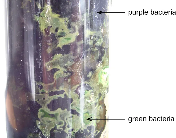
The sulfur bacteria perform anoxygenic photosynthesis, using sulfites as electron donors and releasing free elemental sulfur. Nonsulfur bacteria use organic substrates, such as succinate and malate, as donors of electrons.
The purple sulfur bacteria oxidize hydrogen sulfide into elemental sulfur and sulfuric acid and get their purple color from the pigments bacteriochlorophylls and carotenoids. Bacteria of the genus Chromatium are purple sulfur Gammaproteobacteria. These microorganisms are strict anaerobes and live in water. They use carbon dioxide as their only source of carbon, but their survival and growth are possible only in the presence of sulfites, which they use as electron donors. Chromatium has been used as a model for studies of bacterial photosynthesis since the 1950s (R.C. Fuller et al., “Carbon Metabolism in Chromatium,” Journal of Biological Chemistry 236 [1961]: 2140–2149).
The green sulfur bacteria use sulfide for oxidation and produce large amounts of green bacteriochlorophyll. The genus Chlorobium is a green sulfur bacterium. These bacteria use at least four types of chlorophyll for photosynthesis. The most prevalent of these, bacteriochlorophyll, is stored in special vesicle-like organelles called chlorosomes.
Purple nonsulfur bacteria are similar to purple sulfur bacteria, except that they use hydrogen rather than hydrogen sulfide for oxidation. Among the purple nonsulfur bacteria is the genus Rhodospirillum. These microorganisms are facultative anaerobes, which are actually pink rather than purple, and can metabolize (“fix”) nitrogen. They may be valuable in the field of biotechnology because of their potential ability to produce biological plastic and hydrogen fuel (T.T. Selao et al., “Comparative Proteomic Studies in Rhodospirillum rubrum Grown Under Different Nitrogen Conditions,” Journal of Proteome Research 7, no. 8 [2008]: 3267–3275).
The green nonsulfur bacteria are similar to green sulfur bacteria but they use substrates other than sulfides for oxidation. Chloroflexus is an example of a green nonsulfur bacterium. It often has an orange color when it grows in the dark, but it becomes green when it grows in sunlight. It stores bacteriochlorophyll in chlorosomes, similar to Chlorobium, and performs anoxygenic photosynthesis, using organic sulfites (low concentrations) or molecular hydrogen as electron donors, so it can survive in the dark if oxygen is available. Chloroflexus does not have flagella but can glide, like Cytophaga. It grows at a wide range of temperatures, from 35 °C to 70 °C, thus can be thermophilic.
Another large, diverse group of phototrophic bacteria compose the phylum Cyanobacteria; they get their blue-green color from the chlorophyll contained in their cells. Species of this group perform oxygenic photosynthesis, producing megatons of gaseous oxygen. Scientists hypothesize that cyanobacteria played a critical role in the change of our planet’s anoxic atmosphere 1–2 billion years ago to the oxygen-rich environment we have today (A. De los Rios et al., “Ultrastructural and Genetic Characteristics of Endolithic Cyanobacterial Biofilms Colonizing Antarctic Granite Rocks,” FEMS Microbiology Ecology 59, no. 2 [2007]: 386–395).
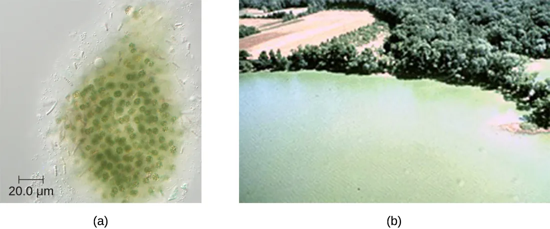
Cyanobacteria have other remarkable properties. Amazingly adaptable, they thrive in many habitats, including marine and freshwater environments, soil, and even rocks. Roseli Ocampo-Friedmann and Imre Friedman identified photosynthetic cyanobacteria living within rocks in Antarctica’s Dry Valleys, a barren, snowless region with extremely low precipitation and an average temperature of -15 degrees Celsius. The discovery was the basis for new theories and practices in the area of astrobiology (both researchers went on to work for NASA), considering that Earth’s polar deserts have conditions resembling those on Mars, which may provide a home to organisms similar to cyanobacteria. They can live as unicellular organisms or in colonies, and they can be filamentous, forming sheaths or biofilms. Many of them fix nitrogen, converting molecular nitrogen into ammonia that other bacteria, plants, and animals can use. The reactions of nitrogen fixation occur in specialized cells called heterocysts.
(Source note: the source says nitrogen fixation converts molecular nitrogen into nitrites and nitrates. Sections 4.1 and 8.7 of the same book identify ammonia as the product of nitrogen fixation and distinguish the later oxidation to nitrite and nitrate as nitrification; this page uses that distinction.)
Photosynthesis in Cyanobacteria is oxygenic, using the same type of chlorophyll a found in plants and algae as the primary photosynthetic pigment. Cyanobacteria also use phycocyanin, a secondary photosynthetic pigment that gives them their characteristic blue color. Phycocyanin occurs in phycobilisomes associated with folds of the cellular membrane called thylakoids, which are remarkably similar to the photosynthetic apparatus of plants. Cyanophycin, by contrast, is a carbon-and-nitrogen storage polymer rather than a photosynthetic pigment. Scientists hypothesize that plants originated from endosymbiosis of ancestral eukaryotic cells and ancestral photosynthetic bacteria (T. Cavalier-Smith, “Membrane Heredity and Early Chloroplast Evolution,” Trends in Plant Science 5, no. 4 [2000]: 174–182). Cyanobacteria are also an interesting object of research in biochemistry (S. Zhang and D.A. Bryant, “The Tricarboxylic Acid Cycle in Cyanobacteria,” Science 334, no. 6062 [2011]: 1551–1553), with studies investigating their potential as biosorbents (A. Cain et al., “Cyanobacteria as a Biosorbent for Mercuric Ion,” Bioresource Technology 99, no. 14 [2008]: 6578–6586) and products of human nutrition (C.S. Ku et al., “Edible Blue-Green Algae Reduce the Production of Pro-Inflammatory Cytokines by Inhibiting NF-κB Pathway in Macrophages and Splenocytes,” Biochimica et Biophysica Acta 1830, no. 4 [2013]: 2981–2988).
(Source note: the source groups cyanophycin with phycocyanin as a secondary photosynthetic pigment. The book’s photosynthesis chapter lists phycocyanin—but not cyanophycin—among photosynthetic pigments, and experimental microbiology identifies cyanophycin as a carbon-and-nitrogen storage polymer; this page corrects the distinction.)
Unfortunately, cyanobacteria can sometimes have a negative impact on human health. Genera such as Microcystis can form harmful cyanobacterial blooms, forming dense mats on bodies of water and producing large quantities of toxins that can harm wildlife and humans. These toxins have been implicated in tumors of the liver and diseases of the nervous system in animals and humans (I. Stewart et al., “Cyanobacterial Poisoning in Livestock, Wild Mammals and Birds – an Overview,” Advances in Experimental Medicine and Biology 619 [2008]: 613–637).
The table below summarizes the characteristics of important phototrophic bacteria.
| Phylum | Class | Example Genus or Species | Common Name | Oxygenic or Anoxygenic | Sulfur Deposition |
|---|---|---|---|---|---|
| Cyanobacteria | Cyanophyceae | Microcystis aeruginosa | Blue-green bacteria | Oxygenic | None |
| Chlorobi | Chlorobia | Chlorobium | Green sulfur bacteria | Anoxygenic | Outside the cell |
| Chloroflexi (Division) | Chloroflexi | Chloroflexus | Green nonsulfur bacteria | Anoxygenic | None |
| Proteobacteria | Alphaproteobacteria | Rhodospirillum | Purple nonsulfur bacteria | Anoxygenic | None |
| Proteobacteria | Betaproteobacteria | Rhodocyclus | Purple nonsulfur bacteria | Anoxygenic | None |
| Proteobacteria | Gammaproteobacteria | Chromatium | Purple sulfur bacteria | Anoxygenic | Inside the cell |
Check Your Understanding
What characteristic makes phototrophic bacteria different from other prokaryotes?
Focus on the energy source that defines this group, rather than on a shared taxonomic rank.Summary
- Gram-negative nonproteobacteria include the taxa spirochetes; the Chlamydia, Cytophaga, Fusobacterium, Bacteroides group; Planctomycetes; and many representatives of phototrophic bacteria.
- Spirochetes are motile, spiral bacteria with a long, narrow body; they are difficult or impossible to culture.
- Several genera of spirochetes contain human pathogens that cause such diseases as syphilis and Lyme disease.
- Cytophaga, Fusobacterium, and Bacteroides are classified together as a phylum called the CFB group. They are rod-shaped anaerobic organoheterotrophs and avid fermenters. Cytophaga are aquatic bacteria with the gliding motility. Fusobacteria inhabit the human mouth and may cause severe infectious diseases. Bacteroides are present in vast numbers in the human gut, most of them being mutualistic but some are pathogenic.
- Planctomycetes are aquatic bacteria that reproduce by budding; they may form large colonies, and develop a holdfast.
- Phototrophic bacteria are not a taxon but, rather, a group categorized by their ability to use the energy of sunlight. They include Proteobacteria and nonproteobacteria, as well as sulfur and nonsulfur bacteria colored purple or green.
- Sulfur bacteria perform anoxygenic photosynthesis, using sulfur compounds as donors of electrons, whereas nonsulfur bacteria use organic compounds (succinate, malate) as donors of electrons.
- Some phototrophic bacteria are able to fix nitrogen, providing the usable forms of nitrogen to other organisms.
- Cyanobacteria are oxygen-producing bacteria thought to have played a critical role in the forming of the earth’s atmosphere.
Key terms
- trachoma — a type of conjunctivitis, caused by Chlamydia trachomatis, that is a major cause of preventable blindness.
- lymphogranuloma venereum — infection caused by Chlamydia trachomatis in tropical regions.
- elementary bodies — metabolically and reproductively inactive, endospore-like form of intracellular bacteria that spreads infection outside of cells.
- spirochetes — a group of long, thin, spiral-shaped fastidious bacteria that includes the human pathogens that cause syphilis, Lyme disease, and leptospirosis.
- CFB group — phylum consisting of the gram-negative, rod-shaped nonproteobacteria genera Cytophaga, Fusobacterium, and Bacteroides.
- phototrophic bacteria — nontaxonomic group of bacteria that use sunlight as their primary source of energy.
- bacteriochlorophylls — green, purple, or blue pigments of bacteria; they are similar to chlorophyll of plants.
- purple sulfur bacteria — phototrophic bacteria that oxidize hydrogen sulfide into elemental sulfur and sulfuric acid; their purple color is due to the pigments bacteriochlorophylls and carotenoids.
- green sulfur bacteria — phototrophic, anaerobic bacteria that use sulfide for oxidation and produce large amounts of green bacteriochlorophyll.
- purple nonsulfur bacteria — phototrophic bacteria that are similar to purple sulfur bacteria except they use hydrogen rather than hydrogen sulfide for oxidation.
- green nonsulfur bacteria — similar to green sulfur bacteria but use substrates other than sulfides for oxidation.
- Cyanobacteria — phototrophic, chlorophyll-containing bacteria that produce large amounts of gaseous oxygen.
Practice
Describe the unique features of nonproteobacteria gram-negative bacteria
Which structure do spirochetes use to propel themselves?
Use the structure that wraps around the cell in the periplasmic space and produces a twisting motion.Sort each characteristic under the Planctomycete cell form it describes.
Swarmer cells
Sessile cells
Explain the term CFB group and name the genera that this group includes.
Show model answer
Did your answer mention:
Give an example of a nonproteobacteria bacterium in each category
Sort each characteristic under the nonproteobacteria genus described by the comparison table.
Chlamydia
Bacteroides
Cytophaga
Leptospira
Which bacteria are the most prevalent in the human gut?
Use the genus described as making up about 30% of the entire gut microbiome.The bacterium that causes syphilis is called ________.
Look for the named subspecies of the genus associated with this sexually transmitted infection.Name and briefly describe the bacterium that causes Lyme disease.
Use the genus and species named just before the list of tick genera, then identify the illness features given there.Describe the unique features of phototrophic bacteria
Which term refers to photosynthesis performed by bacteria with the use of water as the donor of electrons?
Compare whether the process produces oxygen with whether it does not.Bacteria in the genus Rhodospirillum that use hydrogen for oxidation and fix nitrogen are ________ bacteria.
Use the color-and-sulfur category contrasted with purple sulfur bacteria.How do sulfur bacteria and nonsulfur bacteria differ in the electron donors they use?
Compare the two donor categories stated directly after the tube figure.Identify phototrophic bacteria
Sort each bacterium by the photosynthesis type listed in the phototrophic-bacteria table.
Oxygenic
Anoxygenic
What are the specialized cells in which the reactions of nitrogen fixation occur in many cyanobacteria?
Look for the named specialized cells in the paragraph on cyanobacteria’s adaptable forms and habitats.Characterize the phylum Cyanobacteria.
Show model answer
Did your answer mention:
This section is adapted from Microbiology, Section 4.3: Nonproteobacteria Gram-Negative Bacteria and Phototrophic Bacteria by Nina Parker, Mark Schneegurt, Anh-Hue Thi Tu, Philip Lister, Brian M. Forster, and OpenStax, © OpenStax, licensed under CC BY-NC-SA 4.0. Access the original for free at openstax.org. Changes: all six source figures re-encoded as WebP and rendered as mediafigures after image and PDF inspection, with explicit kinds — two diagrams (the Chlamydia life-cycle and the composite spirochete figure) and four photographs or micrographs; alts rewritten to describe the rendered images, and long descriptions added for the two labeled diagrams; the two source tables transcribed as Markdown tables from their cells and each represented by a sort-bins activity in Practice, with the source’s malformed “form; biofilms” corrected to “form biofilms” and the table-summary discrepancies recorded separately; source footnotes rendered as inline parenthetical citations, with bare access URLs absent from the source; same-module figure and table references rendered as describing phrases; Check Your Understanding questions retained in body order and converted to graded items where one module sentence fixes the answer; the source’s three Multiple Choice and two Fill in the Blank items adapted into Practice, and its three unkeyed Short Answer items adapted into a self-check (CFB group), a multiple choice (B. burgdorferi as the Lyme-disease bacterium), and a self-check (Cyanobacteria), respectively, from this module’s text; author-written Practice items, including both table sorts and the Planctomycete cell-form sort, are built only from this module’s sentences and tables to provide three items per objective; key terms compiled from the module’s twelve defined terms and the book’s Glossary appendix, with all twelve definitions taken directly from the Glossary. The source’s claims that nitrogen fixation produces nitrites and nitrates and that cyanophycin is a photosynthetic pigment are corrected with visible Source notes. No source exercise was omitted.