Unicellular Eukaryotic Parasites
By the end of this section, you will be able to:
- Summarize the general characteristics of unicellular eukaryotic parasites
- Describe the general life cycles and modes of reproduction in unicellular eukaryotic parasites
- Identify challenges associated with classifying unicellular eukaryotes
- Explain the taxonomic scheme used for unicellular eukaryotes
- Give examples of infections caused by unicellular eukaryotes
Clinical Focus. Part 1
Upon arriving home from school, 7-year-old Sarah complains that a large spot on her arm will not stop itching. She keeps scratching at it, drawing the attention of her parents. Looking more closely, they see that it is a red circular spot with a raised red edge (the photographs below). The next day, Sarah’s parents take her to their doctor, who examines the spot using a Wood’s lamp. A Wood’s lamp produces ultraviolet light that causes the spot on Sarah’s arm to fluoresce, which confirms what the doctor already suspected: Sarah has a case of ringworm.
Sarah’s mother is mortified to hear that her daughter has a “worm.” How could this happen?
- What are some likely ways that Sarah might have contracted ringworm?
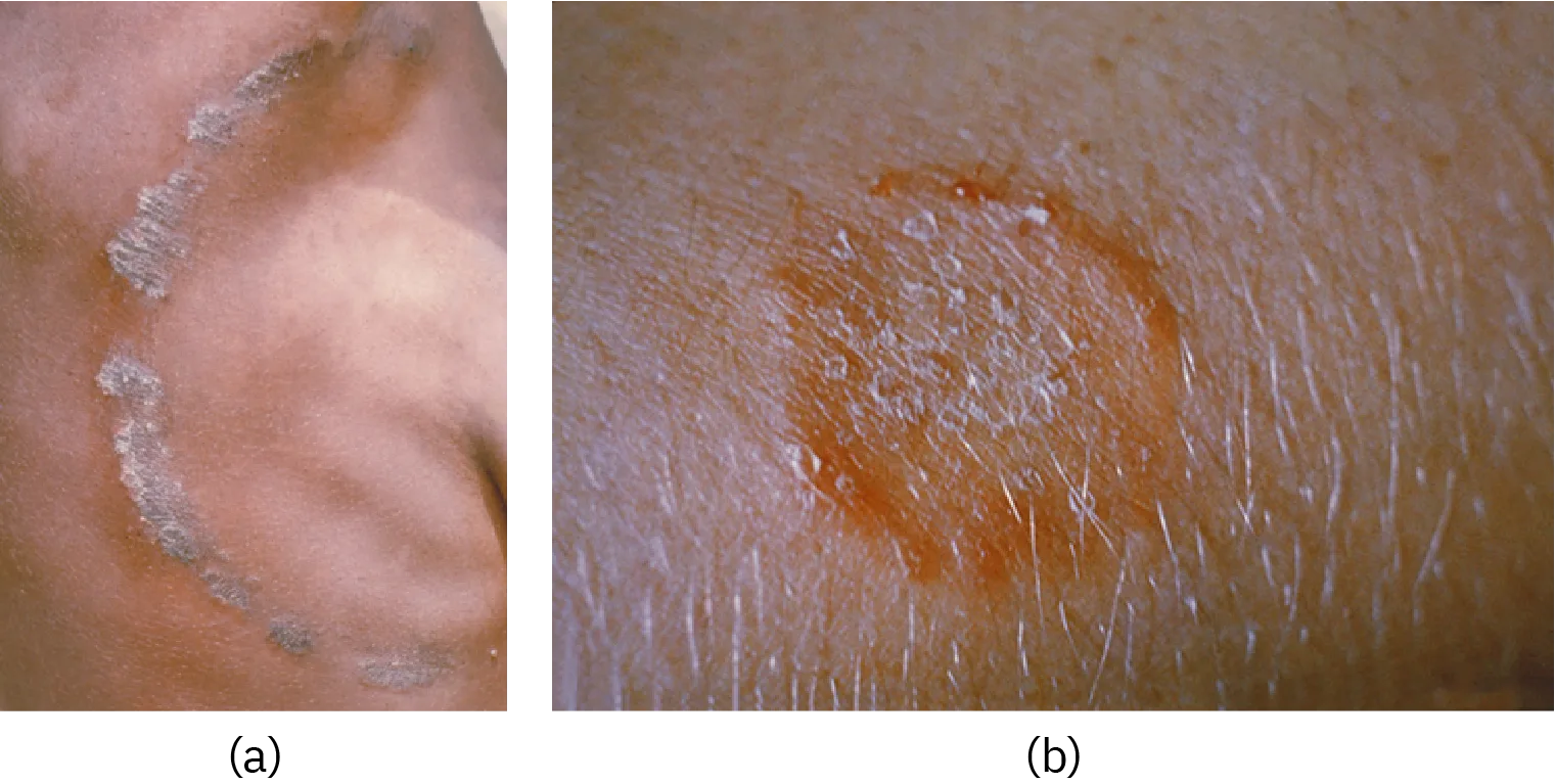
The case continues in Parasitic Helminths.
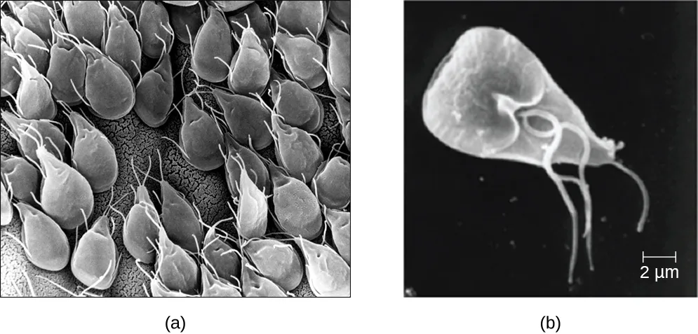
Eukaryotic microbes are an extraordinarily diverse group, including species with a wide range of life cycles, morphological specializations, and nutritional needs. Although more diseases are caused by viruses and bacteria than by microscopic eukaryotes, these eukaryotes are responsible for some diseases of great public health importance. For example, the protozoal disease malaria was responsible for 584,000 deaths worldwide (primarily children in Africa) in 2013, according to the World Health Organization (WHO). The protist parasite Giardia causes a diarrheal illness (giardiasis) that is easily transmitted through contaminated water supplies. In the United States, Giardia is the most common human intestinal parasite (the micrograph below). Although it may seem surprising, parasitic worms are included within the study of microbiology because identification depends on observation of microscopic adult worms or eggs. Even in developed countries, these worms are important parasites of humans and of domestic animals. There are fewer fungal pathogens, but these are important causes of illness, as well. On the other hand, fungi have been important in producing antimicrobial substances such as penicillin. In this chapter, we will examine characteristics of protists, worms, and fungi while considering their roles in causing disease.
Characteristics of Protists
The word protist is a historical term that is now used informally to refer to a diverse group of microscopic eukaryotic organisms. It is not considered a formal taxonomic term because the organisms it describes do not have a shared evolutionary origin. Historically, the protists were informally grouped into the “animal-like” protozoans, the “plant-like” algae, and the “fungus-like” protists such as water molds. These three groups of protists differ greatly in terms of their basic characteristics. For example, algae are photosynthetic organisms that can be unicellular or multicellular. Protozoa, on the other hand, are nonphotosynthetic, motile organisms that are always unicellular. Other informal terms may also be used to describe various groups of protists. For example, microorganisms that drift or float in water, moved by currents, are referred to as plankton. Types of plankton include zooplankton, which are motile and nonphotosynthetic, and phytoplankton, which are photosynthetic.
Protozoans inhabit a wide variety of habitats, both aquatic and terrestrial. Many are free-living, while others are parasitic, carrying out a life cycle within a host or hosts and potentially causing illness. There are also beneficial symbionts that provide metabolic services to their hosts. During the feeding and growth part of their life cycle, they are called trophozoites; these feed on small particulate food sources such as bacteria. While some types of protozoa exist exclusively in the trophozoite form, others can develop from trophozoite to an encapsulated cyst stage when environmental conditions are too harsh for the trophozoite. A cyst is a cell with a protective wall, and the process by which a trophozoite becomes a cyst is called encystment. When conditions become more favorable, these cysts are triggered by environmental cues to become active again through excystment.
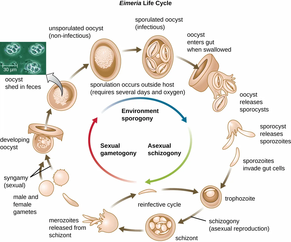
One protozoan genus capable of encystment is Eimeria, which includes some human and animal pathogens. The life-cycle diagram below illustrates the life cycle of Eimeria.
Extended description
The cycle begins with an oocyst shed in feces, which is non-infectious and unsporulated. Sporulation occurs outside the host, requiring several days and oxygen, and produces an infectious sporulated oocyst. The oocyst enters the gut when swallowed and releases sporocysts, which release sporozoites. The sporozoites invade gut cells and form trophozoites. Trophozoites undergo asexual schizogony to form a schizont, which releases merozoites. From there, merozoites either reinfect gut cells and repeat the trophozoite stage (the reinfective cycle) or undergo sexual gametogony to form male and female gametes. The gametes undergo syngamy to form a developing oocyst, which matures into a new unsporulated, non-infectious oocyst that is shed in feces, restarting the cycle.
Protozoans have a variety of reproductive mechanisms. Some protozoans reproduce asexually and others reproduce sexually; still others are capable of both sexual and asexual reproduction. In protozoans, asexual reproduction occurs by binary fission, budding, or schizogony. In schizogony, the nucleus of a cell divides multiple times before the cell divides into many smaller cells. The products of schizogony are called merozoites and they are stored in structures known as schizonts. Protozoans may also reproduce sexually, which increases genetic diversity and can lead to complex life cycles. Protozoans can produce haploid gametes that fuse through syngamy. However, they can also exchange genetic material by joining to exchange DNA in a process called conjugation. This is a different process than the conjugation that occurs in bacteria. The term protist conjugation refers to a true form of eukaryotic sexual reproduction between two cells of different mating types. It is found in ciliates, a group of protozoans, and is described later in this subsection.
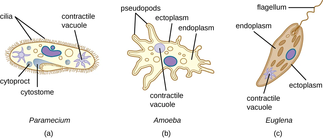
All protozoans have a plasma membrane, or plasmalemma, and some have bands of protein just inside the membrane that add rigidity, forming a structure called the pellicle. Some protists, including protozoans, have distinct layers of cytoplasm under the membrane. In these protists, the outer gel layer (with microfilaments of actin) is called the ectoplasm. Inside this layer is a sol (fluid) region of cytoplasm called the endoplasm. These structures contribute to complex cell shapes in some protozoans, whereas others (such as amoebas) have more flexible shapes (the diagram below).
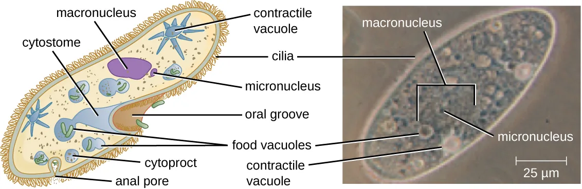
Different groups of protozoans have specialized feeding structures. They may have a specialized structure for taking in food through phagocytosis, called a cytostome, and a specialized structure for the exocytosis of wastes called a cytoproct. Oral grooves leading to cytostomes are lined with hair-like cilia to sweep in food particles. Protozoans are heterotrophic. Protozoans that are holozoic ingest whole food particles through phagocytosis. Forms that are saprozoic ingest small, soluble food molecules.
Many protists have whip-like flagella or hair-like cilia made of microtubules that can be used for locomotion (the diagram below). Other protists use cytoplasmic extensions known as pseudopodia (“false feet”) to attach the cell to a surface; they then allow cytoplasm to flow into the extension, thus moving themselves forward.
Protozoans have a variety of unique organelles and sometimes lack organelles found in other cells. Some have contractile vacuoles, organelles that can be used to move water out of the cell for osmotic regulation (salt and water balance) (the diagram below). Mitochondria may be absent in parasites or altered to kinetoplastids (modified mitochondria) or hydrogenosomes (see Unique Characteristics of Eukaryotic Cells for more discussion of these structures).
Check Your Understanding
What is the sequence of events in reproduction by schizogony, and what are the cells produced called?
Locate the sentence that defines schizogony, just after the discussion of the Eimeria life cycle.Taxonomy of Protists
The protists are a polyphyletic group, meaning they lack a shared evolutionary origin. Since the current taxonomy is based on evolutionary history (as determined by biochemistry, morphology, and genetics), protists are scattered across many different taxonomic groups within the domain Eukarya. Eukarya is currently divided into six supergroups that are further divided into subgroups, as illustrated in the tree diagram below. In this section, we will primarily be concerned with the supergroups Amoebozoa, Excavata, and Chromalveolata; these supergroups include many protozoans of clinical significance. The supergroups Opisthokonta and Rhizaria also include some protozoans, but few of clinical significance. In addition to protozoans, Opisthokonta also includes animals and fungi, some of which we will discuss in Parasitic Helminths and Fungi. Some examples of the Archaeplastida will be discussed in Algae. The tree diagram and table below summarize the characteristics of each supergroup and subgroup and list representatives of each.
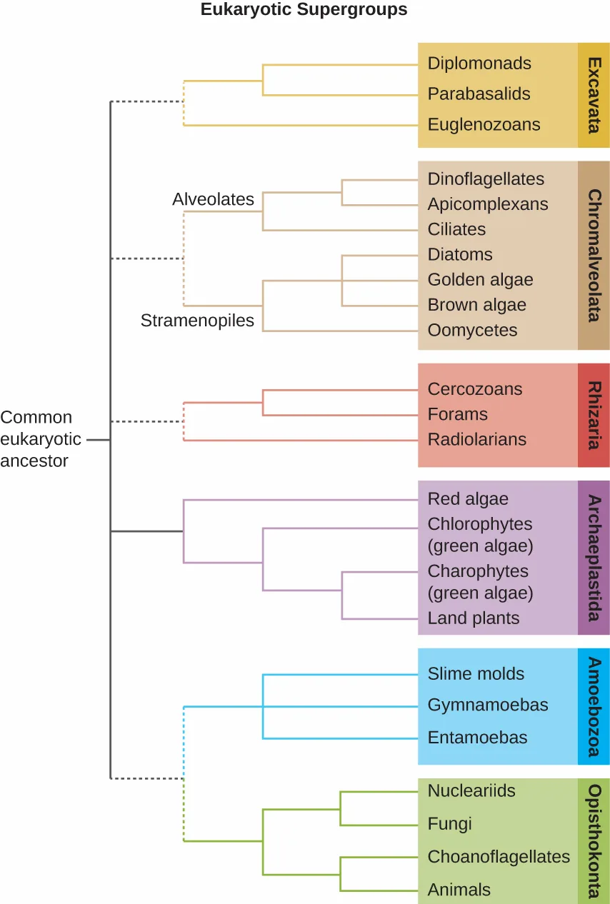
Extended description
Reading left to right from the common eukaryotic ancestor: Excavata divides into Diplomonads, Parabasalids, and Euglenozoans. Chromalveolata divides into Alveolates (Dinoflagellates, Apicomplexans, Ciliates) and Stramenopiles (Diatoms, Golden algae, Brown algae, Oomycetes). Rhizaria divides into Cercozoans, Forams, and Radiolarians. Archaeplastida divides into Red algae, Chlorophytes, Charophytes, and Land plants. Amoebozoa divides into Slime molds, Gymnamoebas, and Entamoebas. Opisthokonta divides into Nucleariids, Fungi, Choanoflagellates, and Animals.
| Supergroup | Subgroup | Distinguishing Features | Examples | Clinical Notes |
|---|---|---|---|---|
| Excavata | Fornicata | Form cysts; pair of equal nuclei; no mitochondria; often parasitic; four free flagella | Giardia lamblia | Giardiasis |
| Excavata | Parabasalids | No mitochondria; four free flagella; one attached flagellum; no cysts; parasitic or symbiotic; basal bodies; kinetoplastids | Trichomonas | Trichomoniasis |
| Excavata | Euglenozoans | Photosynthetic or heterotrophic; flagella | Euglena | N/a |
| Excavata | Euglenozoans | Photosynthetic or heterotrophic; flagella | Trypanosoma | African sleeping sickness, Chagas disease |
| Excavata | Euglenozoans | Photosynthetic or heterotrophic; flagella | Leishmania | Leishmaniasis |
| Chromalveolata | Dinoflagellates | Cellulose theca; two dissimilar flagella | Gonyaulax | Red tides |
| Chromalveolata | Dinoflagellates | Cellulose theca; two dissimilar flagella | Alexandrium | Paralytic shellfish poisoning |
| Chromalveolata | Dinoflagellates | Cellulose theca; two dissimilar flagella | Pfiesteria | Harmful algal blooms |
| Chromalveolata | Apicomplexans | Intracellular parasite; apical organelles | Plasmodium | Malaria |
| Chromalveolata | Apicomplexans | Intracellular parasite; apical organelles | Cryptosporidium | Cryptosporidiosis |
| Chromalveolata | Apicomplexans | Intracellular parasite; apical organelles | Theileria (Babesia) | Babesiosis |
| Chromalveolata | Apicomplexans | Intracellular parasite; apical organelles | Toxoplasma | Toxoplasmosis |
| Chromalveolata | Ciliates | Cilia | Balantidium | Balantidiasis |
| Chromalveolata | Ciliates | Cilia | Paramecium | N/a |
| Chromalveolata | Ciliates | Cilia | Stentor | N/a |
| Chromalveolata | Öomycetes/peronosporomycetes | “Water molds”; generally diploid; cellulose cell walls | Phytophthora | Diseases in crops |
| Rhizaria | Foraminifera | Amoeboid; threadlike pseudopodia; calcium carbonate shells | Astrolonche | N/a |
| Rhizaria | Radiolaria | Amoeboid; threadlike pseudopodia; silica shells | Actinomma | N/a |
| Rhizaria | Cercozoa | Amoeboid; threadlike pseudopodia; complex shells; parasitic forms | Spongospora subterranea | Powdery scab (potato disease) |
| Rhizaria | Cercozoa | Amoeboid; threadlike pseudopodia; complex shells; parasitic forms | Plasmodiophora brassicae | Cabbage clubroot |
| Archaeplastida | Red algae | Chlorophyll a; phycoerythrin; phycocyanin; floridean starch; agar in cell walls | Gelidium | Source of agar |
| Archaeplastida | Red algae | Chlorophyll a; phycoerythrin; phycocyanin; floridean starch; agar in cell walls | Gracilaria | Source of agar |
| Archaeplastida | Chlorophytes | Chlorophyll a; chlorophyll b; cellulose cell walls; starch storage | Acetabularia | N/a |
| Archaeplastida | Chlorophytes | Chlorophyll a; chlorophyll b; cellulose cell walls; starch storage | Ulva | N/a |
| Amoebozoa | Slime molds | Plasmodial and cellular forms | Dictyostelium | N/a |
| Amoebozoa | Entamoebas | Trophozoites; form cysts | Entamoeba | Amoebiasis |
| Amoebozoa | Entamoebas | Trophozoites; form cysts | Naegleria | Primary amoebic meningoencephalitis |
| Amoebozoa | Entamoebas | Trophozoites; form cysts | Acanthamoeba | Keratitis, granulomatous amoebic encephalitis |
| Opisthokonta | Fungi | Chitin cell walls; unicellular or multicellular; often hyphae | Zygomycetes | Zygomycosis |
| Opisthokonta | Fungi | Chitin cell walls; unicellular or multicellular; often hyphae | Ascomycetes | Candidiasis |
| Opisthokonta | Fungi | Chitin cell walls; unicellular or multicellular; often hyphae | Basidiomycetes | Cryptococcosis |
| Opisthokonta | Fungi | Chitin cell walls; unicellular or multicellular; often hyphae | Microsporidia | Microsporidiosis |
| Opisthokonta | Animals | Multicellular heterotrophs; no cell walls | Nematoda | Trichinosis; hookworm and pinworm infections |
| Opisthokonta | Animals | Multicellular heterotrophs; no cell walls | Trematoda | Schistosomiasis |
| Opisthokonta | Animals | Multicellular heterotrophs; no cell walls | Cestoda | Tapeworm infections |
Check Your Understanding
Which supergroups contain the clinically significant protists? Sort each supergroup discussed in this section under whether it is described as including many clinically significant protists or only a few.
Includes many clinically significant protists
Includes few or none
Amoebozoa
The supergroup Amoebozoa includes protozoans that use amoeboid movement. Actin microfilaments produce pseudopodia, into which the remainder of the protoplasm flows, thereby moving the organism. The genus Entamoeba includes commensal or parasitic species, including the medically important E. histolytica, which is transmitted by cysts in feces and is the primary cause of amoebic dysentery. Another member of this group that is pathogenic to humans is Acanthamoeba, which can cause keratitis (corneal inflammation) and blindness. The notorious “brain eating amoeba,” Naegleria fowleri, is considered a distant relative of the Amoebozoa and is classified in the phylum Percolozoa.
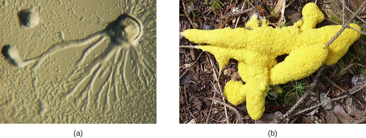
The Eumycetozoa are an unusual group of organisms called slime molds, which have previously been classified as animals, fungi, and plants (the photographs below). Slime molds can be divided into two types: cellular slime molds and plasmodial slime molds. The cellular slime molds exist as individual amoeboid cells that periodically aggregate into a mobile slug. The aggregate then forms a fruiting body that produces haploid spores. Plasmodial slime molds exist as large, multinucleate amoeboid cells that form reproductive stalks to produce spores that divide into gametes. One cellular slime mold, Dictyostelium discoideum, has been an important study organism for understanding cell differentiation, because it has both single-celled and multicelled life stages, with the cells showing some degree of differentiation in the multicelled form. The two life-cycle diagrams below illustrate the life cycles of cellular and plasmodial slime molds, respectively.
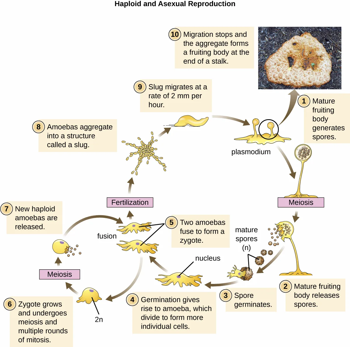
Extended description
Stage 1: a mature fruiting body generates spores by meiosis. Stage 2: the mature fruiting body releases spores. Stage 3: a spore germinates. Stage 4: germination gives rise to amoebas, which divide to form more individual cells. Stage 5: two amoebas fuse to form a zygote. Stage 6: the zygote (2n) grows and undergoes meiosis and multiple rounds of mitosis. Stage 7: new haploid amoebas are released. Stage 8: amoebas aggregate into a structure called a slug. Stage 9: the slug migrates at a rate of 2 mm per hour. Stage 10: migration stops and the aggregate forms a fruiting body at the end of a stalk, returning to stage 1. A photograph beside stage 1 shows a fruiting body growing on bark.
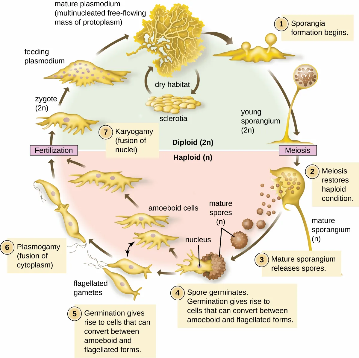
Extended description
In the diploid half of the cycle: a feeding plasmodium begins sporangia formation (stage 1), producing a young sporangium. Meiosis restores the haploid condition (stage 2) as the mature sporangium releases spores (stage 3). In the haploid half of the cycle: a spore germinates (stage 4), giving rise to cells that can convert between amoeboid and flagellated forms (stage 5). Plasmogamy, the fusion of cytoplasm of two cells (stage 6), produces amoeboid cells that combine in fertilization to form a zygote. Karyogamy, the fusion of nuclei (stage 7), returns the cycle to the diploid half, where the zygote divides to form a multinucleated feeding plasmodium. In a dry habitat, the mature plasmodium can instead form dormant sclerotia.
Chromalveolata
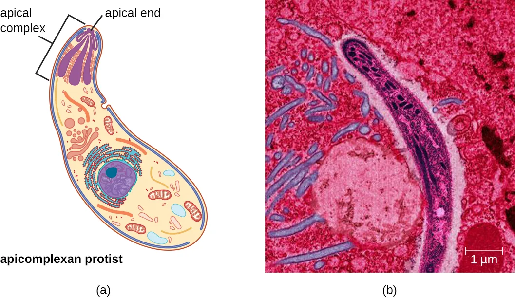
The supergroup Chromalveolata is united by similar origins of its members’ plastids and includes the apicomplexans, ciliates, diatoms, and dinoflagellates, among other groups (we will cover the diatoms and dinoflagellates in Algae). The apicomplexans are intra- or extracellular parasites that have an apical complex at one end of the cell. The apical complex is a concentration of organelles, vacuoles, and microtubules that allows the parasite to enter host cells (the images below). Apicomplexans have complex life cycles that include an infective sporozoite that undergoes schizogony to make many merozoites (see the example in the Eimeria life-cycle diagram above). Many are capable of infecting a variety of animal cells, from insects to livestock to humans, and their life cycles often depend on transmission between multiple hosts. The genus Plasmodium is an example of this group.
Other apicomplexans are also medically important. Cryptosporidium parvum causes intestinal symptoms and can cause epidemic diarrhea when the cysts contaminate drinking water. Theileria (Babesia) microti, transmitted by the tick Ixodes scapularis, causes recurring fever that can be fatal and is becoming a common transfusion-transmitted pathogen in the United States (Theileria and Babesia are closely related genera and there is some debate about the best classification). Finally, Toxoplasma gondii causes toxoplasmosis and can be transmitted from cat feces, unwashed fruit and vegetables, or from undercooked meat. Because toxoplasmosis can be associated with serious birth defects, pregnant people need to be aware of this risk and use caution if they are exposed to the feces of potentially infected cats. A national survey found the frequency of individuals with antibodies for toxoplasmosis (and thus who presumably have a current latent infection) in the United States to be 11% (J. Flegr et al., “Toxoplasmosis—A Global Threat. Correlation of Latent Toxoplasmosis With Specific Disease Burden in a Set of 88 Countries,” PloS ONE 9, no. 3 [2014]: e90203). Rates are much higher in other countries, including some developed countries. There is also evidence and a good deal of theorizing that the parasite may be responsible for altering infected humans’ behavior and personality traits (J. Flegr, “Effects of Toxoplasma on Human Behavior,” Schizophrenia Bull 33, no. 3 [2007]: 757–760).
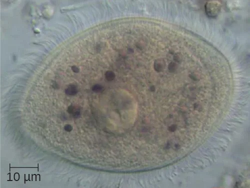
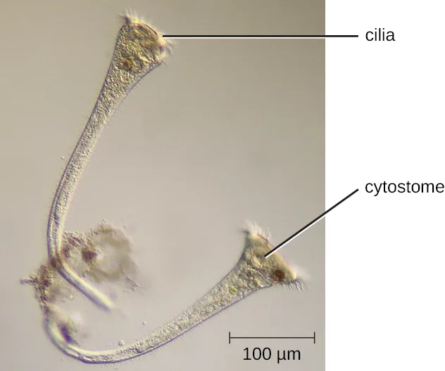
The ciliates (Ciliophora), also within the Chromalveolata, are a large, very diverse group characterized by the presence of cilia on their cell surface. Although the cilia may be used for locomotion, they are often used for feeding, as well, and some forms are nonmotile. Balantidium coli (the micrograph below) is the only parasitic ciliate that affects humans by causing intestinal illness, although it rarely causes serious medical issues except in the immunocompromised (those having a weakened immune system). Perhaps the most familiar ciliate is Paramecium, a motile organism with a clearly visible cytostome and cytoproct that is often studied in biology laboratories (the diagram below). Another ciliate, Stentor, is sessile and uses its cilia for feeding (the micrograph below). Generally, these organisms have a micronucleus that is diploid, somatic, and used for sexual reproduction by conjugation. They also have a macronucleus that is derived from the micronucleus; the macronucleus becomes polyploid (multiple sets of duplicate chromosomes), and has a reduced set of metabolic genes.
Ciliates are able to reproduce through conjugation, in which two cells attach to each other. In each cell, the diploid micronuclei undergo meiosis, producing eight haploid nuclei each. Then, all but one of the haploid micronuclei and the macronucleus disintegrate; the remaining (haploid) micronucleus undergoes mitosis. The two cells then exchange one micronucleus each, which fuses with the remaining micronucleus present to form a new, genetically different, diploid micronucleus. The diploid micronucleus undergoes two mitotic divisions, so each cell has four micronuclei, and two of the four combine to form a new macronucleus. The chromosomes in the macronucleus then replicate repeatedly, the macronucleus reaches its polyploid state, and the two cells separate. The two cells are now genetically different from each other and from their previous versions.
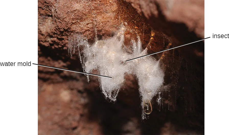
Öomycetes have similarities to fungi and were once classified with them. They are also called water molds. However, they differ from fungi in several important ways. Öomycetes have cell walls of cellulose (unlike the chitinous cell walls of fungi) and they are generally diploid, whereas the dominant life forms of fungi are typically haploid. Phytophthora, the plant pathogen found in the soil that caused the Irish potato famine, is classified within this group (the photograph below).
Link to Learning
Explore the procedures for detecting the presence of an apicomplexan in a public water supply, at this website describing detection procedures for Cryptosporidium and Giardia. This video of a feeding Stentor shows the feeding of Stentor.
Excavata
The third and final supergroup to be considered in this section is the Excavata, which includes primitive eukaryotes and many parasites with limited metabolic abilities. These organisms have complex cell shapes and structures, often including a depression on the surface of the cell called an excavate. The group Excavata includes the subgroups Fornicata, Parabasalia, and Euglenozoa. The Fornicata lack mitochondria but have flagella. This group includes Giardia lamblia (also known as G. intestinalis or G. duodenalis), a widespread pathogen that causes diarrheal illness and can be spread through cysts from feces that contaminate water supplies (the micrograph above). Parabasalia are frequent animal endosymbionts; they live in the guts of animals like termites and cockroaches. They have basal bodies and modified mitochondria (kinetoplastids). They also have a large, complex cell structure with an undulating membrane and often have many flagella. The trichomonads (a subgroup of the Parabasalia) include pathogens such as Trichomonas vaginalis, which causes the human sexually transmitted disease trichomoniasis. Trichomoniasis often does not cause symptoms in males, but they are able to transmit the infection. In females, it causes vaginal discomfort and discharge and may cause complications in pregnancy if left untreated.
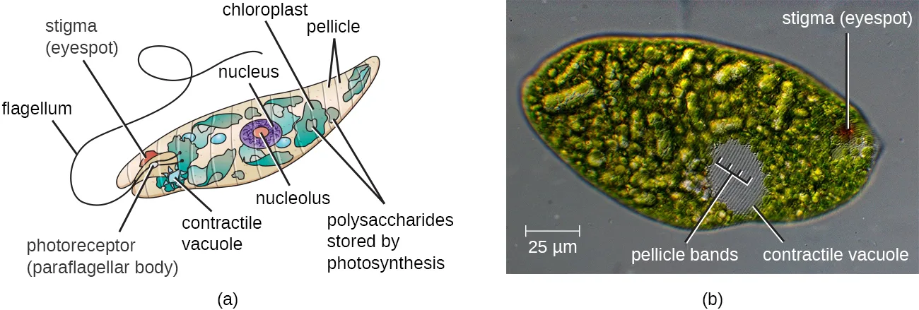
The Euglenozoa are common in the environment and include photosynthetic and nonphotosynthetic species. Members of the genus Euglena are typically not pathogenic. Their cells have two flagella, a pellicle, a stigma (eyespot) to sense light, and chloroplasts for photosynthesis (the diagram below). The pellicle of Euglena is made of a series of protein bands surrounding the cell; it supports the cell membrane and gives the cell shape.
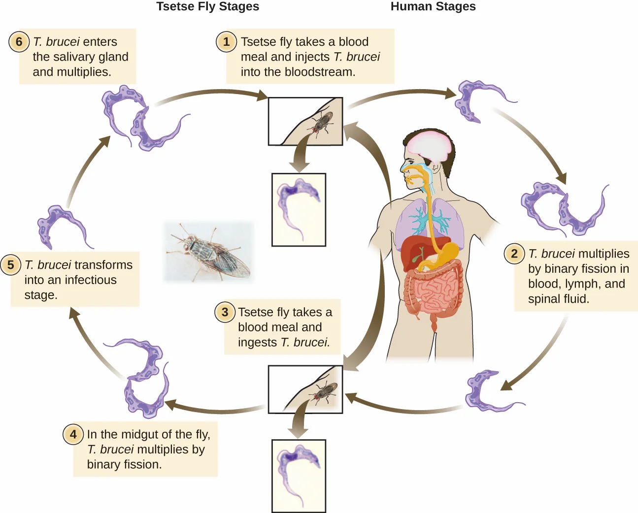
The Euglenozoa also include the trypanosomes, which are parasitic pathogens. The genus Trypanosoma includes T. brucei, which causes African trypanosomiasis (African sleeping sickness) and T. cruzi, which causes American trypanosomiasis (Chagas disease). These tropical diseases are spread by insect bites. In African sleeping sickness, T. brucei colonizes the blood and the brain after being transmitted via the bite of a tsetse fly (Glossina spp.) (the life-cycle diagram below). The early symptoms include confusion, difficulty sleeping, and lack of coordination. Left untreated, it is fatal.
Extended description
Stage 1: a tsetse fly takes a blood meal and injects T. brucei into a human’s bloodstream. Stage 2: T. brucei multiplies by binary fission in the human’s blood, lymph, and spinal fluid. Stage 3: another tsetse fly takes a blood meal and ingests T. brucei from an infected human. Stage 4: in the fly’s midgut, T. brucei multiplies by binary fission. Stage 5: T. brucei transforms into an infectious stage. Stage 6: T. brucei enters the fly’s salivary gland and multiplies, ready to be injected into another human at the fly’s next blood meal, restarting the cycle.
Chagas’ disease originated and is most common in Latin America. The disease is transmitted by Triatoma spp., insects often called “kissing bugs,” and affects either the heart tissue or tissues of the digestive system. Untreated cases can eventually lead to heart failure or significant digestive or neurological disorders.
The genus Leishmania includes trypanosomes that cause disfiguring skin disease and sometimes systemic illness as well.
Eye on Ethics. Neglected Parasites
The Centers for Disease Control and Prevention (CDC) is responsible for identifying public health priorities in the United States and developing strategies to address areas of concern. As part of this mandate, the CDC has officially identified five parasitic diseases it considers to have been neglected (i.e., not adequately studied). These neglected parasitic infections (NPIs) include toxoplasmosis, Chagas disease, toxocariasis (a nematode infection transmitted primarily by infected dogs), cysticercosis (a disease caused by a tissue infection of the tapeworm Taenia solium), and trichomoniasis (a sexually transmitted disease caused by the parabasalid Trichomonas vaginalis).
The decision to name these specific diseases as NPIs means that the CDC will devote resources toward improving awareness and developing better diagnostic testing and treatment through studies of available data. The CDC may also advise on treatment of these diseases and assist in the distribution of medications that might otherwise be difficult to obtain (Centers for Disease Control and Prevention, “Neglected Parasitic Infections (NPIs) in the United States,” last updated July 10, 2014).
Of course, the CDC does not have unlimited resources, so by prioritizing these five diseases, it is effectively deprioritizing others. Given that many Americans have never heard of many of these NPIs, it is fair to ask what criteria the CDC used in prioritizing diseases. According to the CDC, the factors considered were the number of people infected, the severity of the illness, and whether the illness can be treated or prevented. Although several of these NPIs may seem to be more common outside the United States, the CDC argues that many cases in the United States likely go undiagnosed and untreated because so little is known about these diseases (Centers for Disease Control and Prevention, “Fact Sheet: Neglected Parasitic Infections in the United States”).
What criteria should be considered when prioritizing diseases for purposes of funding or research? Are those identified by the CDC reasonable? What other factors could be considered? Should government agencies like the CDC have the same criteria as private pharmaceutical research labs? What are the ethical implications of deprioritizing other potentially neglected parasitic diseases such as leishmaniasis?
Summary
- Protists are a diverse, polyphyletic group of eukaryotic organisms.
- Protists may be unicellular or multicellular. They vary in how they get their nutrition, morphology, method of locomotion, and mode of reproduction.
- Important structures of protists include contractile vacuoles, cilia, flagella, pellicles, and pseudopodia; some lack organelles such as mitochondria.
- Taxonomy of protists is changing rapidly as relationships are reassessed using newer techniques.
- The protists include important pathogens and parasites.
Key terms
- plankton — microscopic organisms that float in the water and are carried by currents; they may be autotrophic (phytoplankton) or heterotrophic (zooplankton).
- zooplankton — heterotrophic plankton.
- phytoplankton — photosynthetic plankton.
- trophozoites — a life cycle phase in which protists are actively feeding and growing.
- cyst — microbial cells surrounded by a protective outer covering; some microbial cysts are formed to help the microbe survive harsh conditions, whereas others are a normal part of the life cycle.
- encystment — the process of forming a cyst.
- excystment — the process of emerging from a cyst.
- schizogony — asexual reproduction in protozoans that is characterized by multiple cell divisions (one cell dividing to form many smaller cells).
- syngamy — process in which haploid gametes fuse.
- ciliates — protists with cilia (Ciliophora), including Paramecium and Stentor, classified within the Chromalveolata.
- plasmalemma — protist plasma membrane.
- pellicle — structure that underlies the plasma membrane in protists, providing additional support.
- ectoplasm — outer, more gelatinous layer of cytoplasm under a protist cell membrane.
- endoplasm — inner, more fluid layer of cytoplasm under a protist cell membrane (inside of the ectoplasm).
- cytostome — a protozoan cell structure that is specialized for phagocytosis (i.e., to take in food).
- cytoproct — a protozoan cell structure that is specialized for excretion.
- holozoic — refers to protozoans that consume food particles through phagocytosis.
- saprozoic — refers to protozoans that ingest small, soluble food molecules.
- contractile vacuoles — organelles found in some cells, especially in some protists, that take up water and then move the water out of the cell for osmoregulatory purposes (i.e., to maintain an appropriate salt and water balance).
- polyphyletic — refers to a grouping of organisms that is not descended from a single common ancestor.
- micronucleus — smaller nucleus in ciliate protists that have two nuclei; diploid, somatic, and used for sexual reproduction through conjugation.
- macronucleus — larger nucleus in ciliate protists that have two nuclei; polyploid with a reduced genome of metabolic genes and derived from the micronucleus.
- stigma — light-sensing eyespot found in Euglena.
Practice
Summarize the general characteristics of unicellular eukaryotic parasites
The plasma membrane of a protist is called the ________.
This is the same protective term used for the whole-cell membrane discussed at the start of the section.What are kinetoplastids?
Look for the parenthetical definition beside the term in the discussion of protozoan organelles.What is the function of the ciliate macronucleus?
Show model answer
Did your answer mention:
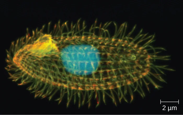
The protist shown has which of the following?
Count how many short projections cover the entire cell surface, rather than looking for a single whip-like structure, lobed extensions, or a hard outer covering.Describe the general life cycles and modes of reproduction in unicellular eukaryotic parasites
Sort each reproductive mechanism under whether protozoans use it for asexual or sexual reproduction.
Asexual reproduction
Sexual reproduction
The process by which a trophozoite becomes a cyst is called ________.
Use the term for turning INTO a protective cyst, not the term for emerging from one.Cysts triggered by favorable environmental cues become active again through a process called ________.
Use the term for emerging from a cyst, the opposite of the process in the item above.Identify challenges associated with classifying unicellular eukaryotes
Protist taxonomy has changed greatly in recent years as relationships have been re-examined using newer approaches. How do newer approaches differ from older approaches?
Show model answer
Did your answer mention:
The protists are described as a ________ group, meaning they lack a shared evolutionary origin.
Use the term for a group whose members do not share a single common ancestor.Current taxonomy of protists is based on evolutionary history, determined by biochemistry, morphology, and ________.
This is the field of evidence most associated with DNA and heredity, alongside biochemistry and morphology.Explain the taxonomic scheme used for unicellular eukaryotes
Animals belong to the same supergroup as the kingdom ________.
Look for the supergroup whose table row lists mushrooms, molds, and yeasts as one subgroup alongside Animals.Eukarya is currently divided into six ________ that are further divided into subgroups.
Use the term introduced at the start of the Taxonomy of Protists section for these six major groups.Sort each subgroup under the supergroup the table above assigns it to.
Excavata
Chromalveolata
Rhizaria
Archaeplastida
Give examples of infections caused by unicellular eukaryotes
Which genus includes the causative agent for malaria?
This apicomplexan genus is named directly in the Chromalveolata discussion of medically important genera.Which protist is a concern because of its ability to contaminate water supplies and cause diarrheal illness?
This Fornicata pathogen is named in the section’s opening paragraph and again in the Excavata discussion.Aside from a risk of birth defects, what other effect might a toxoplasmosis infection have?
Look for the sentence about theorizing and evidence that follows the birth-defect risk in the Toxoplasma gondii discussion.What characteristics might make you think a protist could be pathogenic? Are certain nutritional characteristics, methods of locomotion, or morphological differences likely to be associated with the ability to cause disease?
Show model answer
Did your answer mention:
Sort each genus or species under whether the table above lists it as causing human disease.
Causes disease listed in the table
No disease listed (N/a) in the table
This section is adapted from Microbiology, Section 5.1: Unicellular Eukaryotic Parasites by Nina Parker, Mark Schneegurt, Anh-Hue Thi Tu, Philip Lister, Brian M. Forster, and OpenStax, © OpenStax, licensed under CC BY-NC-SA 4.0. Access the original for free at openstax.org. Changes: all 16 vendored figures are re-encoded as WebP with kind set explicitly after inspecting each image (correcting the media manifest’s guess of “diagram” to “photo” for the ringworm figure); alts are rewritten from the served images rather than the source alt text, which is heavily typo-ridden in several places (most severely the Eimeria life-cycle alt: “Envronment,” “occruing,” “proess,” “Oocsts realease sporocyts,” “invate,” “trphozoites,” “sezual gametogon,” “maerozoites,” “gamets,” “gamees,” “mautres,” reported to the parent as a source defect); the four life-cycle diagrams (Eimeria, the cellular and plasmodial slime molds, and Trypanosoma brucei) each carry a stage-by-stage longdesc walking the numbered or arrow-ordered stages the caption does not name; the two supergroup-and-example table images (OSC_Microbio_05_01_groups1 and groups2), which carry no micrograph column, are transcribed as one combined Markdown table from the images (checked against PDF pages 173–174) rather than vendored, following the book’s rule for a table image whose content a table can fully carry; the Clinical Focus box is rendered as a callout, its “Jump to the next Clinical Focus box” link replaced with a plain sentence naming that the case continues in Parasitic Helminths; the Link to Learning and Eye on Ethics boxes are rendered as callouts with their URLs kept and their closing discussion questions kept as unanswered prose; the section’s four footnotes are rendered as inline parenthetical citations, with the two CDC citations’ bare access URLs dropped (no DOI to keep) and the two journal citations’ DOI-bearing details kept; both body Check Your Understanding notes are rendered as graded body items (a multiplechoice on the schizogony sequence and a sortbins on which supergroups are clinically significant) because one module passage fixes each whole answer; of the section’s ten end-of-section exercises (2 Multiple Choice, 2 Fill in the Blank, 3 Short Answer, 3 Critical Thinking), all are used and none is omitted — the 2 Multiple Choice and 2 Fill in the Blank items are source-keyed; one Short Answer (“What are kinetoplastids?”) converts to a textin keyed by the module’s own parenthetical; one Short Answer (the toxoplasmosis question) converts to a multiplechoice keyed by the module’s own sentence, its three distractors drawn from the module’s own descriptions of other pathogens’ effects; one Short Answer (the ciliate macronucleus question) and two Critical Thinking questions (the taxonomy-approaches question and the pathogenicity-characteristics question) remain self-checks with model answers and rubrics assembled only from this module’s text, because their honest answers need several sentences assembled or ask the learner to speculate; the Critical Thinking Art Connection item is graded as a mediafigure + multiplechoice keyed from the image (the pictured protist is covered in short hair-like projections, i.e., cilia), with an author-written caption and an alt that describes the projections without naming any of the four answer choices; 8 filler items (2 term-recall textins from the section’s own defining sentences on reproduction, 2 on classification vocabulary, 1 on the taxonomic-scheme vocabulary, 1 sortbins on reproductive mechanisms, and 2 sortbins built from the transcribed supergroup table) fill out three items per objective group; key terms are compiled from the module’s 23 defined terms, all of which have Glossary appendix entries (the appendix’s holozoic entry prints “phagoctytosis”; the bullet prints “phagocytosis”, a one-word source typo logged as an erratum); the misspelled phylum name “Ciliaphora” is corrected to “Ciliophora” (matching the book’s own Glossary entry), the extraneous “a” in “is a considered a distant relative” is removed, and a missing closing parenthesis after “African sleeping sickness” is added — all three are one-word or single-character corrections, logged as suspected source defects rather than disclosed inline; the cross-reference to m58793 is rendered as a link to that module’s actual title, “Unique Characteristics of Eukaryotic Cells” (the source’s own link text names it “Unique Characteristics of Prokaryotic Cells,” which is that module’s neighboring section, not its own title — logged as a suspected source defect); and cross-references to Sections 5.2–5.4 are rendered as absolute site-root links to their landing pages.