Parasitic Helminths
By the end of this section, you will be able to:
- Explain why we include the study of parasitic worms within the discipline of microbiology
- Compare the basic morphology of the major groups of parasitic helminthes
- Describe the characteristics of parasitic nematodes, and give an example of infective eggs and infective larvae
- Describe the characteristics of parasitic trematodes and cestodes, and give examples of each
- Identify examples of the primary causes of infections due to nematodes, trematodes, and cestodes
- Classify parasitic worms according to major groups
Parasitic helminths are animals that are often included within the study of microbiology because many species of these worms are identified by their microscopic eggs and larvae. There are two major groups of parasitic helminths: the roundworms (Nematoda) and flatworms (Platyhelminthes). Of the many species that exist in these groups, about half are parasitic and some are important human pathogens. As animals, they are multicellular and have organ systems. However, the parasitic species often have limited digestive tracts, nervous systems, and locomotor abilities. Parasitic forms may have complex reproductive cycles with several different life stages and more than one type of host. Some are monoecious, having both male and female reproductive organs in a single individual, while others are dioecious, each having either male or female reproductive organs.
Nematoda (Roundworms)
Phylum Nematoda (the roundworms) is a diverse group containing more than 15,000 species, of which several are important human parasites (the micrograph below). These unsegmented worms have a full digestive system even when parasitic. Some are common intestinal parasites, and their eggs can sometimes be identified in feces or around the anus of infected individuals. Ascaris lumbricoides is the largest nematode intestinal parasite found in humans; females may reach lengths of 20–35 cm. (Source note: the source says females “may reach lengths greater than 1 meter.” The CDC’s DPDx reference on ascariasis gives adult females as 20–35 cm long; this page uses that range.) A. lumbricoides is also very widespread, even in developed nations, although it is now a relatively uncommon problem in the United States. It may cause symptoms ranging from relatively mild (such as a cough and mild abdominal pain) to severe (such as intestinal blockage and impaired growth).
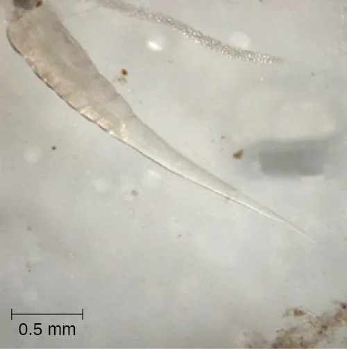
Of all nematode infections in the United States, pinworm (caused by Enterobius vermicularis) is the most common. Pinworm causes sleeplessness and itching around the anus, where the female worms lay their eggs during the night. Toxocara canis and T. cati are nematodes found in dogs and cats, respectively, that can be transmitted to humans, causing toxocariasis. Antibodies to these parasites have been found in approximately 13.9% of the U.S. population, suggesting that exposure is common (Won K, Kruszon-Moran D, Schantz P, Jones J. “National seroprevalence and risk factors for zoonotic Toxocara spp. infection.” In: Abstracts of the 56th American Society of Tropical Medicine and Hygiene; Philadelphia, Pennsylvania; 2007 Nov 4-8.). Infection can cause larval migrans, which can result in vision loss and eye inflammation, or fever, fatigue, coughing, and abdominal pain, depending on whether the organism infects the eye or the viscera. Another common nematode infection is hookworm, which is caused by Necator americanus (the New World or North American hookworm) and Ancylostoma duodenale (the Old World hookworm). Symptoms of hookworm infection can include abdominal pain, diarrhea, loss of appetite, weight loss, fatigue, and anemia.
Trichinellosis, also called trichinosis, caused by Trichinella spiralis, is contracted by consuming undercooked meat, which releases the larvae and allows them to encyst in muscles. Infection can cause fever, muscle pains, and digestive system problems; severe infections can lead to lack of coordination, breathing and heart problems, and even death. Finally, heartworm in dogs and other animals is caused by the nematode Dirofilaria immitis, which is transmitted by mosquitoes. Symptoms include fatigue and cough; when left untreated, death may result.
Clinical Focus. Part 2
The physician explains to Sarah’s mother that ringworm can be transferred between people through touch. “It’s common in school children, because they often come in close contact with each other, but anyone can become infected,” he adds. “Because you can transfer it through objects, locker rooms and public pools are also a potential source of infection. It’s very common among wrestlers and athletes in other contact sports.”
Looking very uncomfortable, Sarah says to her mother “I want this worm out of me.”
The doctor laughs and says, “Sarah, you’re in luck because ringworm is just a name; it is not an actual worm. You have nothing wriggling around under your skin.”
“Then what is it?” asks Sarah.
- What type of pathogen causes ringworm?
The case continues in Fungi. The case began in Unicellular Eukaryotic Parasites.
Check Your Understanding
What is the most common nematode infection in the United States?
Name the infection whose female worms lay eggs around the anus at night, described just after this section introduces roundworm infections.Platyhelminths (Flatworms)
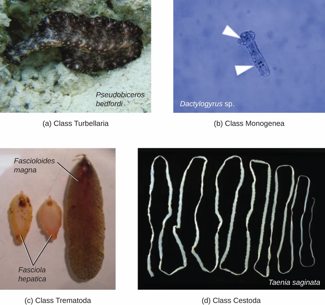
Phylum Platyhelminthes (the platyhelminths) are flatworms. This group includes the flukes, tapeworms, and the turbellarians, which include planarians. The flukes and tapeworms are medically important parasites (the figure below).
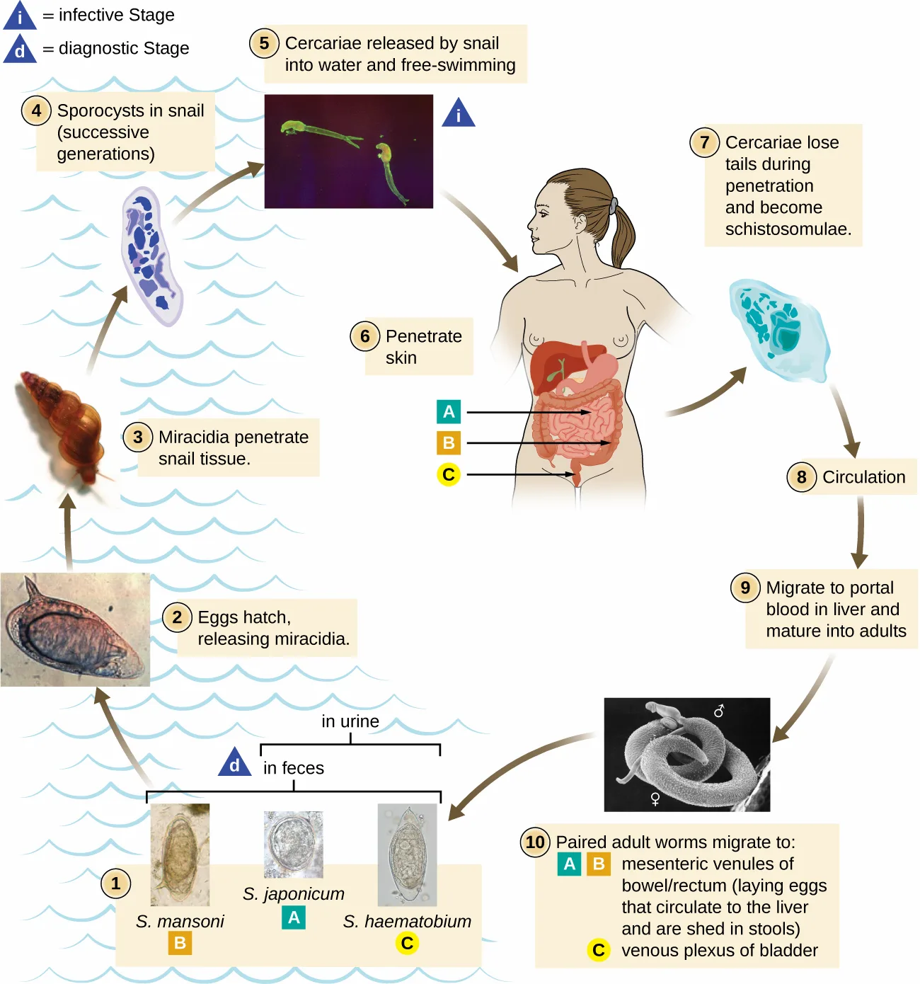
The flukes (trematodes) are nonsegmented flatworms that have an oral sucker (the micrograph below) (and sometimes a second ventral sucker) and attach to the inner walls of intestines, lungs, large blood vessels, or the liver. Trematodes have complex life cycles, often with multiple hosts. Several important examples are the liver flukes (Clonorchis and Opisthorchis), the intestinal fluke (Fasciolopsis buski), and the oriental lung fluke (Paragonimus westermani). Schistosomiasis is a serious parasitic disease, considered second in the scale of its impact on human populations only to malaria. The parasites Schistosoma mansoni, S. haematobium, and S. japonicum, which are found in freshwater snails, are responsible for schistosomiasis (the figure below). Immature forms burrow through the skin into the blood. They migrate to the lungs, then to the liver and, later, other organs. Symptoms include anemia, malnutrition, fever, abdominal pain, fluid buildup, and sometimes death.
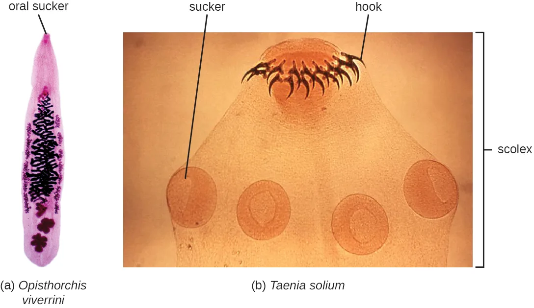
The other medically important group of platyhelminths are commonly known as tapeworms (cestodes) and are segmented flatworms that may have suckers or hooks at the scolex (head region) (see the micrograph above). Tapeworms use these suckers or hooks to attach to the wall of the small intestine. The body of the worm is made up of segments called proglottids that contain reproductive structures; these detach when the gametes are fertilized, releasing gravid proglottids with eggs. Tapeworms often have an intermediate host that consumes the eggs, which then hatch into a larval form called an oncosphere. The oncosphere migrates to a particular tissue or organ in the intermediate host, where it forms cysticerci. After being eaten by the definitive host, the cysticerci develop into adult tapeworms in the host’s digestive system (the figure below). Taenia saginata (the beef tapeworm) and T. solium (the pork tapeworm) enter humans through ingestion of undercooked, contaminated meat. The adult worms develop and reside in the intestine, but the larval stage may migrate and be found in other body locations such as skeletal and smooth muscle. The beef tapeworm is relatively benign, although it can cause digestive problems and, occasionally, allergic reactions. The pork tapeworm can cause more serious problems when the larvae leave the intestine and colonize other tissues, including those of the central nervous system. Diphyllobothrium latum is the largest human tapeworm and can be ingested in undercooked fish. It can grow to a length of 15 meters. Echinococcus granulosus, the dog tapeworm, can parasitize humans and uses dogs as an important host.
Extended description
The numbered stages are: (1) eggs are passed in feces (S. mansoni, S. japonicum) or urine (S. haematobium) — the diagnostic stage; (2) eggs hatch, releasing miracidia; (3) miracidia penetrate snail tissue; (4) sporocysts develop in the snail across successive generations; (5) cercariae are released by the snail into the water and are free-swimming — the infective stage; (6) cercariae penetrate the skin; (7) cercariae lose their tails during penetration and become schistosomulae; (8) schistosomulae enter circulation; (9) the parasites migrate to portal blood in the liver and mature into adults; (10) paired adult worms migrate — S. japonicum and S. mansoni to the mesenteric venules of the bowel and rectum, laying eggs that circulate to the liver and are shed in stools, and S. haematobium to the venous plexus of the bladder.
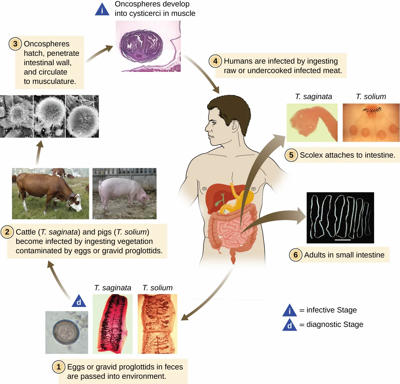
Extended description
The numbered stages are: (1) eggs or gravid proglottids in feces are passed into the environment — the diagnostic stage; (2) cattle (T. saginata) and pigs (T. solium) become infected by ingesting vegetation contaminated by eggs or gravid proglottids; (3) oncospheres hatch, penetrate the intestinal wall, and circulate to the musculature; the oncospheres develop into cysticerci in the muscles and become infective; (4) humans are infected by ingesting raw or undercooked infected meat; (5) the scolex attaches to the intestine; (6) adults are found in the small intestine.
Check Your Understanding
What group of medically important flatworms is segmented and what group is unsegmented? Sort each description under the class it fits.
Segmented flatworms
Unsegmented flatworms
Micro Connection. Food for Worms?
For residents of temperate, developed countries, it may be difficult to imagine just how common helminth infections are in the human population. In fact, they are quite common and even occur frequently in the United States. Worldwide, approximately 807–1,221 million people are infected with Ascaris lumbricoides (perhaps one-sixth of the human population) and far more are infected if all nematode species are considered (Fenwick, A. “The global burden of neglected tropical diseases.” Public health 126 no.3 (Mar 2012): 233–6.). Rates of infection are relatively high even in industrialized nations. Approximately 604–795 million people are infected with whipworm (Trichuris) worldwide (Trichuris can also infect dogs), and 576–740 million people are infected with hookworm (Necator americanus and Ancylostoma duodenale) (de Silva, N., et. al. (2003). “Soil-transmitted helminth infections: updating the global picture”. Trends in Parasitology 19 (December 2003): 547–51.). Toxocara, a nematode parasite of dogs and cats, is also able to infect humans. It is widespread in the United States, with about 10,000 symptomatic cases annually. However, one study found 14% of the population (more than 40 million Americans) was seropositive, meaning they had been exposed to the parasite at one time. More than 200 million people have schistosomiasis worldwide. Most of the World Health Organization (WHO) neglected tropical diseases are helminths. In some cases, helminths may cause subclinical illnesses, meaning the symptoms are so mild that they go unnoticed. In other cases, the effects may be more severe or chronic, leading to fluid accumulation and organ damage. With so many people affected, these parasites constitute a major global public health concern.
Micro Connection. Eradicating the Guinea Worm
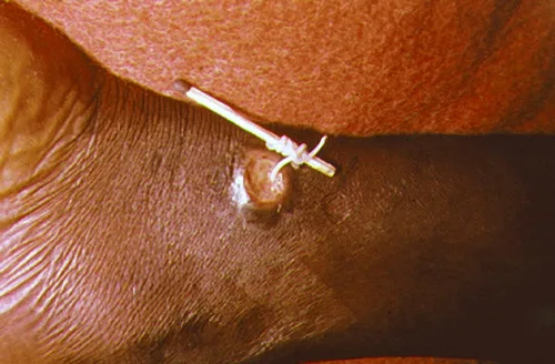
Dracunculiasis, or Guinea worm disease, is caused by a nematode called Dracunculus medinensis. When people consume contaminated water, water fleas (small crustaceans) containing the nematode larvae may be ingested. These larvae migrate out of the intestine, mate, and move through the body until females eventually emerge (generally through the feet). While Guinea worm disease is rarely fatal, it is extremely painful and can be accompanied by secondary infections and edema (the photo below).
An eradication campaign led by WHO, the CDC, the United Nations Children’s Fund (UNICEF), and the Carter Center (founded by former U.S. president Jimmy Carter) has been extremely successful in reducing cases of dracunculiasis. This has been possible because diagnosis is straightforward, there is an inexpensive method of control, there is no animal reservoir, the water fleas are not airborne (they are restricted to still water), the disease is geographically limited, and there has been a commitment from the governments involved. Additionally, no vaccines or medication are required for treatment and prevention. In 1986, 3.5 million people were estimated to be affected. After the eradication campaign, which included helping people in affected areas learn to filter water with cloth, only four countries continue to report the disease (Chad, Mali, South Sudan, and Ethiopia) with a total of 126 cases reported to WHO in 2014 (World Health Organization. “South Sudan Reports Zero Cases of Guinea-Worm Disease for Seventh Consecutive Month.” 2016. Accessed May 2, 2016.).
Summary
- Helminth parasites are included within the study of microbiology because they are often identified by looking for microscopic eggs and larvae.
- The two major groups of helminth parasites are the roundworms (Nematoda) and the flatworms (Platyhelminthes).
- Nematodes are common intestinal parasites often transmitted through undercooked foods, although they are also found in other environments.
- Platyhelminths include tapeworms and flukes, which are often transmitted through undercooked meat.
Key terms
- monoecious — refers to sexually reproducing organisms in which individuals have both male and female reproductive organs.
- dioecious — refers to sexually reproducing organisms in which individuals have either male or female reproductive organs (not both).
- Nematoda — phylum comprising roundworms.
- Platyhelminthes — phylum comprising flatworms.
- flukes — any of the parasitic nonsegmented flatworms (trematodes) that have an oral sucker and sometimes a second ventral sucker; they attach to the inner walls of intestines, lungs, large blood vessels, or the liver in human hosts.
- tapeworms — segmented, hermaphroditic, parasitic flatworms (Platyhelminthes).
- scolex — the head region of a cestode (tapeworm), which typically has suckers and/or hooks for attachment to the host.
- proglottids — body segment of a cestode (tapeworm).
Practice
Explain why we include the study of parasitic worms within the discipline of microbiology
Helminth parasites are included within the study of microbiology because they are often identified by looking for their microscopic ________.
Use the life stages named in the summary’s first bullet, not the specific life-cycle stage names used later for tapeworms or Schistosoma.Parasitic forms may have complex reproductive cycles with several different life stages and more than one type of ________.
Name what a parasite’s larval and adult stages each require to develop, as stated in this section’s opening paragraph.As animals, parasitic helminths are ________ and have organ systems.
Name the trait shared by all animals, stated at the start of this section, before the sentence about limited digestive tracts and nervous systems.Compare the basic morphology of the major groups of parasitic helminthes
A species of worm in which there are distinct male and female individuals is described as ________.
Use the term contrasted with an organism having both sexes’ reproductive organs in one individual.Some parasitic helminths are ________, having both male and female reproductive organs in a single individual.
Use the term for a worm that carries both sexes’ reproductive organs, defined at the opening of this section.The two major groups of helminth parasites are the roundworms and the flatworms (________).
Name the phylum introduced alongside ‘flatworms’ at the start of this section’s second heading.Describe the characteristics of parasitic nematodes, and give an example of infective eggs and infective larvae
Some nematodes are common intestinal parasites, and their eggs can sometimes be identified in ________ or around the anus of infected individuals.
Name where a stool sample would reveal these infective eggs.Infection with the nematodes Toxocara canis and T. cati can cause larval migrans, with symptoms depending on whether the organism infects the eye or the ________.
Contrast the two possible infection sites named in the toxocariasis sentence, not the sites named for trichinosis or hookworm.Trichinellosis is contracted by consuming undercooked meat, which releases Trichinella spiralis larvae and allows them to encyst in ________.
Name the tissue where these infective larvae become dormant after release from undercooked meat.Describe the characteristics of parasitic trematodes and cestodes, and give examples of each
A segmented worm has male and female reproductive organs in each segment. Some use hooks to attach to the intestinal wall. Which type of worm is this?
Match the body segmentation and hooked attachment to the group named for tapeworms.Flukes are in class ________.
Use the class name given as a synonym for flukes earlier in this section.Given the life cycle of the Schistosoma parasite, suggest a method of prevention of the disease.
Show model answer
Did your answer mention:
Identify examples of the primary causes of infections due to nematodes, trematodes, and cestodes
A nonsegmented worm is found during a routine colonoscopy of an individual who reported having abdominal cramps, nausea, and vomiting. This worm is likely which of the following?
Match the intestinal site and the symptoms of abdominal cramps, nausea, and vomiting to the group this section calls common intestinal parasites.What is the best defense against tapeworm infection?
Show model answer
Did your answer mention:
Heartworm in dogs and other animals is caused by the nematode Dirofilaria immitis, which is transmitted by ________.
Name the insect vector responsible for transmitting this nematode between hosts.Classify parasitic worms according to major groups
A fluke is classified within which of the following?
Name the phylum that includes flukes and tapeworms, introduced at the start of this section’s second heading.Sort each parasite into the phylum that includes it.
Nematoda
Platyhelminthes
Phylum ________ comprises the roundworms, a diverse group containing more than 15,000 species.
Name the phylum introduced at the start of this section’s first heading.This section is adapted from Microbiology, Section 5.2: Parasitic Helminths by Nina Parker, Mark Schneegurt, Anh-Hue Thi Tu, Philip Lister, Brian M. Forster, and OpenStax, © OpenStax, licensed under CC BY-NC-SA 4.0. Access the original for free at openstax.org. Changes: all six figures re-encoded as WebP and rendered as mediafigures with an explicit, image-checked kind (four photos, two life-cycle diagrams); the Schistosoma and tapeworm life-cycle alts and longdescs are author-written from the rendered artwork rather than the source alts, which misspell “schistosomulae,” double the word “the,” and attribute distinct intestinal segments to each Schistosoma species that the diagram does not show — the diagram instead groups S. mansoni and S. japonicum together at the bowel/rectum and S. haematobium alone at the bladder; the tapeworm life-cycle alt’s “proplottidis” is corrected to “proglottids” with no inline note (one-word typo); the pinworm micrograph’s alt is rewritten because the source’s claimed length (“approximately 30 mm”) is an order of magnitude larger than what the pictured 0.5 mm scale bar supports. The Clinical Focus box is rendered as a callout with its “Jump to/Go back to” links replaced by plain sentences naming Fungi (Section 5.3) as where the case continues and Unicellular Eukaryotic Parasites (Section 5.1) as where it began; both target pages land in this run. The two Check Your Understanding bullets are rendered as body items at their note’s position: the pinworm question as a textin graded from this section’s “most common nematode infection” sentence, and the segmented/unsegmented flatworm question as a sortbins graded from the cestode and trematode descriptions earlier in this section. The two Micro Connection boxes are rendered as callouts with their citations kept as parenthetical text after the sentences they support (bare access URLs dropped, one duplicated “that that” silently corrected as a one-word-class typo); the WHO footnote’s bare URL is dropped and its “Accessed May 2, 2016” date is kept. All three source footnotes in the body and Micro Connection boxes are rendered as inline parenthetical citations. The three source Multiple Choice items and two Fill in the Blank items are adapted into Practice unchanged and keyed as printed. The two unkeyed end-of-section questions — a Short Answer (“What is the best defense against tapeworm infection?”) and a Critical Thinking item (the Schistosoma prevention question) — remain self-checks whose model answers and rubrics are assembled from this section’s own transmission sentences, because the source prints no key for them and no single sentence states a prevention method outright. Eleven filler items (seven textin, three multiplechoice, one sortbins) fill the six objective groups to this book’s three-per-group floor, each built from one body or summary sentence of this module with no new claim; no source item was omitted. Key terms are compiled from the module’s eight defined terms and the book’s Glossary appendix (all eight from the appendix; none sentence-derived); the proglottid bullet is printed as the plural proglottids to match its bolded form in the body. Author-built multiple-choice items vary their key position rather than defaulting to the first option; the three source-keyed items keep their printed option order. The source’s “Diphylobothrium latum” is printed as Diphyllobothrium latum, the spelling the book’s own taxonomy and glossary appendices use (a one-word source typo, logged as an erratum). The Ascaris lumbricoides female length is corrected from the source’s “greater than 1 meter” to 20–35 cm with a visible Source note citing the CDC DPDx reference, per this book’s prose claim pass. The four-class platyhelminth figure’s alt is rewritten from the artwork because the source alt misspells “Monogenea” as “Monegena,” describes panel (b) as a rectangular cell with a bulb where the artwork shows an elongated worm with anchor hooks, and calls panel (d) a single tapeworm where several specimens are pictured.