Fungi
By the end of this section, you will be able to:
- Explain why the study of fungi such as yeast and molds is within the discipline of microbiology
- Describe the unique characteristics of fungi
- Describe examples of asexual and sexual reproduction of fungi
- Compare the major groups of fungi in this chapter, and give examples of each
- Identify examples of the primary causes of infections due to yeasts and molds
- Identify examples of toxin-producing fungi
- Classify fungal organisms according to major groups
The fungi comprise a diverse group of organisms that are heterotrophic and typically saprotrophic. (Source note: the source says “saprozoic” here; the module’s own Summary says the fungi are “saprotrophic,” and the book’s Glossary appendix defines “saprozoic” as a feeding mode of protozoans, so this page uses the Summary’s word.) In addition to the well-known macroscopic fungi (such as mushrooms and molds), many unicellular yeasts and spores of macroscopic fungi are microscopic. For this reason, fungi are included within the field of microbiology.
Fungi are important to humans in a variety of ways. Both microscopic and macroscopic fungi have medical relevance, with some pathogenic species that can cause mycoses (illnesses caused by fungi). Some pathogenic fungi are opportunistic, meaning that they mainly cause infections when the host’s immune defenses are compromised and do not normally cause illness in healthy individuals. Fungi are important in other ways. They act as decomposers in the environment, and they are critical for the production of certain foods such as cheeses. Fungi are also major sources of antibiotics, such as penicillin from the fungus Penicillium.
Characteristics of Fungi
Fungi have well-defined characteristics that set them apart from other organisms. Most multicellular fungal bodies, commonly called molds, are made up of filaments called hyphae. Hyphae can form a tangled network called a mycelium and form the thallus (body) of fleshy fungi. Hyphae that have walls between the cells are called septate hyphae; hyphae that lack walls and cell membranes between the cells are called nonseptate or coenocytic hyphae (shown below).
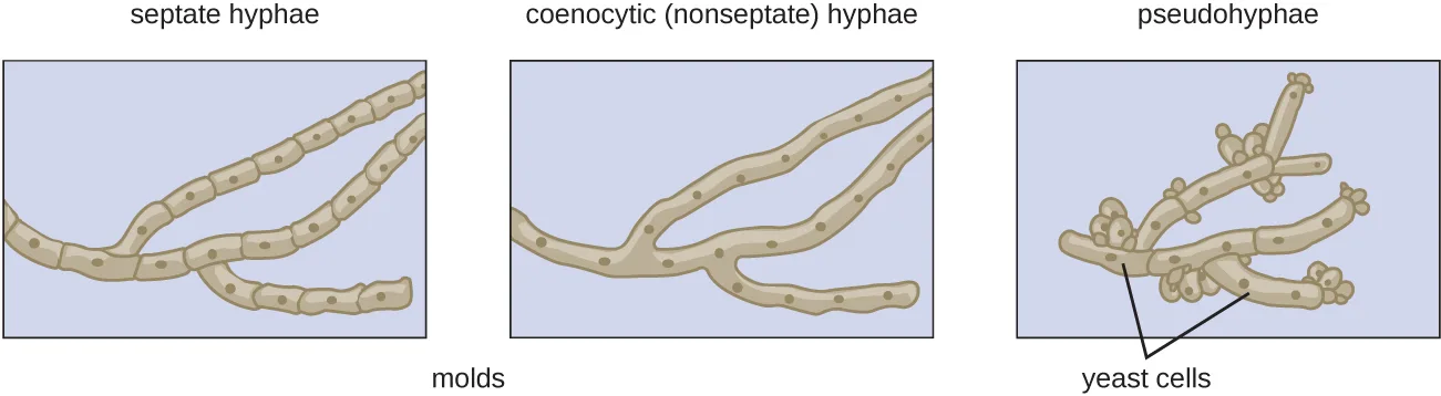
In contrast to molds, yeasts are unicellular fungi. The budding yeasts reproduce asexually by budding off a smaller daughter cell; the resulting cells may sometimes stick together as a short chain or pseudohypha (shown above).
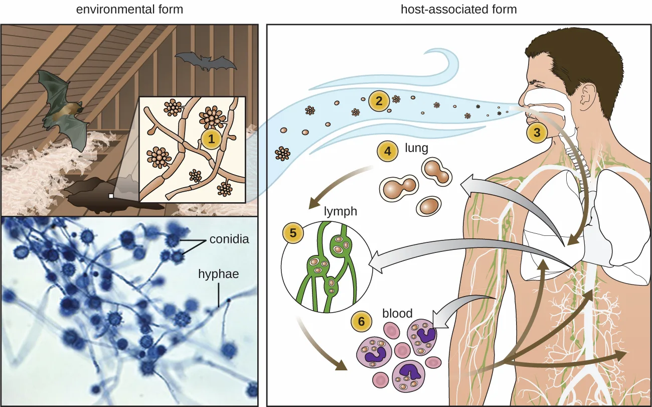
Some fungi are dimorphic, having more than one appearance during their life cycle. These dimorphic fungi may be able to appear as yeasts or molds, which can be important for infectivity. They are capable of changing their appearance in response to environmental changes such as nutrient availability or fluctuations in temperature, growing as a mold, for example, at 25 °C (77 °F), and as yeast cells at 37 °C (98.6 °F). This ability helps dimorphic fungi to survive in diverse environments. Two examples of dimorphic yeasts are the human pathogens Histoplasma capsulatum and Candida albicans. H. capsulatum causes the lung disease histoplasmosis, and C. albicans is associated with vaginal yeast infections, oral thrush, and candidiasis of the skin (shown below).
Extended description
The figure walks through Histoplasma capsulatum’s dimorphic life cycle in six numbered stages. (1) In the environmental form, fungal filaments bearing round spore clusters called conidia grow in soil enriched with bird or bat guano, shown in an attic setting and confirmed by the micrograph below it. (2) Spores become airborne. (3) A person inhales the airborne spores through the nose. (4) The spores reach the lungs, where they appear as small, paired, budding structures. (5) The organism travels to the lymphatic system, shown as budding cells within a lymph vessel. (6) The organism reaches the bloodstream, shown as budding cells within blood cells.
There are notable unique features in fungal cell walls and membranes. Fungal cell walls contain chitin, as opposed to the cellulose found in the cell walls of plants and many protists. Additionally, whereas animals have cholesterol in their cell membranes, fungal cell membranes have different sterols called ergosterols. Ergosterols are often exploited as targets for antifungal drugs.
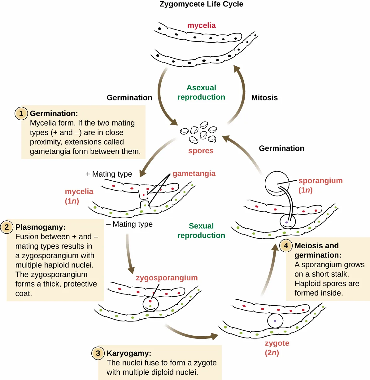
Fungal life cycles are unique and complex. Fungi reproduce sexually either through cross- or self-fertilization. Haploid fungi form hyphae that have gametes at the tips. Two different mating types (represented as “+ type” and “– type”) are involved. The cytoplasms of the + and – type gametes fuse (in an event called plasmogamy), producing a cell with two distinct nuclei (a dikaryotic cell). Later, the nuclei fuse (in an event called karyogamy) to create a diploid zygote. The zygote undergoes meiosis to form spores that germinate to start the haploid stage, which eventually creates more haploid mycelia (shown below). Depending on the taxonomic group, these sexually produced spores are known as zygospores (in Zygomycota), ascospores (in Ascomycota), or basidiospores (in Basidiomycota) (shown below).
Extended description
Four numbered stages of sexual reproduction. (1) Germination: mycelia form; if the plus and minus mating types are close together, extensions called gametangia form between them. (2) Plasmogamy: fusion between the plus and minus mating types produces a zygosporangium with multiple haploid nuclei, which forms a thick, protective coat. (3) Karyogamy: the nuclei fuse to form a zygote with multiple diploid nuclei. (4) Meiosis and germination: a sporangium grows on a short stalk, and haploid spores form inside it, then release to begin the cycle again.
Fungi may also exhibit asexual reproduction by mitosis, mitosis with budding, fragmentation of hyphae, and formation of asexual spores by mitosis. These spores are specialized cells that, depending on the organism, may have unique characteristics for survival, reproduction, and dispersal. Fungi exhibit several types of asexual spores and these can be important in classification.
Check Your Understanding
Is a dimorphic fungus a yeast or a mold? Explain.
Show model answer
Did your answer mention:
Fungal Diversity
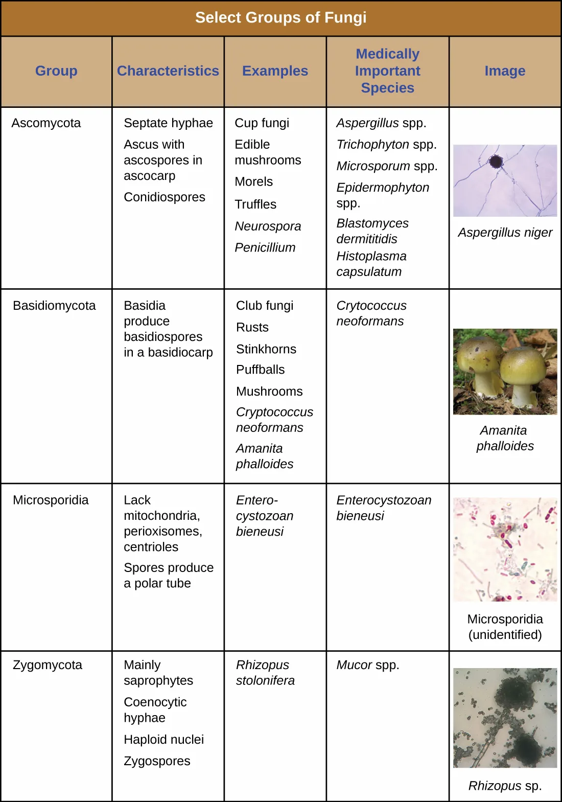
The fungi are very diverse, comprising seven major groups. Not all of the seven groups contain pathogens. Some of these groups are generally associated with plants and include plant pathogens. For example, Urediniomycetes and Ustilagomycetes include the plant rusts and smuts, respectively. These form reddish or dark masses, respectively, on plants as rusts (red) or smuts (dark). Some species have substantial economic impact because of their ability to reduce crop yields. Glomeromycota includes the mycorrhizal fungi, important symbionts with plant roots that can promote plant growth by acting like an extended root system. The Glomeromycota are obligate symbionts, meaning that they can only survive when associated with plant roots; the fungi receive carbohydrates from the plant and the plant benefits from the increased ability to take up nutrients and minerals from the soil. The Chytridiomycetes (chytrids) are small fungi, but are extremely ecologically important. Chytrids are generally aquatic and have flagellated, motile gametes; specific types are implicated in amphibian declines around the world. Because of their medical importance, we will focus on Zygomycota, Ascomycota, Basidiomycota, and Microsporidia. The table below summarizes the characteristics of these medically important groups of fungi.
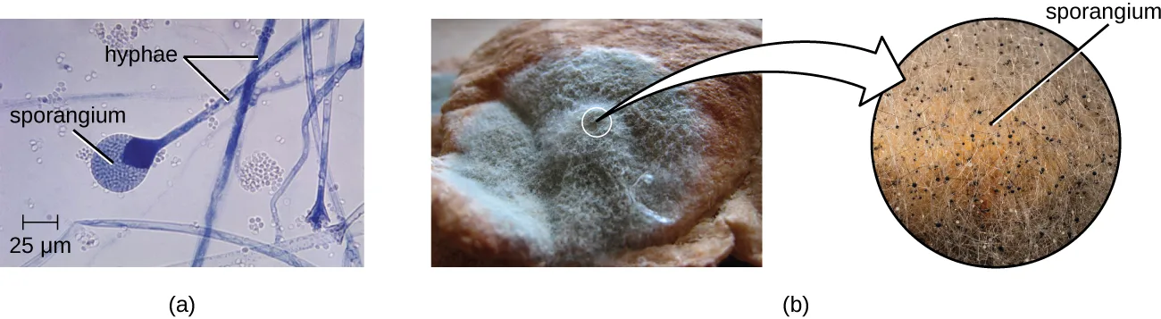
The Zygomycota (zygomycetes) are mainly saprophytes with coenocytic hyphae and haploid nuclei. They use sporangiospores for asexual reproduction. The group name comes from the zygospores that they use for sexual reproduction (shown above), which have hard walls formed from the fusion of reproductive cells from two individuals. Zygomycetes are important for food science and as crop pathogens. One example is Rhizopus stolonifer (shown above), an important bread mold that also causes rice seedling blight. Mucor is a genus of fungi that can potentially cause necrotizing infections in humans, although most species are intolerant of temperatures found in mammalian bodies (shown above).
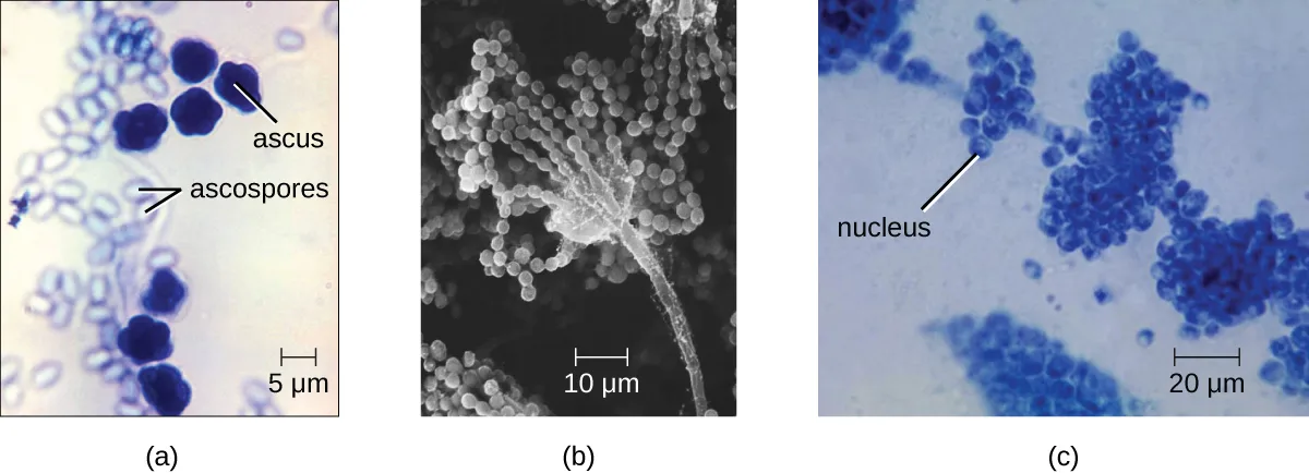
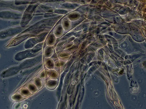
The Ascomycota include fungi that are used as food (edible mushrooms, morels, and truffles), others that are common causes of food spoilage (bread molds and plant pathogens), and still others that are human pathogens. Ascomycota may have septate hyphae and cup-shaped fruiting bodies called ascocarps. Some genera of Ascomycota use sexually produced ascospores as well as asexual spores called conidia, but sexual phases have not been discovered or described for others. Some produce an ascus containing ascospores within an ascocarp (shown below).
Examples of the Ascomycota include several bread molds and minor pathogens, as well as species capable of causing more serious mycoses. Species in the genus Aspergillus are important causes of allergy and infection, and are useful in research and in the production of certain fermented alcoholic beverages such as Japanese sake. The fungus Aspergillus flavus, a contaminant of nuts and stored grains, produces an aflatoxin that is both a toxin and the most potent known natural carcinogen. Neurospora crassa is of particular use in genetics research because the spores produced by meiosis are kept inside the ascus in a row that reflects the cell divisions that produced them, giving a direct view of segregation and assortment of genes (shown below). Penicillium produces the antibiotic penicillin (shown below).
Many species of ascomycetes are medically important. A large number of species in the genera Trichophyton, Microsporum, and Epidermophyton are dermatophytes, pathogenic fungi capable of causing skin infections such as athlete’s foot, jock itch, and ringworm. Blastomyces dermatitidis is a dimorphic fungus that can cause blastomycosis, a respiratory infection that, if left untreated, can become disseminated to other body sites, sometimes leading to death. Another important respiratory pathogen is the dimorphic fungus Histoplasma capsulatum (shown above), which is associated with birds and bats in the Ohio and Mississippi river valleys. Coccidioides immitis causes the serious lung disease Valley fever. Candida albicans, the most common cause of vaginal and other yeast infections, is also an ascomycete fungus; it is a part of the normal microbiota of the skin, intestine, genital tract, and ear (shown below). Ascomycetes also cause plant diseases, including ergot infections, Dutch elm disease, and powdery mildews.
Saccharomyces yeasts, including the baker’s yeast S. cerevisiae, are unicellular ascomycetes with haploid and diploid stages (shown below). This and other Saccharomyces species are used for brewing beer.
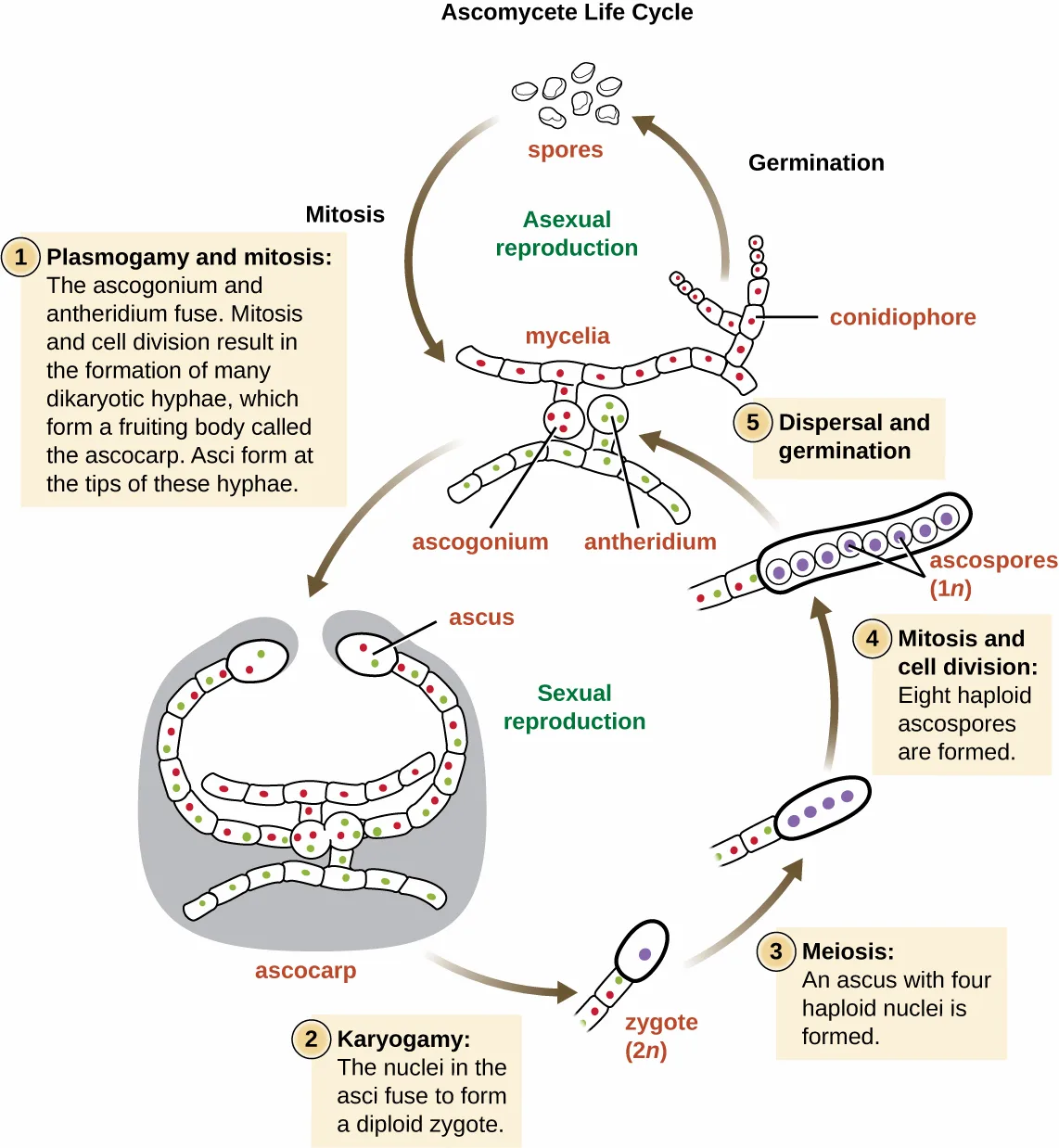
Extended description
Five numbered stages of sexual reproduction. (1) Plasmogamy and mitosis: the ascogonium and antheridium fuse; mitosis and cell division form many dikaryotic hyphae, which form a fruiting body called the ascocarp, with asci forming at the tips of these hyphae. (2) Karyogamy: the nuclei in the asci fuse to form a diploid zygote. (3) Meiosis: an ascus with four haploid nuclei is formed. (4) Mitosis and cell division: eight haploid ascospores are formed. (5) Dispersal and germination: the ascospores disperse and germinate into new mycelia, which can also form conidiophores that release asexual spores.
The Basidiomycota (basidiomycetes) are fungi that have basidia (club-shaped structures) that produce basidiospores (spores produced through budding) within fruiting bodies called basidiocarps (shown below). They are important as decomposers and as food. This group includes rusts, stinkhorns, puffballs, and mushrooms. Several species are of particular importance. Cryptococcus neoformans, a fungus commonly found as a yeast in the environment, can cause serious lung infections when inhaled by individuals with weakened immune systems. The edible meadow mushroom, Agaricus campestris, is a basidiomycete, as is the poisonous mushroom Amanita phalloides, known as the death cap. The deadly toxins produced by A. phalloides have been used to study transcription.
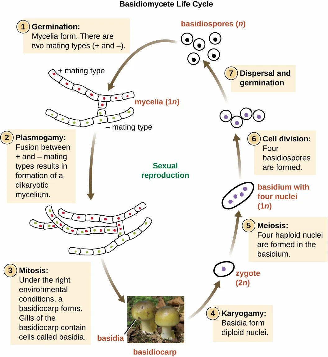
Extended description
Seven numbered stages. (1) Germination: mycelia form; there are two mating types, plus and minus. (2) Plasmogamy: fusion between the plus and minus mating types forms a dikaryotic mycelium. (3) Mitosis: under the right environmental conditions, a basidiocarp forms; gills of the basidiocarp contain cells called basidia, shown in a photograph of two mushrooms. (4) Karyogamy: basidia form diploid nuclei. (5) Meiosis: four haploid nuclei are formed in the basidium. (6) Cell division: four basidiospores are formed. (7) Dispersal and germination: the basidiospores disperse and germinate into new mycelia.
Finally, the Microsporidia are unicellular fungi that are obligate intracellular parasites. They lack mitochondria, peroxisomes, and centrioles, but their spores release a unique polar tubule that pierces the host cell membrane to allow the fungus to gain entry into the cell. A number of microsporidia are human pathogens, and infections with microsporidia are called microsporidiosis. One pathogenic species is Enterocytozoon bieneusi (Source note: the source prints “Enterocystozoan bieneusi”; the accepted name is Enterocytozoon bieneusi — “Etymologia: Enterocytozoon bieneusi,” Emerging Infectious Diseases 27, no. 6 [2021], DOI 10.3201/eid2706.ET2706.), which can cause symptoms such as diarrhea, cholecystitis (inflammation of the gall bladder), and in rare cases, respiratory illness.
Select Groups of Fungi
| Group | Characteristics | Examples | Medically Important Species |
|---|---|---|---|
| Ascomycota | Septate hyphae; ascus with ascospores in ascocarp; conidiospores | Cup fungi; edible mushrooms; morels; truffles; Neurospora; Penicillium | Aspergillus spp.; Trichophyton spp.; Microsporum spp.; Epidermophyton spp.; Blastomyces dermatitidis; Histoplasma capsulatum |
| Basidiomycota | Basidia produce basidiospores in a basidiocarp | Club fungi; rusts; stinkhorns; puffballs; mushrooms; Cryptococcus neoformans; Amanita phalloides | Cryptococcus neoformans |
| Microsporidia | Lack mitochondria, peroxisomes, centrioles; spores produce a polar tube | Enterocytozoon bieneusi | Enterocytozoon bieneusi |
| Zygomycota | Mainly saprophytes; coenocytic hyphae; haploid nuclei; zygospores | Rhizopus stolonifer | Mucor spp. |
Check Your Understanding
Which group of fungi appears to be associated with the greatest number of human diseases?
Compare how many medically important species are listed for each group in the table above.Micro Connection. Eukaryotic Pathogens in Eukaryotic Hosts
When we think about antimicrobial medications, antibiotics such as penicillin often come to mind. Penicillin and related antibiotics interfere with the synthesis of peptidoglycan cell walls, which effectively targets bacterial cells. These antibiotics are useful because humans (like all eukaryotes) do not have peptidoglycan cell walls.
Developing medications that are effective against eukaryotic cells but not harmful to human cells is more difficult. Despite huge morphological differences, the cells of humans, fungi, and protists are similar in terms of their ribosomes, cytoskeletons, and cell membranes. As a result, it is more challenging to develop medications that target protozoans and fungi in the same way that antibiotics target prokaryotes.
Fungicides have relatively limited modes of action. Because fungi have ergosterols (instead of cholesterol) in their cell membranes, the different enzymes involved in sterol production can be a target of some medications. The azole and morpholine fungicides interfere with the synthesis of membrane sterols. These are used widely in agriculture (fenpropimorph) and clinically (e.g., miconazole). Some antifungal medications target the chitin cell walls of fungi. Despite the success of these compounds in targeting fungi, antifungal medications for systemic infections still tend to have more toxic side effects than antibiotics for bacteria.
Clinical Focus. Part 3
Sarah is relieved the ringworm is not an actual worm, but wants to know what it really is. The physician explains that ringworm is a fungus. He tells her that she will not see mushrooms popping out of her skin, because this fungus is more like the invisible part of a mushroom that hides in the soil. He reassures her that they are going to get the fungus out of her too.
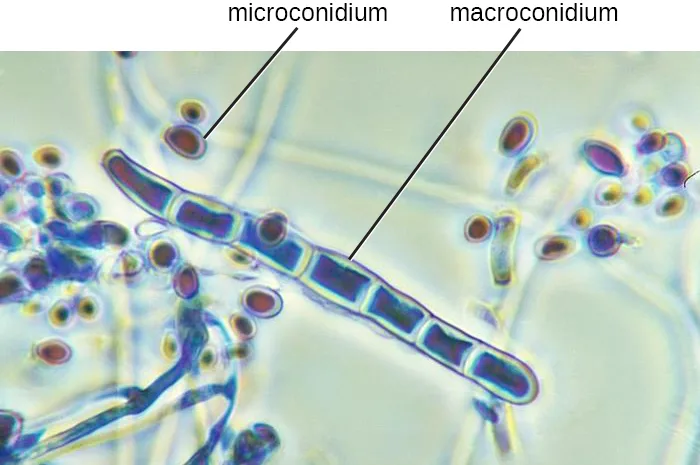
The doctor cleans and then carefully scrapes the lesion to place a specimen on a slide. By looking at it under a microscope, the physician is able to confirm that a fungal infection is responsible for Sarah’s lesion. In the micrograph below, it is possible to see macro- and microconidia in Trichophyton rubrum. Cell walls are also visible. Even if the pathogen resembled a helminth under the microscope, the presence of cell walls would rule out the possibility because animal cells lack cell walls.
The doctor prescribes an antifungal cream for Sarah’s mother to apply to the ringworm. Sarah’s mother asks, “What should we do if it doesn’t go away?”
- Can all forms of ringworm be treated with the same antifungal medication?
The case continues in Lichens. The case began in Unicellular Eukaryotic Parasites.
Summary
- The fungi include diverse saprotrophic eukaryotic organisms with chitin cell walls
- Fungi can be unicellular or multicellular; some (like yeast) and fungal spores are microscopic, whereas some are large and conspicuous
- Reproductive types are important in distinguishing fungal groups
- Medically important species exist in the four fungal groups Zygomycota, Ascomycota, Basidiomycota, and Microsporidia
- Members of Zygomycota, Ascomycota, and Basidiomycota produce deadly toxins
- Important differences in fungal cells, such as ergosterols in fungal membranes, can be targets for antifungal medications, but similarities between human and fungal cells make it difficult to find targets for medications and these medications often have toxic adverse effects
Key terms
- mycoses — refers to diseases caused by fungi.
- hyphae — tubular, filamentous structures that makes up most fungi.
- mycelium — vegetative network of branched, tubular hyphae.
- thallus — body of fleshy fungi (more generally, a body without a root, stem, or leaf).
- septate hyphae — hyphae that contain walls between individual cells; characteristic of some fungi.
- coenocytic hyphae — nonseptate hyphae that are multinucleate and lack cell walls or membranes between cells; characteristic of some fungi.
- budding yeasts — yeasts that divide by budding off of daughter cells.
- pseudohypha — short chains of yeast cells stuck together.
- dimorphic fungi — a fungus that can take the form of a yeast or a mold, depending on environmental conditions.
- chitin — polysaccharide that is an important component of fungal cell walls.
- dikaryotic — having two separate nuclei within one cell.
- spores — specialized cells that may be used for reproduction or may be specialized to withstand harsh conditions.
- zygospores — spores used by Zygomycetes for sexual reproduction; they have hard walls formed from the fusion of reproductive cells from two individuals.
- ascocarps — cup-shaped fruiting bodies of an ascomycete fungus.
- ascospores — sexually produced spores of ascomycete fungi, formed in an ascus.
- conidia — asexual fungal spores not enclosed in a sac; produced in a chain at the end of specialized hyphae called conidiophores.
- ascus — structure of ascomycete fungi containing spores.
- aflatoxin — chemical produced by the fungus Aspergillus flavus; both a toxin and the most potent known natural carcinogen.
- basidia — small club-shaped structures of basidiomycete fungi where basidiospores are produced.
- basidiospores — spores produced sexually via budding in basidiomycete fungi.
- basidiocarps — fruiting bodies of basidiomycete fungi.
- Microsporidia — fungi that lack mitochondria, centrioles, and peroxisomes; some can be human pathogens.
- polar tubule — a tube-like structure produced by spores of parasitic Microsporidia fungi that pierces host cell membranes.
Practice
Explain why the study of fungi such as yeast and molds is within the discipline of microbiology
Although mushrooms and molds are macroscopic, many single-celled fungi and the spores of macroscopic fungi are ________, which is why fungi are studied within microbiology.
Use the opening paragraph’s explanation for why fungi, despite some being macroscopic, belong to microbiology.The general term for diseases caused by fungi is ________.
Look for the term defined in parentheses as ‘illnesses caused by fungi.’Some fungi have proven medically useful because they can be used to produce ________.
The opening paragraphs name penicillin as an example.Describe the unique characteristics of fungi
Nonseptate hyphae are also called ________.
Use the term paired with ’nonseptate’ in the Characteristics of Fungi discussion.Unicellular fungi are called ________.
Contrast this term with the multicellular fungi called molds.Fungal cell walls are composed of the polysaccharide ________, unlike the cellulose found in the cell walls of plants and many protists.
This substance also gives the fungal cell wall its resistance and is a target for some antifungal drugs.Explain the benefit of research into the pathways involved in the synthesis of chitin in fungi.
Show model answer
Did your answer mention:
Describe examples of asexual and sexual reproduction of fungi
What is a dikaryotic cell?
Look for the term introduced in the discussion of plasmogamy, the fusion of the + and – type gametes’ cytoplasm.Ascomycota may produce sexually produced ascospores as well as asexual spores called ________.
These asexual spores are produced at the end of specialized hyphae called conidiophores.Sort each reproductive spore type or characteristic under the fungal group it describes.
Zygomycota
Ascomycota
Basidiomycota
Compare the major groups of fungi in this chapter, and give examples of each
Sort each characteristic, example, or medically important species under the group described in the table above.
Ascomycota
Basidiomycota
Microsporidia
Zygomycota
Which ascomycete genus is noted in this section for its use in genetics research, because the spores produced by meiosis are kept inside the ascus in a row that reflects the cell divisions that produced them?
Look for the genus whose ascus arrangement gives a direct view of gene segregation and assortment.Which fungal group described in this section is made up of obligate symbionts that promote plant growth by acting like an extended root system?
Look for the group described as including the mycorrhizal fungi.Identify examples of the primary causes of infections due to yeasts and molds
Which of the following is the most common cause of human yeast infections?
Look for the yeast described as part of the normal microbiota of the skin, intestine, genital tract, and ear.Which of the following is an ascomycete fungus associated with bat droppings that can cause a respiratory infection if inhaled?
Use the dimorphic fungus discussed with the bat-guano life-cycle figure.Which genera of fungi are common dermatophytes (fungi that cause skin infections)?
Find the sentence naming three genera as common causes of athlete’s foot, jock itch, and ringworm.Identify examples of toxin-producing fungi
Aspergillus flavus produces a ________ that is both a toxin and the most potent known natural carcinogen.
This word names the toxin itself and shares its first syllable with the genus that produces it.Which basidiomycete mushroom produces toxins that have been used to study transcription?
Look for the poisonous mushroom known as the death cap.According to the summary, which of the following fungal groups is not identified as a producer of deadly toxins?
Compare the summary’s list of toxin-producing groups with the four groups discussed in this section.Classify fungal organisms according to major groups
Which classification includes the unicellular yeast Saccharomyces cerevisiae, used with other Saccharomyces species for brewing beer?
This yeast has haploid and diploid stages and produces ascospores like other members of its group.Mushrooms are a type of which of the following?
Use the term for the fruiting body that contains basidia in its gills.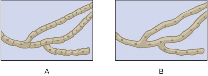
Which of the drawings shows septate hyphae?
Septate hyphae have walls between their cells; compare the two drawings for internal cross-walls.This section is adapted from Microbiology, Section 5.3: Fungi by Nina Parker, Mark Schneegurt, Anh-Hue Thi Tu, Philip Lister, Brian M. Forster, and OpenStax, © OpenStax, licensed under CC BY-NC-SA 4.0. Access the original for free at openstax.org. Changes: all eleven source images (ten figures plus the bare Critical Thinking media) are re-encoded as WebP and rendered as mediafigures after image and PDF inspection; six are set kind="diagram" against the manifest’s default photo guess because they are wholly or partly drawn (the hyphae comparison, the Histoplasma transmission diagram, the three numbered life-cycle diagrams, and the Critical Thinking hyphae comparison), and five stay kind="photo" (the two- and three-panel micrograph composites, the sexual-spore micrographs, the fungal-groups table image, and the Trichophyton rubrum micrograph); the zygospore figure is placed between the two Zygomycota paragraphs, at the sentence that refers to it, rather than after both as the source floats it; alts are rewritten from the served images, correcting the source alt’s fabricated per-object measurements in the ascospore/conidia figure (it claims separate 10 µm and 2 µm object sizes that the artwork does not print, only a single scale bar per panel); the three numbered life-cycle figures (zygomycete, ascomycete, basidiomycete) and the Histoplasma transmission diagram each carry a stage-by-stage longdesc, one figure beyond the three life-cycle diagrams because its numbered transmission stages are not walked in the caption either; the table-as-image (Figure 5.33) is transcribed as a Markdown table checked against the image and the PDF page, correcting four one-word typos the image itself prints (“Blastomyces dermititidis” to Blastomyces dermatitidis, “Crytococcus neoformans” to Cryptococcus neoformans, “perioxisomes” to “peroxisomes”, “Rhizopus stolonifera” to Rhizopus stolonifer, the last also corrected in the source Multiple Choice option that repeats it) and dropping its Image column, whose content becomes the mediafigure immediately after the table with an author-extended credit caption and an alt describing only the micrograph column; the table is kept at its document position, ending the Fungal Diversity subsection, because the sentence that first refers to it (“the table below summarizes…”) opens the subsection it summarizes; one further one-word typo, “Agricus campestris” for Agaricus campestris, is corrected in the body prose; the Micro Connection and Clinical Focus feature boxes are rendered as callouts, the Clinical Focus box’s “Jump to the next / Go back to the previous” links replaced with plain sentences naming the case’s continuation in Lichens and its beginning in Unicellular Eukaryotic Parasites, and the closing question kept as an unanswered bullet at its printed position, before the box’s figure; both Check Your Understanding notes become body interactions, one a self-check because its “explain” answer needs two module sentences assembled and one a multiplechoice graded from the transcribed table’s medically-important-species counts (Ascomycota lists six species or genera against one apiece for the other three groups); the three source Multiple Choice and three Fill in the Blank items are adapted into Practice; the two unkeyed Short Answer questions are graded as multiplechoice from this module’s own dermatophyte-genera sentence and dikaryotic-cell sentence; of the two unkeyed Critical Thinking questions, the bare-media hyphae comparison is rendered as a mediafigure followed by a two-option multiplechoice keyed from the drawings (an author-written caption and alt describe the two drawings without naming septate or coenocytic hyphae), and the chitin-synthesis question remains a self-check whose model answer states only what this module gives about chitin and antifungal drug targets, without speculating on research benefits the module does not discuss; no source exercise is omitted; three summary/body-sentence multiple-choice items (the Glomeromycota symbiont question, the toxin-producing-groups question, and the Saccharomyces cerevisiae classification question), a spore-naming sortbins, a second sortbins built from the transcribed fungal-groups table, a genetics-research multiple-choice, a toxin-producing-mushroom multiple-choice (Amanita phalloides), and five key-term/body-sentence textins fill out the Practice groups to this book’s floor (twelve author-written items in all); key terms are compiled from the module’s 23 defined terms and the book’s Glossary appendix; 21 definitions are taken directly from the glossary, and two are taken from the module’s own defining sentences because the appendix entries are defective — its thallus entry runs on into an unrelated fragment about HIV infection, and its ascospore entry calls the spore “asexual” where this module says ascospores are sexually produced (both logged as errata). The source’s stray closing parenthesis after “coenocytic hyphae” is dropped (logged as an erratum). Two claims are corrected with visible Source notes per this book’s prose claim pass: “saprozoic” is printed as “saprotrophic,” the module’s own Summary word, and “Enterocystozoan bieneusi” is printed as Enterocytozoon bieneusi, the accepted name (both logged as errata).