Isolation, Culture, and Identification of Viruses
By the end of this section, you will be able to:
- Discuss why viruses were originally described as filterable agents
- Describe the cultivation of viruses and specimen collection and handling
- Compare in vivo and in vitro techniques used to cultivate viruses
At the beginning of this chapter, we described how porcelain Chamberland filters with pores small enough to allow viruses to pass through were used to discover TMV. Today, porcelain filters have been replaced with membrane filters and other devices used to isolate and identify viruses.
Isolation of Viruses
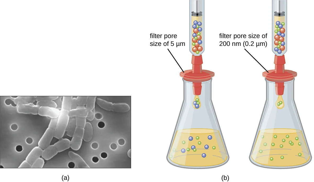
Unlike bacteria, many of which can be grown on an artificial nutrient medium, viruses require a living host cell for replication. Infected host cells (eukaryotic or prokaryotic) can be cultured and grown, and then the growth medium can be harvested as a source of virus. Virions in the liquid medium can be separated from the host cells by either centrifugation or filtration. Filters can physically remove anything present in the solution that is larger than the virions; the viruses can then be collected in the filtrate (see the micrograph and diagram below).
Check Your Understanding
What size filter pore is needed to collect a virus?
Show model answer
Did your answer mention:
Cultivation of Viruses
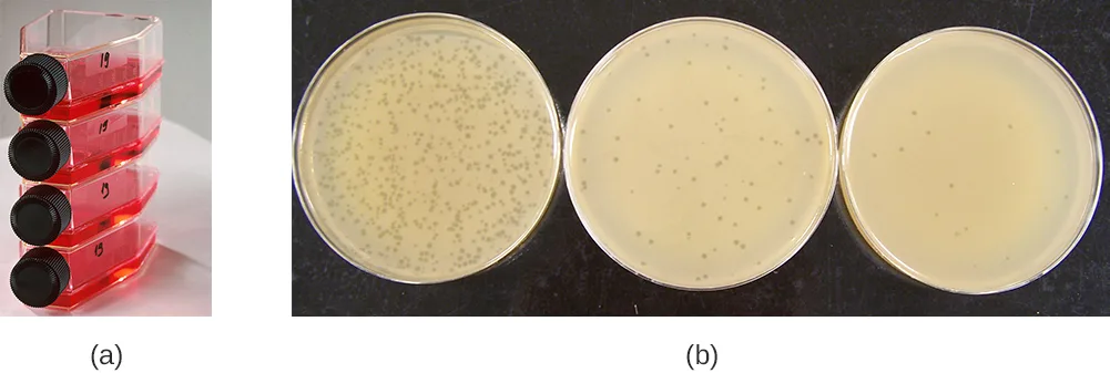
Viruses can be grown in vivo (within a whole living organism, plant, or animal) or in vitro (outside a living organism in cells in an artificial environment). Flat horizontal cell culture flasks (see the flask photo below) are a common vessel used for in vitro work. Bacteriophages can be grown in the presence of a dense layer of bacteria (also called a bacterial lawn) grown in a 0.7 % soft agar in a Petri dish or flat (horizontal) flask (see the plate photo below). As the phage kills the bacteria, many plaques are observed among the cloudy bacterial lawn.
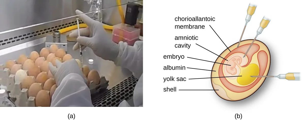
Animal viruses require cells within a host animal or tissue-culture cells derived from an animal. Animal virus cultivation is important for 1) identification and diagnosis of pathogenic viruses in clinical specimens, 2) production of vaccines, and 3) basic research studies. In vivo host sources can be a developing embryo in an embryonated bird’s egg (e.g., chicken, turkey) or a whole animal. For example, most of the influenza vaccine manufactured for annual flu vaccination programs is cultured in hens’ eggs.
The embryo or host animal serves as an incubator for viral replication (see the diagram below). Location within the embryo or host animal is important. Many viruses have a tissue tropism, and must therefore be introduced into a specific site for growth. Within an embryo, target sites include the amniotic cavity, the chorioallantoic membrane, or the yolk sac. Viral infection may damage tissue membranes, producing lesions called pox; disrupt embryonic development; or cause the death of the embryo.
Extended description
The cutaway diagram labels, moving from the shell inward: the shell; the albumin surrounding the inner contents; the chorioallantoic membrane, where the first syringe needle is inserted near the shell; the amniotic cavity, which surrounds the embryo and receives the second needle; the yolk sac, a large yellow region that receives the third needle; and the embryo itself, shown connected to the yolk sac and enclosed within the amniotic cavity.
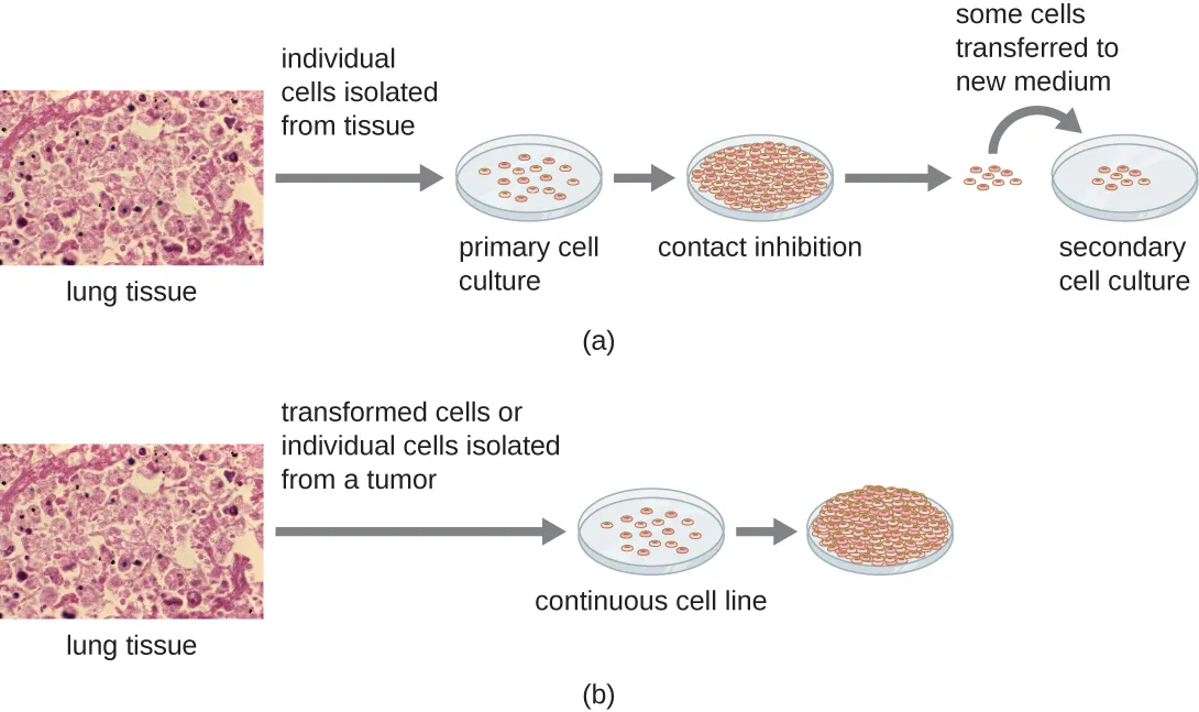
For in vitro studies, various types of cells can be used to support the growth of viruses. A primary cell culture is freshly prepared from animal organs or tissues. Cells are extracted from tissues by mechanical scraping or mincing to release cells or by an enzymatic method using trypsin or collagenase to break up tissue and release single cells into suspension. Because of anchorage-dependence requirements, primary cell cultures require a liquid culture medium in a Petri dish or tissue-culture flask so cells have a solid surface such as glass or plastic for attachment and growth. Primary cultures usually have a limited life span. When cells in a primary culture undergo mitosis and a sufficient density of cells is produced, cells come in contact with other cells. When this cell-to-cell-contact occurs, mitosis is triggered to stop. This is called contact inhibition and it prevents the density of the cells from becoming too high. To prevent contact inhibition, cells from the primary cell culture must be transferred to another vessel with fresh growth medium. This is called a secondary cell culture. Periodically, cell density must be reduced by pouring off some cells and adding fresh medium to provide space and nutrients to maintain cell growth. In contrast to primary cell cultures, continuous cell lines, usually derived from transformed cells or tumors, are often able to be subcultured many times or even grown indefinitely (in which case they are called immortal). Continuous cell lines may not exhibit anchorage dependency (they will grow in suspension) and may have lost their contact inhibition. As a result, continuous cell lines can grow in piles or lumps resembling small tumor growths (see the diagram below).
Extended description
Row (a): a micrograph of lung tissue is labeled individual cells isolated from tissue, and an arrow leads to a plate of sparse, separated round cells labeled primary cell culture. An arrow leads to a second plate where the cells have grown to cover the entire surface and touch one another, labeled contact inhibition. An arrow labeled some cells transferred to new medium leads from a few of those cells to a new plate of sparse cells labeled secondary cell culture. Row (b): the same lung-tissue micrograph is labeled transformed cells or individual cells isolated from a tumor, and an arrow leads to a plate of sparse cells labeled continuous cell line. A further arrow leads to a plate where the cells have piled up on top of one another rather than stopping at a single layer.
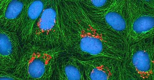
An example of an immortal cell line is the HeLa cell line, which was originally cultivated from tumor cells obtained from Henrietta Lacks, a patient who died of cervical cancer in 1951. HeLa cells were the first continuous tissue-culture cell line and were used to establish tissue culture as an important technology for research in cell biology, virology, and medicine. Prior to the discovery of HeLa cells, scientists were not able to establish tissue cultures with any reliability or stability. More than six decades later, this cell line is still alive and being used for medical research. See the Eye on Ethics feature below to read more about this important cell line and the controversial means by which it was obtained.
Check Your Understanding
What property of cells makes periodic dilutions of primary cell cultures necessary?
Identify the process, named earlier in this subsection, that halts mitosis once cells grow dense enough to touch one another.Eye on Ethics. The Immortal Cell Line of Henrietta Lacks
In January 1951, Henrietta Lacks, a 30-year-old African American woman from Baltimore, was diagnosed with cervical cancer at Johns Hopkins Hospital. We now know her cancer was caused by the human papillomavirus (HPV). Cytopathic effects of the virus altered the characteristics of her cells in a process called transformation, which gives the cells the ability to divide continuously. This ability, of course, resulted in a cancerous tumor that eventually killed Mrs. Lacks in October at age 31. Before her death, samples of her cancerous cells were taken without her knowledge or permission. The samples eventually ended up in the possession of Dr. George Gey, a biomedical researcher at Johns Hopkins University. Gey was able to grow some of the cells from Lacks’s sample, creating what is known today as the immortal HeLa cell line. These cells have the ability to live and grow indefinitely and, even today, are still widely used in many areas of research.
According to Lacks’s husband, neither Henrietta nor the family gave the hospital permission to collect her tissue specimen. Indeed, the family was not aware until 20 years after Lacks’s death that her cells were still alive and actively being used for commercial and research purposes. Yet HeLa cells have been pivotal in numerous research discoveries related to polio, cancer, and AIDS, among other diseases. The cells have also been commercialized, although they have never themselves been patented. Despite this, Henrietta Lacks’s estate has never benefited from the use of the cells, although, in 2013, the Lacks family was given control over the publication of the genetic sequence of her cells. In 2023, her family reached a settlement with biotech company Thermo Fisher, whom they had sued for knowingly profiting from the cell line without compensating the Lacks family.
This case raises several bioethical issues surrounding patients’ informed consent and the right to know. At the time Lacks’s tissues were taken, there were no laws or guidelines about informed consent. Does that mean she was treated fairly at the time? Certainly by today’s standards, the answer would be no. Harvesting tissue or organs from a dying patient without consent is not only considered unethical but illegal, regardless of whether such an act could save other patients’ lives. Is it ethical, then, for scientists to continue to use Lacks’s tissues for research, even though they were obtained illegally by today’s standards?
Ethical or not, Lacks’s cells are widely used today for so many applications that it is impossible to list them all. Is this a case in which the ends justify the means? Would Lacks be pleased to know about her contribution to science and the millions of people who have benefited? Would she want her family to be compensated for the commercial products that have been developed using her cells? Or would she feel violated and exploited by the researchers who took part of her body without her consent? Because she was never asked, we will never know.
In 2023, the family of Henrietta Lacks settled a historic lawsuit against Thermo Fisher Scientific.
Detection of a Virus
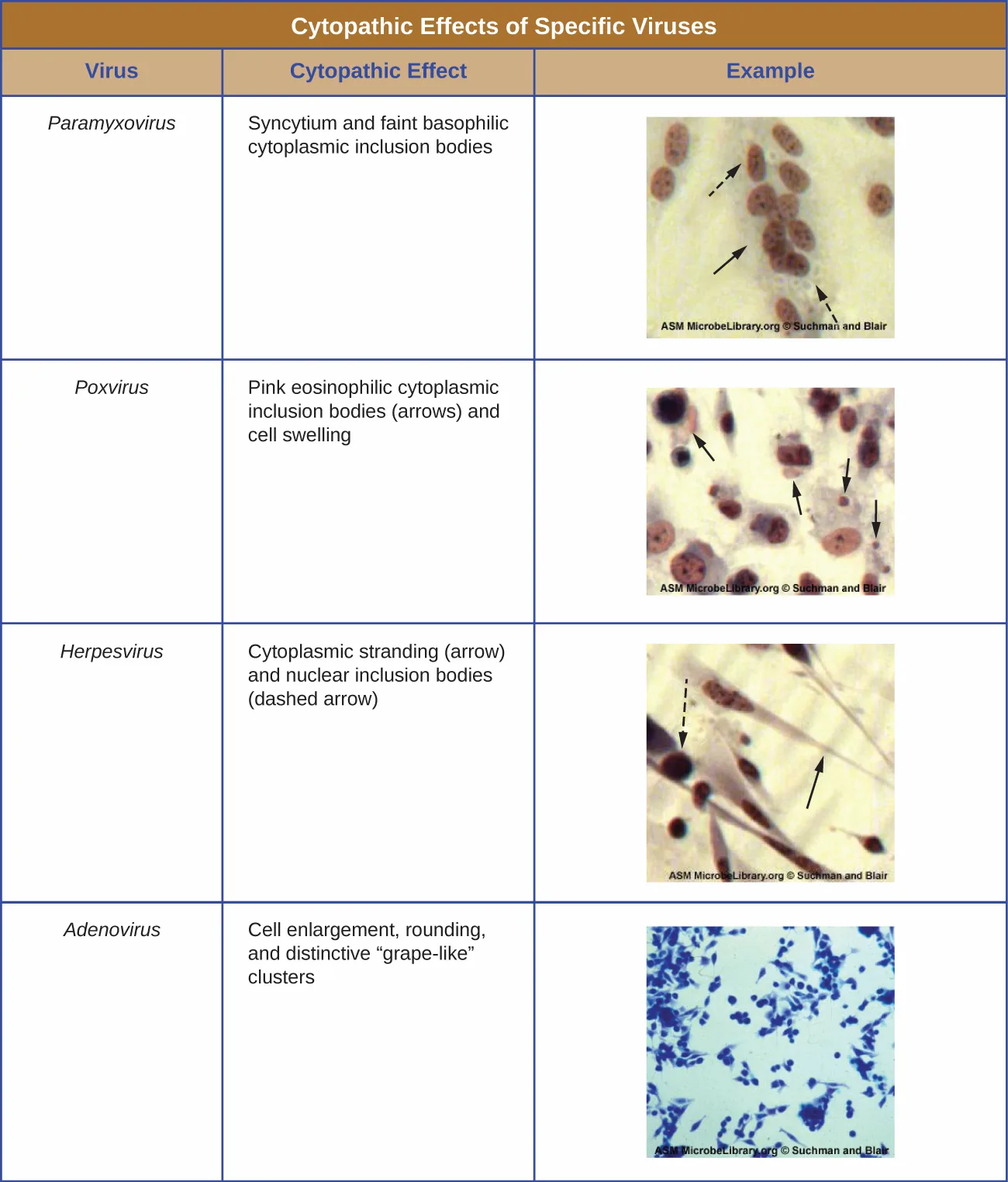
Regardless of the method of cultivation, once a virus has been introduced into a whole host organism, embryo, or tissue-culture cell, a sample can be prepared from the infected host, embryo, or cell line for further analysis under a brightfield, electron, or fluorescent microscope. Cytopathic effects (CPEs) are distinct observable cell abnormalities due to viral infection. CPEs can include loss of adherence to the surface of the container, changes in cell shape from flat to round, shrinkage of the nucleus, vacuoles in the cytoplasm, fusion of cytoplasmic membranes and the formation of multinucleated syncytia, inclusion bodies in the nucleus or cytoplasm, and complete cell lysis (see the table below).
Further pathological changes include viral disruption of the host genome and altering normal cells into transformed cells, which are the types of cells associated with carcinomas and sarcomas. The type or severity of the CPE depends on the type of virus involved. The table below lists CPEs for specific viruses.
| Virus | Cytopathic Effect | Example |
|---|---|---|
| Paramyxovirus | Syncytium and faint basophilic cytoplasmic inclusion bodies | (micrograph, below) |
| Poxvirus | Pink eosinophilic cytoplasmic inclusion bodies (arrows) and cell swelling | (micrograph, below) |
| Herpesvirus | Cytoplasmic stranding (arrow) and nuclear inclusion bodies (dashed arrow) | (micrograph, below) |
| Adenovirus | Cell enlargement, rounding, and distinctive “grape-like” clusters | (micrograph, below) |
Link to Learning
Watch this video on the effects of viruses on cells to learn about the effects of viruses on cells.
Hemagglutination Assay
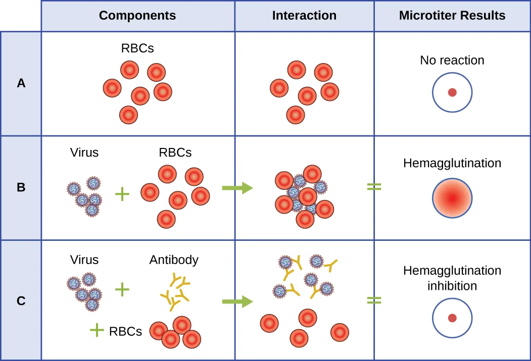
A serological assay is used to detect the presence of certain types of viruses in patient serum. Serum is the straw-colored liquid fraction of blood plasma from which clotting factors have been removed. Serum can be used in a direct assay called a hemagglutination assay to detect specific types of viruses in the patient’s sample. Hemagglutination is the agglutination (clumping) together of erythrocytes (red blood cells). Many viruses produce surface proteins or spikes called hemagglutinins that can bind to receptors on the membranes of erythrocytes and cause the cells to agglutinate. Hemagglutination is observable without using the microscope, but this method does not always differentiate between infectious and noninfectious viral particles, since both can agglutinate erythrocytes.
To identify a specific pathogenic virus using hemagglutination, we must use an indirect approach. Proteins called antibodies, generated by the patient’s immune system to fight a specific virus, can be used to bind to components such as hemagglutinins that are uniquely associated with specific types of viruses. The binding of the antibodies with the hemagglutinins found on the virus subsequently prevent erythrocytes from directly interacting with the virus. So when erythrocytes are added to the antibody-coated viruses, there is no appearance of agglutination; agglutination has been inhibited. We call these types of indirect assays for virus-specific antibodies hemagglutination inhibition (HAI) assays. HAI can be used to detect the presence of antibodies specific to many types of viruses that may be causing or have caused an infection in a patient even months or years after infection (see the chart below). This assay is described in greater detail in Agglutination Assays.
Extended description
Row A mixes red blood cells alone, drawn scattered apart from one another; the well shows a single small red dot at its center. Row B mixes red blood cells with virus particles, drawn linked together into a cluster; the well fills evenly with red color. Row C mixes red blood cells, virus particles, and antibody together, with the virus particles and antibody drawn linked to each other while the red blood cells are drawn apart from them; the well again shows a single small red dot at its center.
Check Your Understanding
What is the outcome of a positive HAI test?
Compare what happens when antibody is present against what happens when only the virus and erythrocytes are mixed.Nucleic Acid Amplification Test
Nucleic acid amplification tests (NAAT) are used in molecular biology to detect unique nucleic acid sequences of viruses in patient samples. Polymerase chain reaction (PCR) is an NAAT used to detect the presence of viral DNA in a patient’s tissue or body fluid sample. PCR is a technique that amplifies (i.e., synthesizes many copies) of a viral DNA segment of interest. Using PCR, short nucleotide sequences called primers bind to specific sequences of viral DNA, enabling identification of the virus.
Reverse transcriptase-PCR (RT-PCR) is an NAAT used to detect the presence of RNA viruses. RT-PCR differs from PCR in that the enzyme reverse transcriptase (RT) is used to make a cDNA from the small amount of viral RNA in the specimen. The cDNA can then be amplified by PCR. Both PCR and RT-PCR are used to detect and confirm the presence of the viral nucleic acid in patient specimens.
Case in Point. HPV Scare
Michelle, a 21-year-old nursing student, came to the university clinic worried that she might have been exposed to a sexually transmitted disease (STD). Her sexual partner had recently developed several bumps on the base of his penis. He had put off going to the doctor, but Michelle suspects they are genital warts caused by HPV. She is especially concerned because she knows that HPV not only causes warts but is a prominent cause of cervical cancer. She and her partner always use condoms for contraception, but she is not confident that this precaution will protect her from HPV.
Michelle’s physician finds no physical signs of genital warts or any other STDs, but recommends that Michelle get a Pap smear along with an HPV test. The Pap smear will screen for abnormal cervical cells and the CPEs associated with HPV; the HPV test will test for the presence of the virus. If both tests are negative, Michelle can be more assured that she most likely has not become infected with HPV. However, her doctor suggests it might be wise for Michelle to get vaccinated against HPV to protect herself from possible future exposure.
- Why does Michelle’s physician order two different tests instead of relying on one or the other?
Enzyme Immunoassay
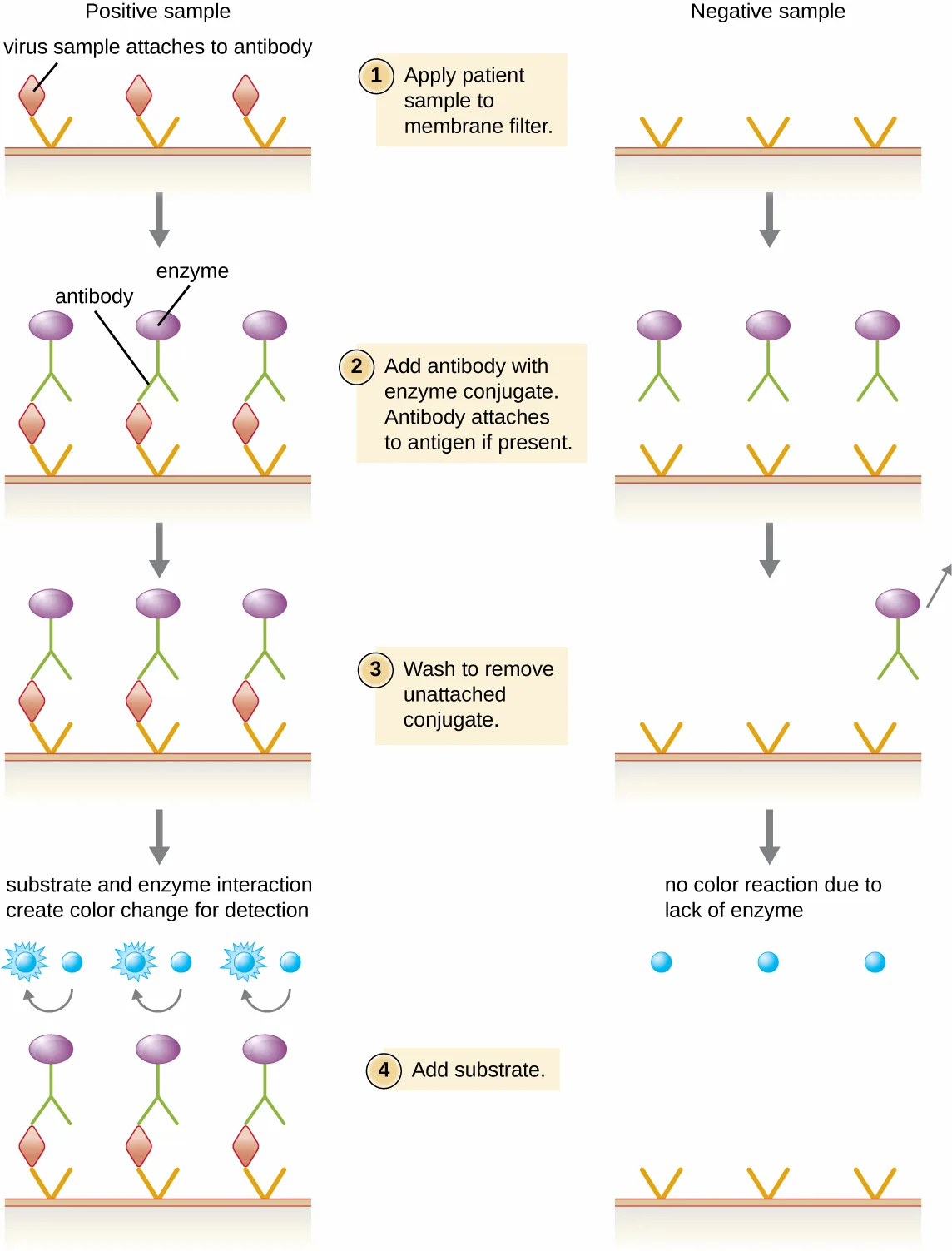
Enzyme immunoassays (EIAs) rely on the ability of antibodies to detect and attach to specific biomolecules called antigens. The detecting antibody attaches to the target antigen with a high degree of specificity in what might be a complex mixture of biomolecules. Also included in this type of assay is a colorless enzyme attached to the detecting antibody. The enzyme acts as a tag on the detecting antibody and can interact with a colorless substrate, leading to the production of a colored end product. EIAs often rely on layers of antibodies to capture and react with antigens, all of which are attached to a membrane filter (see the diagram below). EIAs for viral antigens are often used as preliminary screening tests. If the results are positive, further confirmation will require tests with even greater sensitivity, such as a western blot or an NAAT. EIAs are discussed in more detail in EIAs and ELISAs.
Extended description
Step 1: the patient sample is applied to a membrane filter that already holds antibody bound to its surface; in the positive-sample column a virus particle attaches to that antibody, while in the negative-sample column nothing attaches. Step 2: antibody carrying an enzyme tag is added; in the positive column it attaches to the captured virus, while in the negative column it has nothing to attach to. Step 3: the filter is washed to remove any unattached enzyme-antibody conjugate; the positive column keeps its attached conjugate, and the negative column’s conjugate washes away. Step 4: a substrate is added; blue starburst shapes appear beside the antibody only in the positive-sample column at this step, while the negative-sample column shows plain blue circles with no starburst.
Check Your Understanding
What typically indicates a positive EIA test?
Recall what the enzyme-substrate reaction in the last step of the diagram produces when the antibody stays bound to the filter.Clinical Focus. Part 3
Along with the RT/PCR analysis, David’s saliva was also collected for viral cultivation. In general, no single diagnostic test is sufficient for antemortem diagnosis, since the results will depend on the sensitivity of the assay, the quantity of virions present at the time of testing, and the timing of the assay, since release of virions in the saliva can vary. As it turns out, the result was negative for viral cultivation from the saliva. This is not surprising to David’s doctor, because one negative result is not an absolute indication of the absence of infection. It may be that the number of virions in the saliva is low at the time of sampling. It is not unusual to repeat the test at intervals to enhance the chance of detecting higher virus loads.
- Should David’s doctor modify his course of treatment based on these test results?
The case continues in Viroids, Virusoids, and Prions. The case began in Viruses.
Summary
- Viral cultivation requires the presence of some form of host cell (whole organism, embryo, or cell culture).
- Viruses can be isolated from samples by filtration.
- Viral filtrate is a rich source of released virions.
- Bacteriophages are detected by presence of clear plaques on bacterial lawn.
- Animal and plant viruses are detected by cytopathic effects, molecular techniques (PCR, RT-PCR), enzyme immunoassays, and serological assays (hemagglutination assay, hemagglutination inhibition assay).
Key terms
- in vivo — inside the organism.
- in vitro — outside the organism in a test tube or artificial environment.
- bacterial lawn — layer of confluent bacterial growth on an agar plate.
- cytopathic effects (CPEs) — cell abnormality resulting from a viral infection.
Practice
Discuss why viruses were originally described as filterable agents
Porcelain ________ filters, with pores small enough to let viruses pass through, were used to discover TMV.
Name the filter type mentioned in the section’s opening sentence, before it was replaced by membrane filters.What can a filter physically remove from a solution containing viruses?
Compare the sizes described in the filtration diagram rather than the detection assays discussed later in the section.What does viral filtrate serve as a rich source of?
Use the summary sentence about what filtration yields, not the sentence about what filtration removes.Describe the cultivation of viruses and specimen collection and handling
Which of the following cannot be used to culture viruses?
Compare each option against the requirement, stated at the start of the Cultivation of Viruses subsection, that viruses need a living host cell.Which of the following tests can be used to detect the presence of a specific virus?
Consider whether each named test individually detects viral presence, then decide whether more than one applies.Which of the following is NOT a cytopathic effect?
Recall that CPEs include fusion of cytoplasmic membranes into multinucleated syncytia, not single-nucleus cells.Viruses can be diagnosed and observed using a(n) ________ microscope.
Name the type of microscope needed to see particles as small as viruses.Cell abnormalities resulting from a viral infection are called ________.
Use the term this section defines near the start of the Detection of a Virus heading, then abbreviates for the rest of the section.Briefly explain the various methods of culturing viruses.
Show model answer
Did your answer mention:
What are some characteristics of the viruses that are similar to a computer virus?
Show model answer
Did your answer mention:
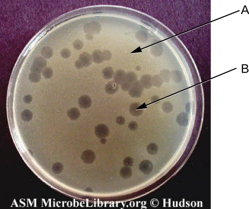
In the figure above, what does the arrow labeled (A) point to?
The lawn is the unbroken area of bacterial growth; a plaque is a distinct clear spot within it.In the figure above, what does the arrow labeled (B) point to?
The lawn is the unbroken area of bacterial growth; a plaque is a distinct clear spot within it.Compare in vivo and in vitro techniques used to cultivate viruses
Growing a virus within a whole living organism, plant, or animal is called growing it ________.
Use the Latin phrase meaning “within the living,” defined at the start of the Cultivation of Viruses subsection.Growing a virus outside a living organism, in cells in an artificial environment, is called growing it ________.
Use the Latin phrase meaning “within glass,” defined alongside the previous term at the start of the Cultivation of Viruses subsection.Which of the following is an example of an in vivo technique for cultivating a virus?
Distinguish the option that uses a whole developing organism from the three that use cells grown outside one.This section is adapted from Microbiology, Section 6.3: Isolation, Culture, and Identification of Viruses by Nina Parker, Mark Schneegurt, Anh-Hue Thi Tu, Philip Lister, Brian M. Forster, and OpenStax, © OpenStax, licensed under CC BY-NC-SA 4.0. Access the original for free at openstax.org. Changes: all 9 figures re-encoded as WebP, with kind set explicitly after inspection — diagram for the filters, egg, culture-flow, and hemagglutination/EIA process figures (each has at least one genuinely drawn panel), photo for the flask/plaque photos, the HeLa fluorescence micrograph, the cytopathic-effects table’s micrograph panels, and the Art Connection plate photo — overriding the media manifest’s JPEG-based guess on the four diagrams; alts rewritten from the served images (the source alt’s “Figure a is… Figure b is…” phrasing tightened to parenthetical (a)/(b) form) and a longdesc added to the four labeled diagrams and process charts, walking each in reading order; the cytopathic-effects table (printed upstream as a table image) is transcribed as a Markdown table from the image, checked against the PDF page, with the vendored figure kept immediately after it, kind="photo", its alt limited to the micrograph column and its credit-only source caption extended with a short author-written phrase naming what the panels show; the Eye on Ethics and Case in Point notes rendered as callouts, each keeping its own bold feature name and italicized title; the Case in Point box’s closing physician’s question kept as an unanswered bulleted prompt inside its callout, as printed; the Link to Learning URL kept, its sentence’s doubled “video” phrasing tightened so the link text alone names the destination; the Clinical Focus Part 3 box’s closing question kept as an unanswered bulleted prompt, and the source’s “Jump to the next / Go back to the previous Clinical Focus box” links replaced with plain sentences naming both the chapter’s earlier part (in Viruses) and its next part (in Viroids, Virusoids, and Prions); the cross-references to Agglutination Assays (m58902) and EIAs and ELISAs (m58903), both in chapter 20, left as plain text because those pages are not yet authored; three of the four Check Your Understanding bullets are graded from this module’s own sentences rather than answered in prose, since the source prints no key for them, and the filtration-pore question stays a self-check whose answer is read from the figure’s own printed pore sizes (the module’s prose never states the value) — a textin from the contact-inhibition sentence, a multiplechoice from the hemagglutination-inhibition sentence (with cytopathic-effect and EIA facts from elsewhere in this module as its distractors), and a textin from the EIA figure’s own caption; the three Multiple Choice and two Fill in the Blank items are adapted into the Practice block under the objective on cultivation and specimen handling, with the source’s “followings” corrected to “following” (a one-word typo, logged); the body Check Your Understanding question about the hemagglutination inhibition assay corrected from the source’s “positive HIA test” to “positive HAI test” (the module defines the assay’s abbreviation as HAI two sentences earlier; a one-word typo, logged), and the Eye on Ethics box’s link text corrected from “Thermo Fischer Scientific” to “Thermo Fisher Scientific,” the company’s real name, which the same box’s own preceding sentence already spells correctly (a one-word typo, logged); a missing closing parenthesis after the “in vitro” definition’s opening sentence silently closed (the source’s parenthetical is never closed before its period; logged); the two unkeyed end-of-section questions remain self-checks with model answers and rubrics assembled only from this module’s own sentences, because the Short Answer question’s honest answer assembles several sentences about different cultivation methods and the Critical Thinking question compares to a subject (a computer virus) this module never discusses, so its model answer states only what the module gives about a biological virus’s host dependency and stops; the Critical Thinking Art Connection figure (a bare <figure> inside the exercise, printing no caption) is rendered as a mediafigure with an author-written caption, immediately followed by two multiplechoice items that label its two lettered arrows from the module’s own “bacterial lawn” and “plaque” vocabulary, keyed from the image; three filler items (one cloze textin from the section’s opening sentence and two select-the-term multiplechoice items from its Isolation and Summary sentences) fill the first objective group, which the source’s own exercise set does not reach, and three further items (two term-recall textins from Key terms and one author-built multiplechoice contrasting an in vivo and three in vitro cultivation examples) fill the third objective group for the same reason; no source exercise was omitted; key terms compiled from the module’s four defined terms and the book’s Glossary appendix, with the cytopathic effects (CPEs) bullet printed lower-case to match how the module’s own Summary sentence uses the term, since its body’s capital “Cytopathic” is only a sentence-initial capital, not a proper noun (correcting the pre-extracted scaffold, reported for the errata file).