Viroids, Virusoids, and Prions
By the end of this section, you will be able to:
- Describe viroids and their unique characteristics
- Describe virusoids and their unique characteristics
- Describe prions and their unique characteristics
Research attempts to discover the causative agents of previously uninvestigated diseases have led to the discovery of nonliving disease agents quite different from viruses. These include particles consisting only of RNA or only of protein that, nonetheless, are able to self-propagate at the expense of a host—a key similarity to viruses that allows them to cause disease conditions. To date, these discoveries include viroids, virusoids, and the proteinaceous prions.
Viroids
In 1971, Theodor Diener, a pathologist working at the Agriculture Research Service, discovered an acellular particle that he named a viroid, meaning “virus-like.” Viroids consist only of a short strand of circular RNA capable of self-replication. The first viroid discovered was found to cause potato tuber spindle disease, which causes slower sprouting and various deformities in potato plants (see the photograph below). Like viruses, potato spindle tuber viroids (PSTVs) take control of the host machinery to replicate their RNA genome. Unlike viruses, viroids do not have a protein coat to protect their genetic information.
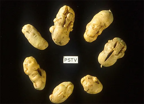
Viroids can result in devastating losses of commercially important agricultural food crops grown in fields and orchards. Since the discovery of PSTV, other viroids have been discovered that cause diseases in plants. Tomato planta macho viroid (TPMVd) infects tomato plants, which causes loss of chlorophyll, disfigured and brittle leaves, and very small tomatoes, resulting in loss of productivity in this field crop. Avocado sunblotch viroid (ASBVd) results in lower yields and poorer-quality fruit. ASBVd is the smallest viroid discovered thus far that infects plants. Peach latent mosaic viroid (PLMVd) can cause necrosis of flower buds and branches, and wounding of ripened fruit, which leads to fungal and bacterial growth in the fruit. PLMVd can also cause similar pathological changes in plums, nectarines, apricots, and cherries, resulting in decreased productivity in these orchards, as well. Viroids, in general, can be dispersed mechanically during crop maintenance or harvesting, vegetative reproduction, and possibly via seeds and insects, resulting in a severe drop in food availability and devastating economic consequences.
Check Your Understanding
What is the genome of a viroid made of?
Look at the shape and the molecule type named in the sentence that first defines a viroid.Virusoids
A second type of pathogenic RNA that can infect commercially important agricultural crops are the virusoids, which are subviral particles best described as non–self-replicating ssRNAs. RNA replication of virusoids is similar to that of viroids but, unlike viroids, virusoids require that the cell also be infected with a specific “helper” virus. There are currently only five described types of virusoids and their associated helper viruses. The helper viruses are all from the family of Sobemoviruses. An example of a helper virus is the subterranean clover mottle virus, which has an associated virusoid packaged inside the viral capsid. Once the helper virus enters the host cell, the virusoids are released and can be found free in plant cell cytoplasm, where they possess ribozyme activity. The helper virus undergoes typical viral replication independent of the activity of the virusoid. The virusoid genomes are small, only 220 to 388 nucleotides long. A virusoid genome does not code for any proteins, but instead serves only to replicate virusoid RNA.
Virusoids belong to a larger group of infectious agents called satellite RNAs, which are similar pathogenic RNAs found in animals. Unlike the plant virusoids, satellite RNAs may encode for proteins; however, like plant virusoids, satellite RNAs must coinfect with a helper virus to replicate. One satellite RNA that infects humans and that has been described by some scientists as a virusoid is the hepatitis delta virus (HDV), which, by some reports, is also called hepatitis delta virusoid. Much larger than a plant virusoid, HDV has a circular, ssRNA genome of 1,700 nucleotides and can direct the biosynthesis of HDV-associated proteins. The HDV helper virus is the hepatitis B virus (HBV). Coinfection with HBV and HDV results in more severe pathological changes in the liver during infection, which is how HDV was first discovered.
Check Your Understanding
What is the main difference between a viroid and a virusoid? Sort each phrase below under the type it describes.
Viroid
Virusoid
Prions
At one time, scientists believed that any infectious particle must contain DNA or RNA. Then, in 1982, Stanley Prusiner, a medical doctor studying scrapie (a fatal, degenerative disease in sheep) discovered that the disease was caused by proteinaceous infectious particles, or prions. Because proteins are acellular and do not contain DNA or RNA, Prusiner’s findings were originally met with resistance and skepticism; however, his research was eventually validated, and he received the Nobel Prize in Physiology or Medicine in 1997.
A prion is a misfolded rogue form of a normal protein (PrPc) found in the cell. This rogue prion protein (PrPsc), which may be caused by a genetic mutation or occur spontaneously, can be infectious, stimulating other endogenous normal proteins to become misfolded, forming plaques (see the diagram below). Today, prions are known to cause various forms of transmissible spongiform encephalopathy (TSE) in human and animals. TSE is a rare degenerative disorder that affects the brain and nervous system. The accumulation of rogue proteins causes the brain tissue to become sponge-like, killing brain cells and forming holes in the tissue, leading to brain damage, loss of motor coordination, and dementia (see the brain scans below). Infected individuals are mentally impaired and become unable to move or speak. There is no cure, and the disease progresses rapidly, eventually leading to death within a few months or years.
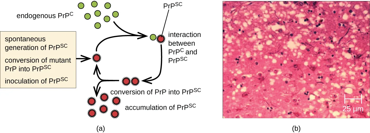
Extended description
The diagram in panel (a) reads left to right and cycles back on itself. At upper left, a cluster of green circles is labeled endogenous PrPC, with an arrow leading right to a step labeled interaction between PrPC and PrPSc, shown as a green circle beside a red spiked shape marked PrPSc. A separate box at left lists three ways PrPSc can arise — spontaneous generation of PrPSc, conversion of mutant PrP into PrPSc, and inoculation of PrPSc — with an arrow from the box to a red spiked shape below the interaction step. An arrow leads from the interaction step down and right to a small cluster of red spiked shapes labeled conversion of PrP into PrPSc, and a final arrow leads left to a larger cluster of red spiked shapes labeled accumulation of PrPSc, completing the cycle.
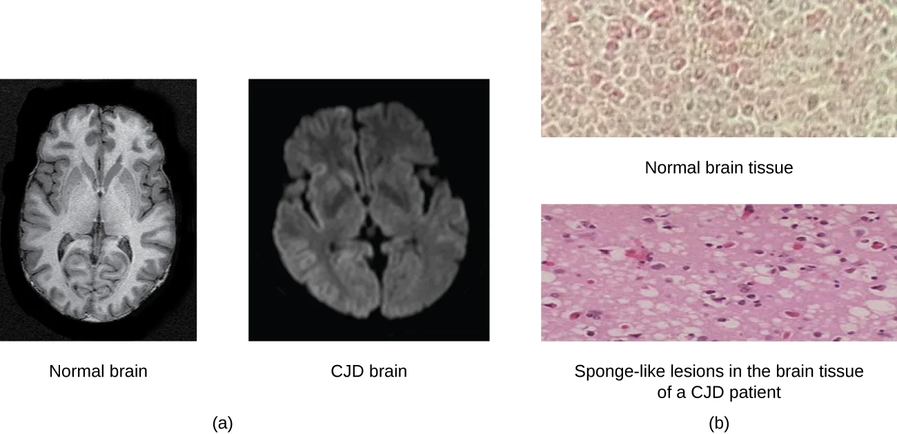
TSEs in humans include kuru, fatal familial insomnia, Gerstmann-Straussler-Scheinker disease, and Creutzfeldt-Jakob disease (see the brain scans above). TSEs in animals include mad cow disease, scrapie (in sheep and goats), and chronic wasting disease (in elk and deer). TSEs can be transmitted between animals and from animals to humans by eating contaminated meat or animal feed. Transmission between humans can occur through heredity (as is often the case with GSS and CJD) or by contact with contaminated tissue, as might occur during a blood transfusion or organ transplant. There is no evidence for transmission via casual contact with an infected person. The table below lists TSEs that affect humans and their modes of transmission (National Institute of Neurological Disorders and Stroke, “Creutzfeldt-Jakob Disease Fact Sheet,” accessed December 31, 2015).
| Disease | Mechanism(s) of Transmission |
|---|---|
| Sporadic CJD (sCJD) | Not known; possibly by alteration of normal prion protein (PrP) to rogue form due to somatic mutation |
| Variant CJD (vCJD) | Eating contaminated cattle products and by secondary bloodborne transmission |
| Familial CJD (fCJD) | Mutation in germline PrP gene |
| Iatrogenic CJD (iCJD) | Contaminated neurosurgical instruments, corneal graft, gonadotrophic hormone, and, secondarily, by blood transfusion |
| Kuru | Eating infected meat through ritualistic cannibalism |
| Gerstmann-Straussler-Scheinker disease (GSS) | Mutation in germline PrP gene |
| Fatal familial insomnia (FFI) | Mutation in germline PrP gene |
Prions are extremely difficult to destroy because they are resistant to heat, chemicals, and radiation. Even standard sterilization procedures do not ensure the destruction of these particles. Currently, there is no treatment or cure for TSE disease, and contaminated meats or infected animals must be handled according to federal guidelines to prevent transmission.
Check Your Understanding
Does a prion have a genome?
Recall what infectious particles are made of, according to the paragraph that introduces prions.Link to Learning
For more information on the handling of animals and prion-contaminated materials, visit the WHO’s guidelines on handling prion-contaminated material.
Clinical Focus. Resolution
A few days later, David’s doctor receives the results of the immunofluorescence test on his skin sample. The test is negative for rabies antigen. A second viral antigen test on his saliva sample also comes back negative. Despite these results, the doctor decides to continue David’s current course of treatment. Given the positive RT-PCR test, it is best not to rule out a possible rabies infection.
Near the site of the bite, David receives an injection of rabies immunoglobulin, which attaches to and inactivates any rabies virus that may be present in his tissues. Over the next 14 days, he receives a series of four rabies-specific vaccinations in the arm. These vaccines activate David’s immune response and help his body recognize and fight the virus. Thankfully, with treatment, David’s symptoms improve and he makes a full recovery.
Not all rabies cases have such a fortunate outcome. In fact, rabies is usually fatal once the patient starts to exhibit symptoms, and postbite treatments are mainly palliative (i.e., sedation and pain management).
The case began in Viruses.
Summary
- Other acellular agents such as viroids, virusoids, and prions also cause diseases. Viroids consist of small, naked ssRNAs that cause diseases in plants. Virusoids are ssRNAs that require other helper viruses to establish an infection. Prions are proteinaceous infectious particles that cause transmissible spongiform encephalopathies.
- Prions are extremely resistant to chemicals, heat, and radiation.
- There are no treatments for prion infection.
Key terms
- viroids — infectious plant pathogen composed of RNA.
- virusoids — small piece of RNA associated with larger RNA of some infectious plant viruses.
- prions — acellular infectious particle consisting of just proteins that can cause progressive diseases in animals and humans.
- transmissible spongiform encephalopathy — degenerative disease caused by prions; leads to the death of neurons in the brain.
Practice
Describe viroids and their unique characteristics
A ________ is an infectious plant pathogen composed of RNA.
Name the acellular RNA particle Theodor Diener discovered and described at the start of this section.In 1971, pathologist Theodor Diener named this newly discovered acellular particle a viroid, meaning “________.”
Recall the literal meaning of the coined term, based on its resemblance to a familiar infectious agent.Which viroid, the first ever discovered, was found to cause potato tuber spindle disease?
Match the viroid named in the same sentence as potato tuber spindle disease, also shown in the photograph above.Describe virusoids and their unique characteristics
Both viroids and virusoids have a(n) ________ genome.
Identify the type of nucleic acid both of these subviral agents are made of, not the shape of that molecule.Unlike viroids, virusoids require a(n) ________ to reproduce.
Name what a virusoid needs to infect a cell, unlike a viroid, described earlier in this section.A ________ is a small piece of RNA associated with the larger RNA of some infectious plant viruses.
Name the subviral RNA particle described at the start of this section, distinct from a viroid.Describe prions and their unique characteristics
Which of these infectious agents do not have nucleic acid?
Recall which of these agents is made only of protein, with no DNA or RNA genome at all.Which of the following is true of prions?
Compare each option against the paragraph naming the two forms of prion protein and the paragraph on how resistant prions are to destruction.Describe the disease symptoms observed in animals infected with prions.
Show model answer
Did your answer mention:
Does a prion replicate? Explain.
Show model answer
Did your answer mention:
This section is adapted from Microbiology, Section 6.4: Viroids, Virusoids, and Prions by Nina Parker, Mark Schneegurt, Anh-Hue Thi Tu, Philip Lister, Brian M. Forster, and OpenStax, © OpenStax, licensed under CC BY-NC-SA 4.0. Access the original for free at openstax.org. Changes: all three source figures re-encoded as WebP and rendered as mediafigures after image and PDF inspection; the PrPc/PrPsc conversion-cycle figure is set kind="diagram" (overriding the manifest’s photo guess) because its panel (a) is a drawn flow diagram, with a longdesc walking that diagram in reading order, while the potato and brain-scan/micrograph figures are kind="photo"; the PSTV alt was expanded from the bare source alt to describe the visible deformities and the PSTV card; the CJD brain-scan alt was rewritten from the image rather than the source alt, which claims the CJD scan shows “larger spaces” and “more black regions” — the rendered scan instead reads as more uniformly hazy and blurred with reduced gray/white contrast, not as darker or more open, a suspected source-alt defect logged in the ledger; same-module figure cross-references rendered as describing prose (“the photograph below,” “the diagram below,” “the brain scans below/above”); the Link to Learning kept its URL and describes its destination; the Clinical Focus box is titled Resolution, and its closing “Go back to the previous Clinical Focus box” link is replaced with a sentence naming where the case began, Viruses; the table’s footnote is rendered as an inline parenthetical citation with its bare access URL dropped; the TSEs-in-Humans table is transcribed as a Markdown table from the CNXML cells (its spanning <thead> title is dropped as a data row, standing in for the sentence that already introduces it), and was evaluated against the table→sortbins rule and skipped: its rows are eight individual named diseases with individual, non-categorical transmission notes, not a 2–4 category axis on either orientation, so no sortbins was built; two one-word source typos are corrected without an inline note — “prior protein” to “prion protein” in the Sporadic CJD row of the TSE table, and the missing possessive in “David symptoms improve” to “David’s symptoms improve” in the Clinical Focus Resolution — both logged as suspected source defects; all three body Check Your Understanding bullets are graded from this module’s own text: the Viroids bullet is a text-recall keyed from the sentence defining a viroid’s genome, the Virusoids bullet (asking the main difference between a viroid and a virusoid) is a sort-into-bins built from this module’s own distinguishing phrases, and the Prions bullet is a multiple-choice keyed from the paragraph stating that proteins contain no DNA or RNA, with distractors built from the viroid and virusoid genome facts stated earlier in this module; the module’s two Multiple Choice and one two-blank Fill in the Blank exercise are adapted into Practice, the Fill in the Blank split into two text-recall items (one per blank), both kept in the Virusoids group; the module’s unkeyed Short Answer and Critical Thinking questions remain self-checks, since each needs several of this module’s own sentences assembled into an explanation, with model answers and rubrics built only from this module’s text; no source exercise was omitted; four author-written items were added to bring the objective groups to this book’s floor — in the Viroids group, two text-recall items from this section’s Key terms and body text and one multiple-choice built from the four viroid names this module names, and in the Virusoids group, one text-recall item keyed from the Key terms definition of a virusoid, disclosed here and in the source ledger; key terms compiled from the module’s four defined terms and the book’s Glossary appendix, all four definitions taken directly from the Glossary, with the plural terms viroids, virusoids, and prions bolded and lower-cased in the Key terms list to match how each is printed at its defining, sentence-initial occurrence in the body.