Using Biochemistry to Identify Microorganisms
By the end of this section, you will be able to:
- Describe examples of biosynthesis products within a cell that can be detected to identify bacteria
Accurate identification of bacterial isolates is essential in a clinical microbiology laboratory because the results often inform decisions about treatment that directly affect patient outcomes. For example, cases of food poisoning require accurate identification of the causative agent so that physicians can prescribe appropriate treatment. Likewise, it is important to accurately identify the causative pathogen during an outbreak of disease so that appropriate strategies can be employed to contain the epidemic.
There are many ways to detect, characterize, and identify microorganisms. Some methods rely on phenotypic biochemical characteristics, while others use genotypic identification. The biochemical characteristics of a bacterium provide many traits that are useful for classification and identification. Analyzing the nutritional and metabolic capabilities of the bacterial isolate is a common approach for determining the genus and the species of the bacterium. Some of the most important metabolic pathways that bacteria use to survive will be discussed in the chapter introduction to Microbial Metabolism. In this section, we will discuss a few methods that use biochemical characteristics to identify microorganisms.
Some microorganisms store certain compounds as granules within their cytoplasm, and the contents of these granules can be used for identification purposes. For example, poly-β-hydroxybutyrate (PHB) is a carbon- and energy-storage compound found in some nonfluorescent bacteria of the genus Pseudomonas. Different species within this genus can be classified by the presence or the absence of PHB and fluorescent pigments. The human pathogen P. aeruginosa and the plant pathogen P. syringae are two examples of fluorescent Pseudomonas species that do not accumulate PHB granules.
Other systems rely on biochemical characteristics to identify microorganisms by their biochemical reactions, such as carbon utilization and other metabolic tests. In small laboratory settings or in teaching laboratories, those assays are carried out using a limited number of test tubes. However, more modern systems, such as the one developed by Biolog, Inc., are based on panels of biochemical reactions performed simultaneously and analyzed by software. Biolog’s system identifies cells based on their ability to metabolize certain biochemicals and on their physiological properties, including pH and chemical sensitivity. It uses all major classes of biochemicals in its analysis. Identifications can be performed manually or with the semi- or fully automated instruments.
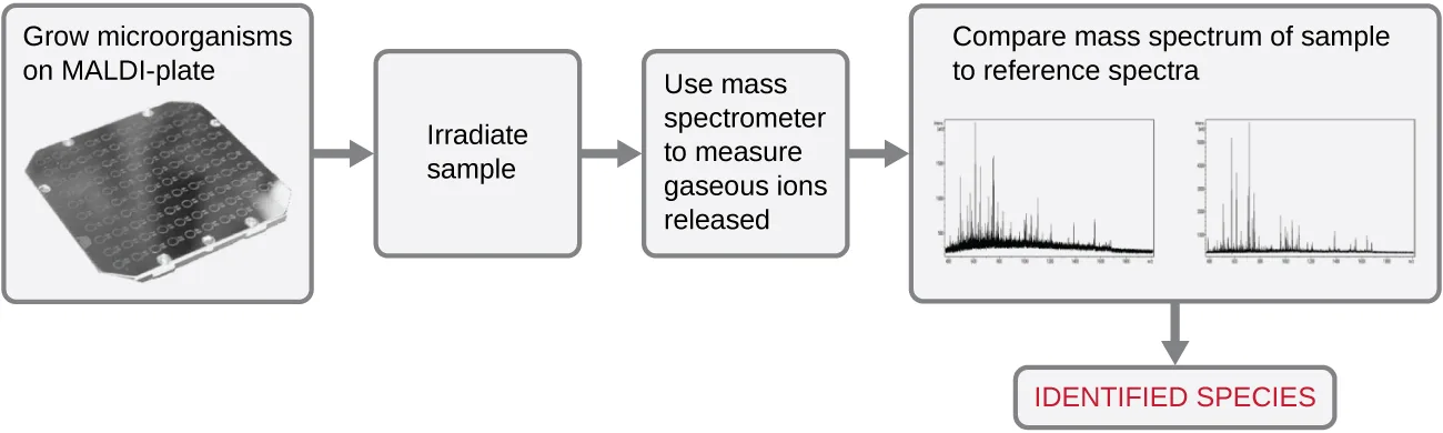
Another automated system identifies microorganisms by determining the specimen’s mass spectrum and then comparing it to a database that contains known mass spectra for thousands of microorganisms. This method is based on matrix-assisted laser desorption/ionization time-of-flight mass spectrometry (MALDI-TOF) and uses disposable MALDI plates on which the microorganism is mixed with a specialized matrix reagent, as shown below. The sample/reagent mixture is irradiated with a high-intensity pulsed ultraviolet laser, resulting in the ejection of gaseous ions generated from the various chemical constituents of the microorganism. These gaseous ions are collected and accelerated through the mass spectrometer, with ions traveling at a velocity determined by their mass-to-charge ratio (m/z), thus, reaching the detector at different times. A plot of detector signal versus m/z yields a mass spectrum for the organism that is uniquely related to its biochemical composition. Comparison of the mass spectrum to a library of reference spectra obtained from identical analyses of known microorganisms permits identification of the unknown microbe.
Extended description
The flowchart proceeds left to right in five stages: (1) a photograph of a square MALDI plate with a grid of small wells, captioned “Grow microorganisms on MALDI-plate”; (2) a box captioned “Irradiate sample”; (3) a box captioned “Use mass spectrometer to measure gaseous ions released”; (4) a box captioned “Compare mass spectrum of sample to reference spectra,” showing two line graphs, each plotting a jagged signal against a horizontal scale; (5) a final box, in red text, captioned “IDENTIFIED SPECIES.”
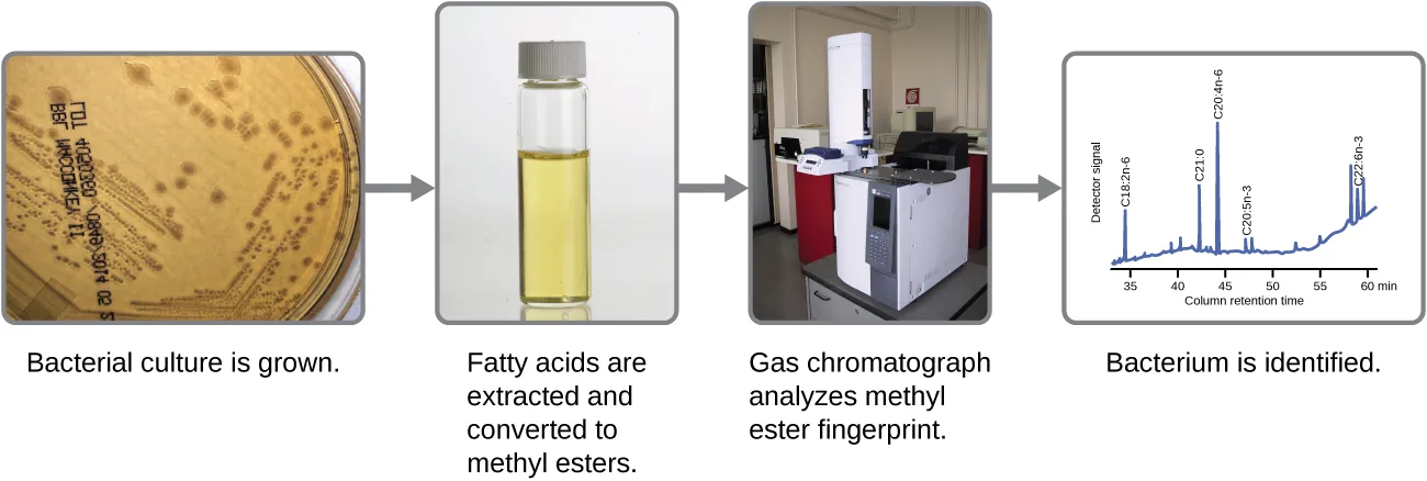
Microbes can also be identified by measuring their unique lipid profiles. As we have learned, fatty acids of lipids can vary in chain length, presence or absence of double bonds, and number of double bonds, hydroxyl groups, branches, and rings. To identify a microbe by its lipid composition, the fatty acids present in their membranes are analyzed. A common biochemical analysis used for this purpose is a technique used in clinical, public health, and food laboratories. It relies on detecting unique differences in fatty acids and is called fatty acid methyl ester (FAME) analysis. In a FAME analysis, fatty acids are extracted from the membranes of microorganisms, chemically altered to form volatile methyl esters, and analyzed by gas chromatography (GC). The resulting GC chromatogram is compared with reference chromatograms in a database containing data for thousands of bacterial isolates to identify the unknown microorganism, as shown below.
Extended description
Four captioned stages run left to right, three photographs and a line graph: (1) a culture plate with scattered colonies, “Bacterial culture is grown”; (2) a vial of pale yellow liquid, “Fatty acids are extracted and converted to methyl esters”; (3) a benchtop gas chromatography instrument, “Gas chromatograph analyzes methyl ester fingerprint”; (4) a graph of detector signal against column retention time with several sharp peaks labeled with fatty-acid designations such as C18:2n-6 and C20:4n-6, “Bacterium is identified.”
A related method for microorganism identification is called phospholipid-derived fatty acids (PLFA) analysis. Membranes are mostly composed of phospholipids, which can be saponified (hydrolyzed with alkali) to release the fatty acids. The resulting fatty acid mixture is then subjected to FAME analysis, and the measured lipid profiles can be compared with those of known microorganisms to identify the unknown microorganism.
Bacterial identification can also be based on the proteins produced under specific growth conditions within the human body. These types of identification procedures are called proteomic analysis. To perform proteomic analysis, proteins from the pathogen are first separated by high-pressure liquid chromatography (HPLC), and the collected fractions are then digested to yield smaller peptide fragments. These peptides are identified by mass spectrometry and compared with those of known microorganisms to identify the unknown microorganism in the original specimen.
Microorganisms can also be identified by the carbohydrates attached to proteins (glycoproteins) in the plasma membrane or cell wall. Antibodies and other carbohydrate-binding proteins can attach to specific carbohydrates on cell surfaces, causing the cells to clump together. Serological tests (e.g., the Lancefield groups tests, which are used for identification of Streptococcus species) are performed to detect the unique carbohydrates located on the surface of the cell.
Clinical Focus. Resolution
Penny stopped using her new sunscreen and applied the corticosteroid cream to her rash as directed. However, after several days, her rash had not improved and actually seemed to be getting worse. She made a follow-up appointment with her doctor, who observed a bumpy red rash and pus-filled blisters around hair follicles, shown below. The rash was especially concentrated in areas that would have been covered by a swimsuit. After some questioning, Penny told the physician that she had recently attended a pool party and spent some time in a hot tub. In light of this new information, the doctor suspected a case of hot tub rash, an infection frequently caused by the bacterium Pseudomonas aeruginosa, an opportunistic pathogen that can thrive in hot tubs and swimming pools, especially when the water is not sufficiently chlorinated. P. aeruginosa is the same bacterium that is associated with infections in the lungs of patients with cystic fibrosis.
The doctor collected a specimen from Penny’s rash to be sent to the clinical microbiology lab. Confirmatory tests were carried out to distinguish P. aeruginosa from enteric pathogens that can also be present in pool and hot-tub water. The test included the production of the blue-green pigment pyocyanin on cetrimide agar and growth at 42 °C. Cetrimide is a selective agent that inhibits the growth of other species of microbial flora and also enhances the production of P. aeruginosa pigments pyocyanin and fluorescein, which are a characteristic blue-green and yellow-green, respectively.
Tests confirmed the presence of P. aeruginosa in Penny’s skin sample, but the doctor decided not to prescribe an antibiotic. Even though P. aeruginosa is a bacterium, Pseudomonas species are generally resistant to many antibiotics. Luckily, skin infections like Penny’s are usually self-limiting; the rash typically lasts about 2 weeks and resolves on its own, with or without medical treatment. The doctor advised Penny to wait it out and keep using the corticosteroid cream. The cream will not kill the P. aeruginosa on Penny’s skin, but it should calm her rash and minimize the itching by suppressing her body’s inflammatory response to the bacteria.

The case began in Organic Molecules and continued in Lipids.
Summary
- Accurate identification of bacteria is essential in a clinical laboratory for diagnostic and management of epidemics, pandemics, and food poisoning caused by bacterial outbreaks.
- The phenotypic identification of microorganisms involves using observable traits, including profiles of structural components such as lipids, biosynthetic products such as sugars or amino acids, or storage compounds such as poly-β-hydroxybutyrate.
- An unknown microbe may be identified from the unique mass spectrum produced when it is analyzed by matrix assisted laser desorption/ionization time of flight mass spectrometry (MALDI-TOF).
- Microbes can be identified by determining their lipid compositions, using fatty acid methyl esters (FAME) or phospholipid-derived fatty acids (PLFA) analysis.
- Proteomic analysis, the study of all accumulated proteins of an organism; can also be used for bacterial identification.
- Glycoproteins in the plasma membrane or cell wall structures can bind to lectins or antibodies and can be used for identification.
Key terms
- fatty acid methyl ester (FAME) analysis — technique in which the microbe’s fatty acids are extracted, converted to volatile methyl esters, and analyzed by gas chromatography, yielding chromatograms that may be compared to reference data for identification purposes.
- phospholipid-derived fatty acids (PLFA) analysis — technique in which membrane phospholipids are saponified to release the fatty acids of the phospholipids, which can be subjected to FAME analysis for identification purposes.
- proteomic analysis — study of all accumulated proteins of an organism.
Practice
Describe examples of biosynthesis products within a cell that can be detected to identify bacteria
Which of the following characteristics/compounds is not considered to be a phenotypic biochemical characteristic used of microbial identification?
Three of the four options are biochemical products or reactions; one is a genotypic (genetic) marker.Proteomic analysis is a methodology that deals with which of the following?
Look for the broadest description of what proteomic analysis studies, rather than one narrower protein category.Which method involves the generation of gas phase ions from intact microorganisms?
Recall which method irradiates a plate of microorganisms with a laser to eject ions directly from the sample.Which method involves the analysis of membrane-bound carbohydrates?
The other three methods all analyze lipids or proteins, not carbohydrates.Which method involves conversion of a microbe’s lipids to volatile compounds for analysis by gas chromatography?
Recall which technique’s name itself refers to a volatile methyl ester.A FAME analysis involves the conversion of ______ to more volatile ______ for analysis using ______. Which set of terms, in order, correctly completes this statement?
Reread the paragraph on lipid profiles: which starting molecules does FAME analysis extract, what are they converted into, and which instrument reads the result?MALDI-TOF relies on obtaining a unique mass spectrum for the bacteria tested and then checking the acquired mass spectrum against the spectrum databases registered in the analysis software to identify the microorganism.
Compare this description with the module’s own explanation of how a mass spectrum is used to identify an unknown microbe.Lancefield group tests can identify microbes using antibodies that specifically bind cell-surface proteins.
Compare what the module says antibodies and other carbohydrate-binding proteins actually detect on the cell surface.Compare MALDI-TOF, FAME, and PLFA, and explain how each technique would be used to identify pathogens.
Show model answer
Did your answer mention:
This section is adapted from Microbiology, Section 7.5: Using Biochemistry to Identify Microorganisms by Nina Parker, Mark Schneegurt, Anh-Hue Thi Tu, Philip Lister, Brian M. Forster, and OpenStax, © OpenStax, licensed under CC BY-NC-SA 4.0. Access the original for free at openstax.org. Changes: all three source figures re-encoded as WebP and rendered as mediafigures; the MALDI-TOF and FAME flowcharts are marked kind="diagram" (each contains drawn boxes or a labelled line-and-axis chart, not only photographs, so the media manifest’s JPEG-derived “photo” guess is overridden) with a longdesc walking each flowchart’s stages, and their alts are rewritten from the source’s own alts, which read awkwardly and, for the MALDI-TOF figure, misspell “specied” for “species”; the hot-tub-rash photo’s alt is lightly expanded from the source’s terse “Skin with red raised bumps.” The Clinical Focus box is rendered as a callout titled Resolution; its closing “go back to the previous Clinical Focus box” link (which pointed only at Section 7.3’s Part 2) is replaced with a sentence naming both earlier parts of the case, Section 7.1’s Part 1 (Organic Molecules) and Section 7.3’s Part 2 (Lipids), since neither earlier box is actually where this box “goes back” to alone. The cross-reference to Section 8’s introduction (Microbial Metabolism, not yet authored) is left as plain text naming the chapter rather than a link. The five source Multiple Choice items are used as printed; the one Fill in the Blank item keys three blanks with a six-word combined answer, too long for a textin, so it is rendered as a multiple-choice question whose four options are term-triples built from this module’s own FAME, PLFA, proteomic-analysis, and glycoprotein/Lancefield sentences; the two True/False items become two-option multiple-choice items, True then False, with hints naming the sentence to compare rather than the verdict; the section’s one unkeyed Short Answer question (“Compare MALDI-TOF, FAME, and PLFA…”) remains a self-check, because its honest answer requires assembling three separate paragraphs of the module rather than one fixing sentence, with a model answer and five-point rubric built only from this module’s MALDI-TOF, FAME, and PLFA paragraphs. No source exercise was omitted. Key terms compiled from the module’s three defined terms and the book’s Glossary appendix, all three definitions taken directly from the Glossary.