Cellular Respiration
By the end of this section, you will be able to:
- Compare and contrast the electron transport system location and function in a prokaryotic cell and a eukaryotic cell
- Compare and contrast the differences between substrate-level and oxidative phosphorylation
- Explain the relationship between chemiosmosis and proton motive force
- Describe the function and location of ATP synthase in a prokaryotic versus eukaryotic cell
- Compare and contrast aerobic and anaerobic respiration
We have just discussed two pathways in glucose catabolism—glycolysis and the Krebs cycle—that generate ATP by substrate-level phosphorylation. Most ATP, however, is generated during a separate process called oxidative phosphorylation, which occurs during cellular respiration. Cellular respiration begins when electrons are transferred from NADH and FADH₂—made in glycolysis, the transition reaction, and the Krebs cycle—through a series of chemical reactions to a final inorganic electron acceptor (either oxygen in aerobic respiration or non-oxygen inorganic molecules in anaerobic respiration). These electron transfers take place on the inner part of the cell membrane of prokaryotic cells or in specialized protein complexes in the inner membrane of the mitochondria of eukaryotic cells. The energy of the electrons is harvested to generate an electrochemical gradient across the membrane, which is used to make ATP by oxidative phosphorylation.
Electron Transport System
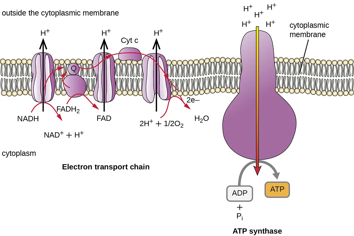
The electron transport system (ETS) is the last component involved in the process of cellular respiration; it comprises a series of membrane-associated protein complexes and associated mobile accessory electron carriers (illustrated below). Electron transport is a series of chemical reactions that resembles a bucket brigade in that electrons from NADH and FADH₂ are passed rapidly from one ETS electron carrier to the next. These carriers can pass electrons along in the ETS because of their redox potential. For a protein or chemical to accept electrons, it must have a more positive redox potential than the electron donor. Therefore, electrons move from electron carriers with more negative redox potential to those with more positive redox potential. The four major classes of electron carriers involved in both eukaryotic and prokaryotic electron transport systems are the cytochromes, flavoproteins, iron-sulfur proteins, and the quinones.
In aerobic respiration, the final electron acceptor (i.e., the one having the most positive redox potential) at the end of the ETS is an oxygen molecule (O₂) that becomes reduced to water (H₂O) by the final ETS carrier. This electron carrier, cytochrome oxidase, differs between bacterial types and can be used to differentiate closely related bacteria for diagnoses. For example, the gram-negative opportunist Pseudomonas aeruginosa and the gram-negative cholera-causing Vibrio cholerae use cytochrome c oxidase, which can be detected by the oxidase test, whereas other gram-negative Enterobacteriaceae, like E. coli, are negative for this test because they produce different cytochrome oxidase types.
There are many circumstances under which aerobic respiration is not possible, including any one or more of the following:
- The cell lacks genes encoding an appropriate cytochrome oxidase for transferring electrons to oxygen at the end of the electron transport system.
- The cell lacks genes encoding enzymes to minimize the severely damaging effects of dangerous oxygen radicals produced during aerobic respiration, such as hydrogen peroxide (H₂O₂) or superoxide (O₂⁻).
- The cell lacks a sufficient amount of oxygen to carry out aerobic respiration.
One possible alternative to aerobic respiration is anaerobic respiration, using an inorganic molecule other than oxygen as a final electron acceptor. There are many types of anaerobic respiration found in bacteria and archaea. Denitrifiers are important soil bacteria that use nitrate (NO₃⁻) and nitrite (NO₂⁻) as final electron acceptors, producing nitrogen gas (N₂). Many aerobically respiring bacteria, including E. coli, switch to using nitrate as a final electron acceptor and producing nitrite when oxygen levels have been depleted.
Microbes using anaerobic respiration commonly have an intact Krebs cycle, so these organisms can access the energy of the NADH and FADH₂ molecules formed. However, anaerobic respirers use altered ETS carriers encoded by their genomes, including distinct complexes for electron transfer to their final electron acceptors. Smaller electrochemical gradients are generated from these electron transfer systems, so less ATP is formed through anaerobic respiration.
Check Your Understanding
Do both aerobic respiration and anaerobic respiration use an electron transport chain?
Compare what the aerobic-respiration and anaerobic-respiration paragraphs each say about the final electron acceptor, not about whether an ETS is used at all.Chemiosmosis, Proton Motive Force, and Oxidative Phosphorylation
In each transfer of an electron through the ETS, the electron loses energy, but with some transfers, the energy is stored as potential energy by using it to pump hydrogen ions (H⁺) across a membrane. In prokaryotic cells, H⁺ is pumped to the outside of the cytoplasmic membrane (called the periplasmic space in gram-negative and gram-positive bacteria), and in eukaryotic cells, they are pumped from the mitochondrial matrix across the inner mitochondrial membrane into the intermembrane space. There is an uneven distribution of H⁺ across the membrane that establishes an electrochemical gradient because H⁺ ions are positively charged (electrical) and there is a higher concentration (chemical) on one side of the membrane. This electrochemical gradient formed by the accumulation of H⁺ (also known as a proton) on one side of the membrane compared with the other is referred to as the proton motive force (PMF). Because the ions involved are H⁺, a pH gradient is also established, with the side of the membrane having the higher concentration of H⁺ being more acidic. Beyond the use of the PMF to make ATP, as discussed in this chapter, the PMF can also be used to drive other energetically unfavorable processes, including nutrient transport and flagella rotation for motility.
The potential energy of this electrochemical gradient generated by the ETS causes the H⁺ to diffuse across a membrane (the plasma membrane in prokaryotic cells and the inner membrane in mitochondria in eukaryotic cells). This flow of hydrogen ions across the membrane, called chemiosmosis, must occur through a channel in the membrane via a membrane-bound enzyme complex called ATP synthase (shown below). The tendency for movement in this way is much like water accumulated on one side of a dam, moving through the dam when opened. ATP synthase (like a combination of the intake and generator of a hydroelectric dam) is a complex protein that acts as a tiny generator, turning by the force of the H⁺ diffusing through the enzyme, down their electrochemical gradient from where there are many mutually repelling H⁺ to where there are fewer H⁺. In prokaryotic cells, H⁺ flows from the outside of the cytoplasmic membrane into the cytoplasm, whereas in eukaryotic mitochondria, H⁺ flows from the intermembrane space to the mitochondrial matrix. The turning of the parts of this molecular machine regenerates ATP from ADP and inorganic phosphate (Pi) by oxidative phosphorylation, a second mechanism for making ATP that harvests the potential energy stored within an electrochemical gradient.
Extended description
Left panel, labeled Electron Transport Chain, with ‘outside the cytoplasmic membrane’ above the membrane and ‘cytoplasm’ below it. Reading left to right: a first membrane complex receives electrons from NADH, which becomes NAD⁺ + H⁺ below it, and pumps H⁺ to the outside (upward arrow). Its electrons pass to ubiquinone (Q), a small carrier shown above the membrane, which also receives electrons from a second complex labeled FAD (formed by the oxidation of FADH₂ flowing up into it); this second complex carries no H⁺ arrow of its own. Q passes electrons to a third complex, which pumps H⁺ to the outside and hands the electrons to cytochrome c (Cyt c), a small carrier sitting atop the membrane between the third and fourth complexes. Cytochrome c carries the electrons to the fourth, rightmost complex, which pumps H⁺ to the outside and combines the incoming electrons (2e⁻) with 2H⁺ and 1/2 O₂ to form H₂O. Right panel, labeled ATP synthase, drawn as a bulb-shaped protein spanning the membrane with four H⁺ ions shown accumulated on the outside in two rows of two; a shaded arrow inside the protein shows H⁺ flowing down through it into the cytoplasm, and a curved arrow beside it shows this turning motion driving ADP plus Pᵢ to ATP.
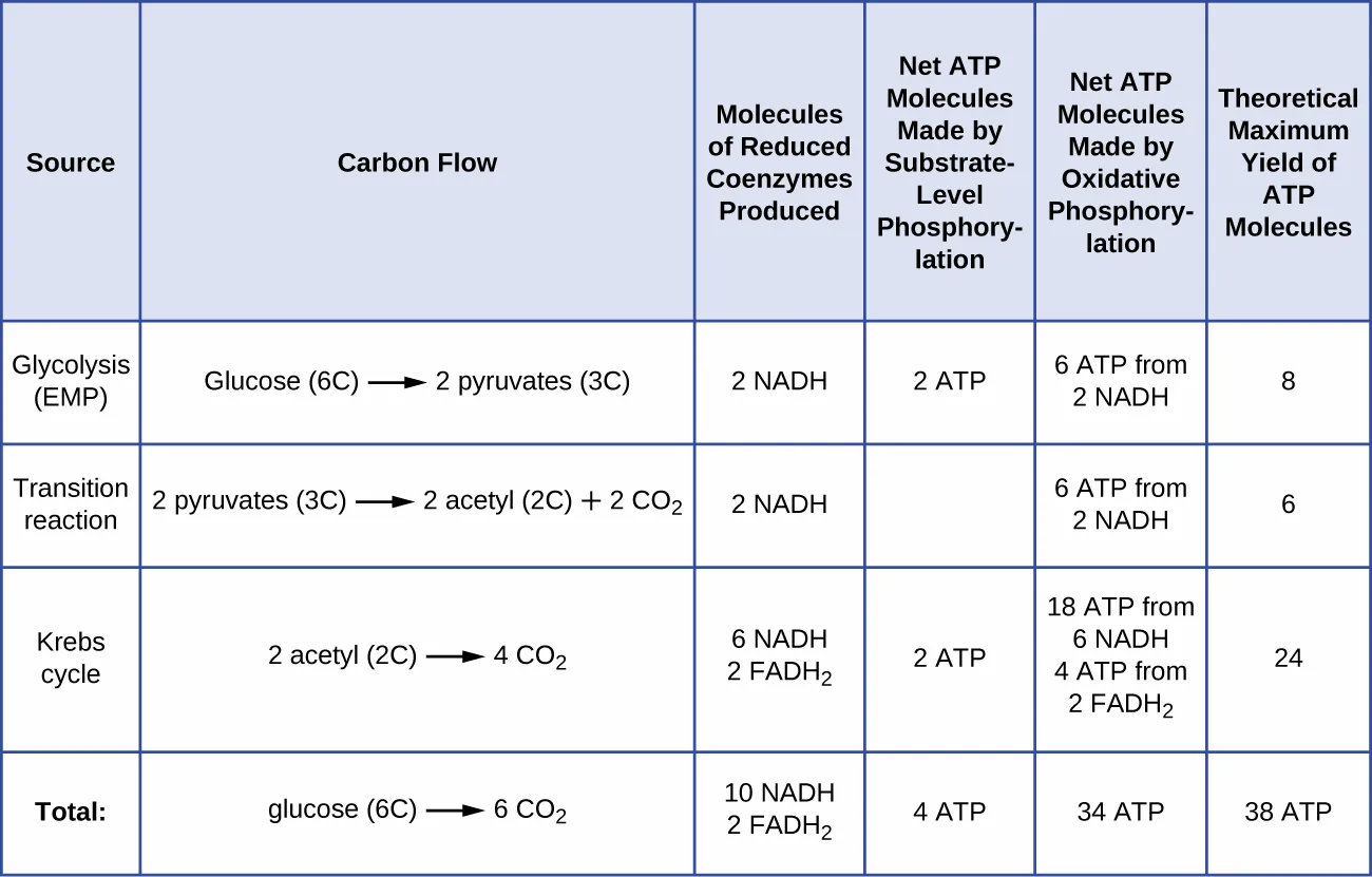
The number of ATP molecules generated from the catabolism of glucose varies. For example, the number of hydrogen ions that the electron transport system complexes can pump through the membrane varies between different species of organisms. In aerobic respiration in mitochondria, the passage of electrons from one molecule of NADH generates enough proton motive force to make three ATP molecules by oxidative phosphorylation, whereas the passage of electrons from one molecule of FADH₂ generates enough proton motive force to make only two ATP molecules. Thus, the 10 NADH molecules made per glucose during glycolysis, the transition reaction, and the Krebs cycle carry enough energy to make 30 ATP molecules, whereas the two FADH₂ molecules made per glucose during these processes provide enough energy to make four ATP molecules. Overall, the theoretical maximum yield of ATP made during the complete aerobic respiration of glucose is 38 molecules, with four being made by substrate-level phosphorylation and 34 being made by oxidative phosphorylation (illustrated in the figure below). In reality, the total ATP yield is usually less, ranging from one to 34 ATP molecules, depending on whether the cell is using aerobic respiration or anaerobic respiration; in eukaryotic cells, some energy is expended to transport intermediates from the cytoplasm into the mitochondria, affecting ATP yield.
The figure below summarizes the theoretical maximum yields of ATP from various processes during the complete aerobic respiration of one glucose molecule.
| Source | Carbon Flow | Molecules of Reduced Coenzymes Produced | Net ATP by Substrate-Level Phosphorylation | Net ATP by Oxidative Phosphorylation | Theoretical Maximum Yield of ATP Molecules |
|---|---|---|---|---|---|
| Glycolysis (EMP) | Glucose (6C) → 2 pyruvates (3C) | 2 NADH | 2 ATP | 6 ATP from 2 NADH | 8 |
| Transition reaction | 2 pyruvates (3C) → 2 acetyl (2C) + 2 CO₂ | 2 NADH | — | 6 ATP from 2 NADH | 6 |
| Krebs cycle | 2 acetyl (2C) → 4 CO₂ | 6 NADH, 2 FADH₂ | 2 ATP | 18 ATP from 6 NADH, 4 ATP from 2 FADH₂ | 24 |
| Total: | Glucose (6C) → 6 CO₂ | 10 NADH, 2 FADH₂ | 4 ATP | 34 ATP | 38 ATP |
Check Your Understanding
What are the functions of the proton motive force?
Look for the sentence that names what the PMF is used for beyond ATP production.Summary
- Most ATP generated during the cellular respiration of glucose is made by oxidative phosphorylation.
- An electron transport system (ETS) is composed of a series of membrane-associated protein complexes and associated mobile accessory electron carriers. The ETS is embedded in the cytoplasmic membrane of prokaryotes and the inner mitochondrial membrane of eukaryotes.
- Each ETS complex has a different redox potential, and electrons move from electron carriers with more negative redox potential to those with more positive redox potential.
- To carry out aerobic respiration, a cell requires oxygen as the final electron acceptor. A cell also needs a complete Krebs cycle, an appropriate cytochrome oxidase, and oxygen detoxification enzymes to prevent the harmful effects of oxygen radicals produced during aerobic respiration.
- Organisms performing anaerobic respiration use alternative electron transport system carriers for the ultimate transfer of electrons to the final non-oxygen electron acceptors.
- Microbes show great variation in the composition of their electron transport systems, which can be used for diagnostic purposes to help identify certain pathogens.
- As electrons are passed from NADH and FADH₂ through an ETS, the electron loses energy. This energy is stored through the pumping of H⁺ across the membrane, generating a proton motive force.
- The energy of this proton motive force can be harnessed by allowing hydrogen ions to diffuse back through the membrane by chemiosmosis using ATP synthase. As hydrogen ions diffuse through down their electrochemical gradient, components of ATP synthase spin, making ATP from ADP and Pi by oxidative phosphorylation.
- Aerobic respiration forms more ATP (a maximum of 34 ATP molecules) during oxidative phosphorylation than does anaerobic respiration (between one and 32 ATP molecules).
Key terms
- oxidative phosphorylation — mechanism for making ATP that uses the potential energy stored within an electrochemical gradient to add Pi to ADP.
- electron transport system (ETS) — series of membrane-associated protein complexes and associated mobile accessory electron carriers important in the generation of the proton motive force required for ATP production by chemiosmosis; the last component involved in the cellular respiration of glucose.
- redox potential — tendency for a molecule to acquire electrons and become reduced; electrons flow from molecules with lower redox potentials to those with higher redox potentials.
- aerobic respiration — use of an oxygen molecule as the final electron acceptor of the electron transport system.
- cytochrome oxidase — final ETS complex used in aerobic respiration that transfers energy-depleted electrons to oxygen to form H₂O.
- anaerobic respiration — use of a non-oxygen inorganic molecule, like CO2, nitrate, nitrite, oxidized iron, or sulfate, as the final electron acceptor at the end of the electron transport system.
- proton motive force — electrochemical gradient formed by the accumulation of hydrogen ions (also known as protons) on one side of a membrane relative to the other.
- chemiosmosis — flow of hydrogen ions across the membrane through ATP synthase.
- ATP synthase — integral membrane protein that harnesses the energy of the proton motive force by allowing hydrogen ions to diffuse down their electrochemical gradient, causing components of this protein to spin, making ATP from ADP and Pi.
Practice
Compare and contrast the electron transport system location and function in a prokaryotic cell and a eukaryotic cell
Which is the location of electron transports systems in prokaryotes?
Prokaryotes have no mitochondria; find the membrane named for prokaryotic electron transport.In prokaryotes, which of the following is true?
Recall which side of the prokaryotic cytoplasmic membrane accumulates H+ as electrons move through the ETS.An ________ is composed of a series of membrane-associated protein complexes and associated mobile accessory electron carriers.
Name the last component of cellular respiration, whose abbreviation is used throughout this section.Compare and contrast the differences between substrate-level and oxidative phosphorylation
Which is the source of the energy used to make ATP by oxidative phosphorylation?
Ask what ATP synthase harnesses to spin and regenerate ATP from ADP and Pi.Most ATP generated during the cellular respiration of glucose is made by ________.
Name the mechanism that harvests the potential energy stored within an electrochemical gradient to add Pi to ADP.How does oxidative phosphorylation differ from substrate-level phosphorylation?
Show model answer
Did your answer mention:
Explain the relationship between chemiosmosis and proton motive force
Because the ions involved are H+, a pH gradient is also established, with the side of the membrane having the higher concentration of H+ being more ________.
Recall what happens to pH on the side of the membrane where H+ ions build up.There is an uneven distribution of H+ across the membrane that establishes an electrochemical gradient because H+ ions are positively charged (________) and there is a higher concentration (chemical) on one side of the membrane.
Name the property of the charged ions themselves that contributes to the gradient, distinct from their higher concentration on one side.What is the relationship between chemiosmosis and the proton motive force?
Show model answer
Did your answer mention:
Describe the function and location of ATP synthase in a prokaryotic versus eukaryotic cell
The passage of hydrogen ions through ________ down their electrochemical gradient harnesses the energy needed for ATP synthesis by oxidative phosphorylation.
Name the membrane-bound enzyme complex whose channel hydrogen ions diffuse through.The final ETS complex used in aerobic respiration that transfers energy-depleted electrons to oxygen to form H₂O is called ________.
Name the final electron carrier of the ETS in aerobic respiration.Which of the following is not an electron carrier within an electron transport system?
Three of these four pass electrons along the ETS; one instead lets H+ diffuse through to make ATP.How does the location of ATP synthase differ between prokaryotes and eukaryotes? Where do protons accumulate as a result of the ETS in each cell type?
Show model answer
Did your answer mention:
Compare and contrast aerobic and anaerobic respiration
A cell might perform anaerobic respiration for which of the following reasons?
Ask which missing component would rule out using oxygen specifically as the final electron acceptor.All organisms that use aerobic cellular respiration have cytochrome oxidase.
Recall which electron carrier reduces oxygen to water at the end of an aerobic ETS.The cell lacks genes encoding enzymes to minimize the severely damaging effects of dangerous oxygen radicals produced during aerobic respiration, such as hydrogen peroxide (H₂O₂) or ________ (O₂⁻).
Name the other dangerous oxygen radical named alongside hydrogen peroxide, from its molecular formula.This section is adapted from Microbiology, Section 8.3: Cellular Respiration by Nina Parker, Mark Schneegurt, Anh-Hue Thi Tu, Philip Lister, Brian M. Forster, and OpenStax, © OpenStax, licensed under CC BY-NC-SA 4.0. Access the original for free at openstax.org. Changes: both source figures re-encoded as WebP and rendered as mediafigures after image and PDF inspection, with explicit kind="diagram"; the electron transport chain figure’s alt and longdesc rewritten from the manifest’s ATP-synthase-only alt to also describe the electron transport chain half of the drawing (the complexes, ubiquinone, and cytochrome c) after inspecting the image, with chemical formulas in Unicode and the H+ ions above ATP synthase counted directly from the image (four, in two rows of two); the ATP-yield figure is a table with no photographic or micrograph content in any cell, so per the book’s “table printed as an image” rule it is transcribed as a Markdown table from the image (checked against PDF page p-0311), kept at the sentence that refers to it, with the vendored figure immediately after as a brief-alt mediafigure and no longdesc (the table already carries the data); both same-module figure cross-references rendered as describing phrases (“illustrated below”, “shown below”); the two body Check Your Understanding bullets rendered as graded multiple-choice items, each keyed from one named module sentence (the source prints no answer key for them), with their option order varied so the key does not sit at the first position on every author-built item; all three unkeyed Short Answer questions — on the relationship between chemiosmosis and PMF, on oxidative versus substrate-level phosphorylation, and on ATP synthase’s location and where protons accumulate in each cell type — remain self-checks with model answers and rubrics assembled only from this module’s own sentences, the last of these needing two named module sentences rather than one; the five Multiple Choice, two Fill in the Blank, and one True/False item are adapted into Practice with the True/False item rendered as a two-option multiple choice, all four options of every source multiple choice kept verbatim in source order; the “which is not an electron carrier” Multiple Choice’s own printed options include both “ATP synthase” and “cytochrome oxidase,” which are also two other textin keys on the page, so the cytochrome oxidase textin (the source’s Fill in the Blank fs-id1167660350553, thematically Objective 5’s) is placed in Objective 4’s group, immediately after the ATP synthase textin and immediately before that multiple choice, so every key it prints has already been typed by the learner rather than read off the option list; Objective 5’s group is filled back to three items with a body-sentence cloze textin (“superoxide,” from the aerobic-respiration bulleted list) in the vacated slot; two summary-sentence cloze textin items (“electron transport system”, “oxidative phosphorylation”) and two body-sentence cloze textin items (“acidic”, “electrical”) fill out the remaining thin objective groups to the section’s three-item floor, since the module’s eleven source exercises alone left them short; the group-3 fillers replace an earlier pair of clozes keyed “chemiosmosis” and “proton motive force” — the first because its own stem printed two sibling items’ keys, the second because the source-keyed multiple choice in the group above already prints “the proton motive force” as its correct option, so a later typed-recall of the same phrase would be answered by a term the learner had already read rather than by understanding it; key terms compiled from the module’s nine defined terms and the book’s Glossary appendix. No source item was omitted.