Microbial Growth
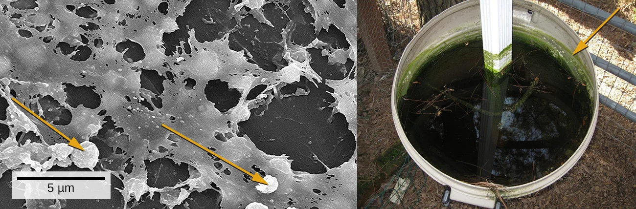
We are all familiar with the slimy layer on a pond surface or that makes rocks slippery. These are examples of biofilms—microorganisms embedded in thin layers of matrix material, as the two photographs above show. Biofilms were long considered random assemblages of cells and had little attention from researchers. Recently, progress in visualization and biochemical methods has revealed that biofilms are an organized ecosystem within which many cells, usually of different species of bacteria, fungi, and algae, interact through cell signaling and coordinated responses. The biofilm provides a protected environment in harsh conditions and aids colonization by microorganisms. Biofilms also have clinical importance. They form on medical devices, resist routine cleaning and sterilization, and cause health-acquired infections. Within the body, biofilms form on the teeth as plaque, in the lungs of patients with cystic fibrosis, and on the cardiac tissue of patients with endocarditis. The slime layer helps protect the cells from host immune defenses and antibiotic treatments.
Biofilms don’t only exist on Earth. They have been frequent occupants (and problems) on the International Space Station. Bacterial and fungal biofilms grow on a number of space station objects and within systems. In some cases, growth has been so significant that hoses and other components had to be completely replaced. While this is difficult on the ISS, it poses even greater risk for longer-range travel planned for the future. As described later in the chapter, a group of scientists has devised a method to prevent growth of biofilms through surface preparation: reducing the ability of the microorganisms to adhere to materials.
Studying biofilms requires new approaches. Because of the cells’ adhesion properties, many of the methods for culturing and counting cells that are explored in this chapter are not easily applied to biofilms. This is the beginning of a new era of challenges and rewarding insight into the ways that microorganisms grow and thrive in nature.
Sections
- How Microbes Grow — binary fission and generation time, the growth curve and its phases, direct and indirect methods of counting cells, alternative patterns of cell division, and biofilms and quorum sensing.
- Oxygen Requirements for Microbial Growth — obligate aerobes and anaerobes, facultative and aerotolerant anaerobes, microaerophiles, reactive oxygen species and the enzymes that detoxify them, and how anaerobes are cultured.
- The Effects of pH on Microbial Growth — minimum, optimum, and maximum growth pH; acidophiles, neutrophiles, and alkaliphiles; and how extreme pH damages macromolecules.
- Temperature and Microbial Growth — psychrophiles, psychrotrophs, mesophiles, thermophiles, and hyperthermophiles, and the adaptations that let cells live at temperature extremes.
- Other Environmental Conditions that Affect Growth — osmotic pressure and salt, pressure, and light as conditions for growth.
- Media Used for Bacterial Growth — chemically defined and complex media, and selective, differential, and enrichment media.
This chapter is adapted from Microbiology, Chapter 9: Microbial Growth by Nina Parker, Mark Schneegurt, Anh-Hue Thi Tu, Philip Lister, Brian M. Forster, and OpenStax, © OpenStax, licensed under CC BY-NC-SA 4.0. Access the original for free at openstax.org. Each section page records its own changes from the source. Changes: the chapter-opening figure is the source’s pair of photographs, re-encoded for the web, with its alt text rewritten to describe both panels; the source’s figure cross-reference is replaced by a describing phrase; the source’s “health-acquired infections” is kept as printed and logged upstream.