Oxygen Requirements for Microbial Growth
By the end of this section, you will be able to:
- Interpret visual data demonstrating minimum, optimum, and maximum oxygen or carbon dioxide requirements for growth
- Identify and describe different categories of microbes with requirements for growth with or without oxygen: obligate aerobe, obligate anaerobe, facultative anaerobe, aerotolerant anaerobe, microaerophile, and capnophile
- Give examples of microorganisms for each category of growth requirements
Ask most people “What are the major requirements for life?” and the answers are likely to include water and oxygen. Few would argue about the need for water, but what about oxygen? Can there be life without oxygen?
The answer is that molecular oxygen (O₂) is not always needed. The earliest signs of life are dated to a period when conditions on earth were highly reducing and free oxygen gas was essentially nonexistent. Only after cyanobacteria started releasing oxygen as a byproduct of photosynthesis and the capacity of iron in the oceans for taking up oxygen was exhausted did oxygen levels increase in the atmosphere. This event, often referred to as the Great Oxygenation Event or the Oxygen Revolution, caused a massive extinction. Most organisms could not survive the powerful oxidative properties of reactive oxygen species (ROS), highly unstable ions and molecules derived from partial reduction of oxygen that can damage virtually any macromolecule or structure with which they come in contact. Singlet oxygen (O₂•), superoxide (O₂⁻), peroxides (H₂O₂), hydroxyl radical (OH•), and hypochlorite ion (OCl⁻), the active ingredient of household bleach, are all examples of ROS. The organisms that were able to detoxify reactive oxygen species harnessed the high electronegativity of oxygen to produce free energy for their metabolism and thrived in the new environment.
Oxygen Requirements of Microorganisms
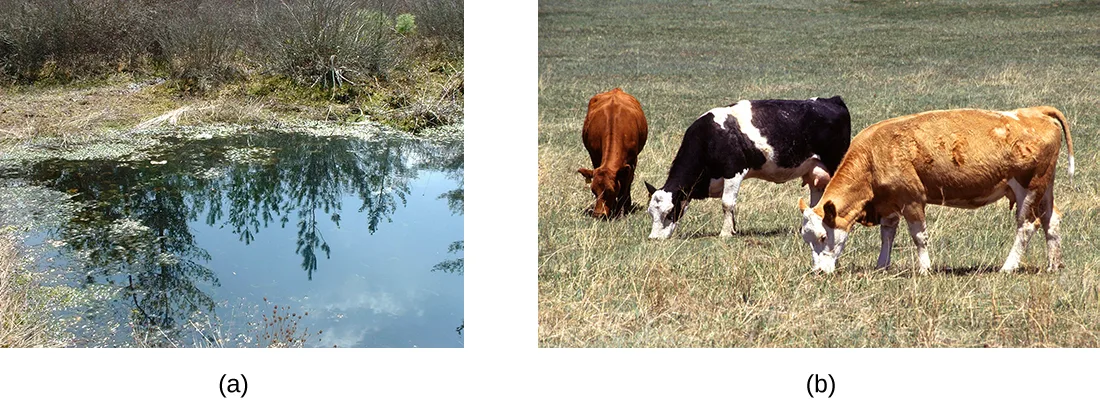
Many ecosystems are still free of molecular oxygen. Some are found in extreme locations, such as deep in the ocean or in earth’s crust; others are part of our everyday landscape, such as marshes, bogs, and sewers. Within the bodies of humans and other animals, regions with little or no oxygen provide an anaerobic environment for microorganisms, as shown below.
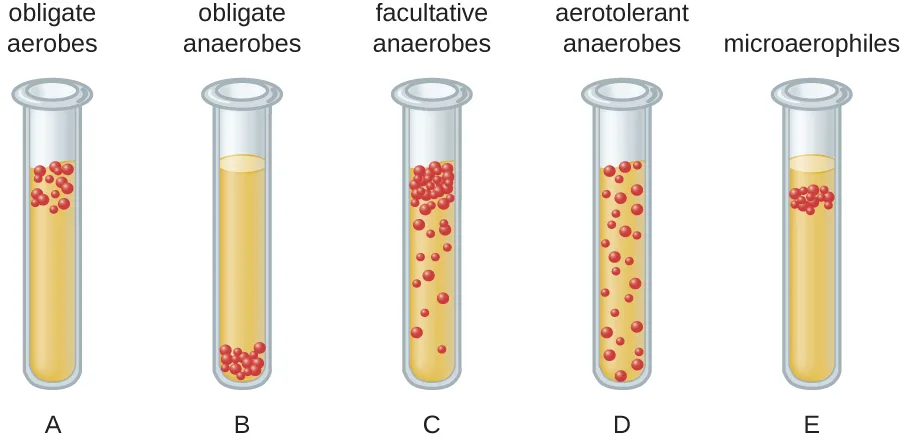
We can easily observe different requirements for molecular oxygen by growing bacteria in thioglycolate tube cultures. A test-tube culture starts with autoclaved thioglycolate medium containing a low percentage of agar to allow motile bacteria to move throughout the medium. Thioglycolate has strong reducing properties and autoclaving flushes out most of the oxygen. The tubes are inoculated with the bacterial cultures to be tested and incubated at an appropriate temperature. Over time, oxygen slowly diffuses throughout the thioglycolate tube culture from the top. Bacterial density increases in the area where oxygen concentration is best suited for the growth of that particular organism.
The growth of bacteria with varying oxygen requirements in thioglycolate tubes is illustrated below. In tube A, all the growth is seen at the top of the tube. The bacteria are obligate (strict) aerobes that cannot grow without an abundant supply of oxygen. Tube B looks like the opposite of tube A. Bacteria grow at the bottom of tube B. Those are obligate anaerobes, which are killed by oxygen. Tube C shows heavy growth at the top of the tube and growth throughout the tube, a typical result with facultative anaerobes. Facultative anaerobes are organisms that thrive in the presence of oxygen but also grow in its absence by relying on fermentation or anaerobic respiration, if there is a suitable electron acceptor other than oxygen and the organism is able to perform anaerobic respiration. The aerotolerant anaerobes in tube D are indifferent to the presence of oxygen. They do not use oxygen because they usually have a fermentative metabolism, but they are not harmed by the presence of oxygen as obligate anaerobes are. Tube E on the right shows a “Goldilocks” culture. The oxygen level has to be just right for growth, not too much and not too little. These microaerophiles are bacteria that require a minimum level of oxygen for growth, about 1%–10%, well below the 21% found in the atmosphere.
Examples of obligate aerobes are Mycobacterium tuberculosis, the causative agent of tuberculosis and Micrococcus luteus, a gram-positive bacterium that colonizes the skin. Neisseria meningitidis, the causative agent of severe bacterial meningitis, and N. gonorrhoeae, the causative agent of sexually transmitted gonorrhea, are also obligate aerobes.
Many obligate anaerobes are found in the environment where anaerobic conditions exist, such as in deep sediments of soil, still waters, and at the bottom of the deep ocean where there is no photosynthetic life. Anaerobic conditions also exist naturally in the intestinal tract of animals. Obligate anaerobes, mainly Bacteroidetes, represent a large fraction of the microbes in the human gut. Transient anaerobic conditions exist when tissues are not supplied with blood circulation; they die and become an ideal breeding ground for obligate anaerobes. Another type of obligate anaerobe encountered in the human body is the gram-positive, rod-shaped Clostridioides and Clostridium spp. Their ability to form endospores allows them to survive in the presence of oxygen. One of the major causes of health-acquired infections is C. difficile, known as C. diff. Prolonged use of antibiotics for other infections increases the probability of a patient developing a secondary C. difficile infection. Antibiotic treatment disrupts the balance of microorganisms in the intestine and allows the colonization of the gut by Clostridioides difficile, causing a significant inflammation of the colon.
Other clostridia responsible for serious infections include C. tetani, the agent of tetanus, and C. perfringens, which causes gas gangrene. In both cases, the infection starts in necrotic tissue (dead tissue that is not supplied with oxygen by blood circulation). This is the reason that deep puncture wounds are associated with tetanus. When tissue death is accompanied by lack of circulation, gangrene is always a danger.
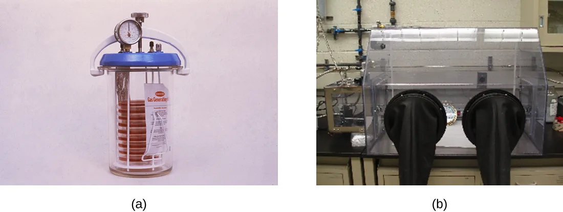
The study of obligate anaerobes requires special equipment. Obligate anaerobic bacteria must be grown under conditions devoid of oxygen. The most common approach is culture in an anaerobic jar, shown below. Anaerobic jars include chemical packs that remove oxygen and release carbon dioxide (CO₂). An anaerobic chamber is an enclosed box from which all oxygen is removed. Gloves sealed to openings in the box allow handling of the cultures without exposing the culture to air.
Staphylococci and Enterobacteriaceae are examples of facultative anaerobes. Staphylococci are found on the skin and upper respiratory tract. Enterobacteriaceae are found primarily in the gut and upper respiratory tract but can sometimes spread to the urinary tract, where they are capable of causing infections. It is not unusual to see mixed bacterial infections in which the facultative anaerobes use up the oxygen, creating an environment for the obligate anaerobes to flourish.
Examples of aerotolerant anaerobes include lactobacilli and streptococci, both found in the oral microbiota. Campylobacter jejuni, which causes gastrointestinal infections, is an example of a microaerophile and is grown under low-oxygen conditions.
The optimum oxygen concentration, as the name implies, is the ideal concentration of oxygen for a particular microorganism. The lowest concentration of oxygen that allows growth is called the minimum permissive oxygen concentration. The highest tolerated concentration of oxygen is the maximum permissive oxygen concentration. The organism will not grow outside the range of oxygen levels found between the minimum and maximum permissive oxygen concentrations.
Check Your Understanding
Would you expect the oldest bacterial lineages to be aerobic or anaerobic?
Recall that oxygen levels rose only after cyanobacteria began releasing oxygen by photosynthesis, long after life first appeared.Which bacteria grow at the top of a thioglycolate tube, and which grow at the bottom of the tube?
Recall which tube (A or B) in the thioglycolate diagram shows all its growth at the top and which shows all its growth at the bottom.Case in Point. An Unwelcome Anaerobe
Charles is a retired bus driver who developed type 2 diabetes over 10 years ago. Since his retirement, his lifestyle has become very sedentary and he has put on a substantial amount of weight. Although he has felt tingling and numbness in his left foot for a while, he has not been worried because he thought his foot was simply “falling asleep.” Recently, a scratch on his foot does not seem to be healing and is becoming increasingly ugly. Because the sore did not bother him much, Charles figured it could not be serious until his daughter noticed a purplish discoloration spreading on the skin and oozing, shown below. When he was finally seen by his physician, Charles was rushed to the operating room. His open sore, or ulcer, is the result of a diabetic foot.
The concern here is that gas gangrene may have taken hold in the dead tissue. The most likely agent of gas gangrene is Clostridium perfringens, an endospore-forming, gram-positive bacterium. It is an obligate anaerobe that grows in tissue devoid of oxygen. Since dead tissue is no longer supplied with oxygen by the circulatory system, the dead tissue provides pockets of ideal environment for the growth of C. perfringens.
A surgeon examines the ulcer and radiographs of Charles’s foot and determines that the bone is not yet infected. The wound will have to be surgically debrided (debridement refers to the removal of dead and infected tissue) and a sample sent for microbiological lab analysis, but Charles will not have to have his foot amputated. Many diabetic patients are not so lucky. In 2008, nearly 70,000 diabetic patients in the United States lost a foot or limb to amputation, according to statistics from the Centers for Disease Control and Prevention (“Living With Diabetes: Keep Your Feet Healthy”).
- Which growth conditions would you recommend for the detection of C. perfringens?

Detoxification of Reactive Oxygen Species
Aerobic respiration constantly generates reactive oxygen species (ROS), byproducts that must be detoxified. Even organisms that do not use aerobic respiration need some way to break down some of the ROS that may form from atmospheric oxygen. Three main enzymes break down those toxic byproducts: superoxide dismutase, peroxidase, and catalase. Each one catalyzes a different reaction. Reactions of type seen in Reaction 1 are catalyzed by peroxidases.
(1) X−(2H⁺) + H₂O₂ → oxidized-X + 2H₂O
In these reactions, an electron donor (reduced compound; e.g., reduced nicotinamide adenine dinucleotide [NADH]) oxidizes hydrogen peroxide, or other peroxides, to water. The enzymes play an important role by limiting the damage caused by peroxidation of membrane lipids. Reaction 2 is mediated by the enzyme superoxide dismutase (SOD) and breaks down the powerful superoxide anions generated by aerobic metabolism:
(2) 2O₂⁻ + 2H⁺ → H₂O₂ + O₂
The enzyme catalase converts hydrogen peroxide to water and oxygen as shown in Reaction 3.
(3) 2H₂O₂ → 2H₂O + O₂
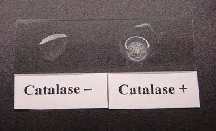
Obligate anaerobes usually lack all three enzymes. Aerotolerant anaerobes do have SOD but no catalase. Reaction 3, shown occurring in the catalase test below, is the basis of a useful and rapid test to distinguish streptococci, which are aerotolerant and do not possess catalase, from staphylococci, which are facultative anaerobes. A sample of culture rapidly mixed in a drop of 3% hydrogen peroxide will release bubbles if the culture is catalase positive.
Bacteria that grow best in a higher concentration of CO₂ and a lower concentration of oxygen than present in the atmosphere are called capnophiles. One common approach to grow capnophiles is to use a candle jar. A candle jar consists of a jar with a tight-fitting lid that can accommodate the cultures and a candle. After the cultures are added to the jar, the candle is lit and the lid closed. As the candle burns, it consumes most of the oxygen present and releases CO₂.
Check Your Understanding
What substance is added to a sample to detect catalase?
Recall the compound a drop of culture is rapidly mixed with in the catalase test, which is also the substrate of Reaction 3.What is the function of the candle in a candle jar?
Recall what happens to the jar’s atmosphere as the lit candle inside it burns with the lid closed.Clinical Focus. Part 2
The health-care provider who saw Jeni was concerned primarily because of her pregnancy. Her condition enhances the risk for infections and makes her more vulnerable to those infections. The immune system is downregulated during pregnancy, and pathogens that cross the placenta can be very dangerous for the fetus. A note on the provider’s order to the microbiology lab mentions a suspicion of infection by Listeria monocytogenes, based on the signs and symptoms exhibited by the patient.
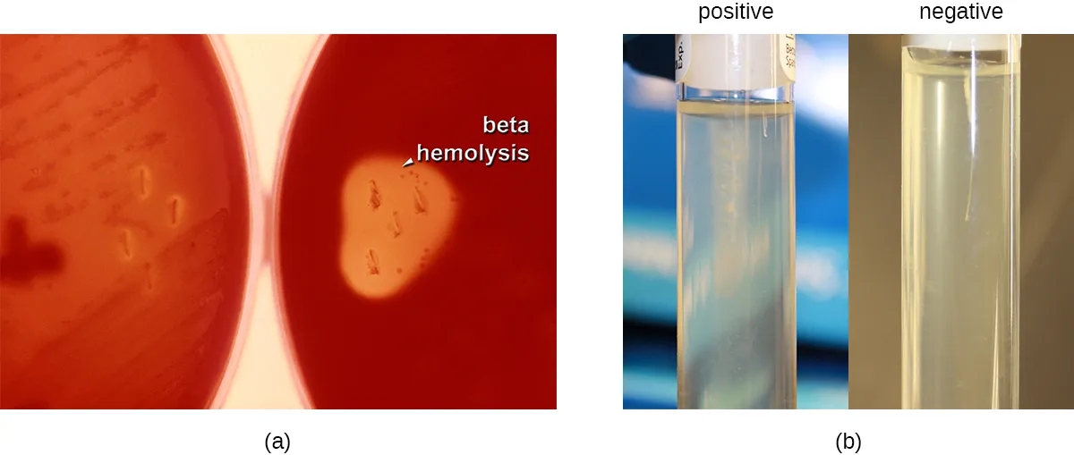
Jeni’s blood samples are streaked directly on sheep blood agar, a medium containing tryptic soy agar enriched with 5% sheep blood. (Blood is considered sterile; therefore, competing microorganisms are not expected in the medium.) The inoculated plates are incubated at 37 °C for 24 to 48 hours. Small grayish colonies surrounded by a clear zone emerge. Such colonies are typical of Listeria and other pathogens such as streptococci; the clear zone surrounding the colonies indicates complete lysis of blood in the medium, referred to as beta-hemolysis, shown below. When tested for the presence of catalase, the colonies give a positive response, eliminating Streptococcus as a possible cause. Furthermore, a Gram stain shows short gram-positive bacilli. Cells from a broth culture grown at room temperature displayed the tumbling motility characteristic of Listeria, also shown below. All of these clues lead the lab to positively confirm the presence of Listeria in Jeni’s blood samples.
- How serious is Jeni’s condition and what is the appropriate treatment?
The case continues in Temperature and Microbial Growth. The case began in How Microbes Grow.
Summary
- Aerobic and anaerobic environments can be found in diverse niches throughout nature, including different sites within and on the human body.
- Microorganisms vary in their requirements for molecular oxygen. Obligate aerobes depend on aerobic respiration and use oxygen as a terminal electron acceptor. They cannot grow without oxygen.
- Obligate anaerobes cannot grow in the presence of oxygen. They depend on fermentation and anaerobic respiration using a final electron acceptor other than oxygen.
- Facultative anaerobes show better growth in the presence of oxygen but will also grow without it.
- Although aerotolerant anaerobes do not perform aerobic respiration, they can grow in the presence of oxygen. Most aerotolerant anaerobes test negative for the enzyme catalase.
- Microaerophiles need oxygen to grow, albeit at a lower concentration than 21% oxygen in air.
- Optimum oxygen concentration for an organism is the oxygen level that promotes the fastest growth rate. The minimum permissive oxygen concentration and the maximum permissive oxygen concentration are, respectively, the lowest and the highest oxygen levels that the organism will tolerate.
- Peroxidase, superoxide dismutase, and catalase are the main enzymes involved in the detoxification of the reactive oxygen species. Superoxide dismutase is usually present in a cell that can tolerate oxygen. All three enzymes are usually detectable in cells that perform aerobic respiration and produce more ROS.
- A capnophile is an organism that requires a higher than atmospheric concentration of CO₂ to grow.
Key terms
- reactive oxygen species — unstable and toxic ions and molecules derived from partial reduction of oxygen.
- thioglycolate tube culture — contains reducing medium through which oxygen diffuses from the tube opening, producing a range of oxygen environments down the length of the tube.
- thioglycolate medium — medium designed to test the aerotolerance of bacteria; it contains a low concentration of agar to allow motile bacteria to move throughout the medium.
- obligate (strict) aerobe — organism that requires oxygen for growth.
- obligate anaerobe — organism that dies in the presence of oxygen.
- facultative anaerobe — organism that grows better in the presence of oxygen but can proliferate in its absence.
- aerotolerant anaerobe — organism that does not use oxygen but tolerates its presence.
- microaerophile — organism that requires oxygen at levels lower than atmospheric concentration.
- anaerobic jar — a container that holds cultures under conditions devoid of oxygen, using chemical packs that remove oxygen and release carbon dioxide.
- anaerobic chamber — an enclosed box from which all oxygen is removed, with gloves sealed to openings in the box to allow handling of cultures inside without exposing them to air.
- optimum oxygen concentration — the ideal concentration of oxygen for a particular microorganism.
- minimum permissive oxygen concentration — the lowest concentration of oxygen that allows an organism’s growth.
- maximum permissive oxygen concentration — the highest concentration of oxygen that an organism tolerates.
- peroxidase — enzyme that catalyzes the detoxification of peroxides.
- superoxide dismutase — enzyme that catalyzes the breakdown of superoxide anions.
- catalase — enzyme that breaks down hydrogen peroxide to water and oxygen.
- capnophiles — organism that requires carbon dioxide levels higher than atmospheric concentration.
- candle jar — container with a tight-fitting lid in which a burning candle consumes oxygen and releases carbon dioxide, thereby creating an environment suitable for capnophiles.
Practice
Interpret visual data demonstrating minimum, optimum, and maximum oxygen or carbon dioxide requirements for growth
An inoculated thioglycolate medium culture tube shows dense growth at the surface and turbidity throughout the rest of the tube. What is your conclusion?
Recall which growth pattern in the thioglycolate diagram shows heavy growth at the top plus growth spread throughout the rest of the tube.An inoculated thioglycolate medium culture tube is clear throughout the tube except for dense growth at the bottom of the tube. What is your conclusion?
Recall which tube in the thioglycolate diagram shows all its growth confined to the bottom, farthest from the oxygen diffusing in at the top.The lowest concentration of oxygen that still allows an organism to grow is called the ________.
Contrast this term with the maximum permissive oxygen concentration, the highest tolerated concentration.Identify and describe different categories of microbes with requirements for growth with or without oxygen: obligate aerobe, obligate anaerobe, facultative anaerobe, aerotolerant anaerobe, microaerophile, and capnophile
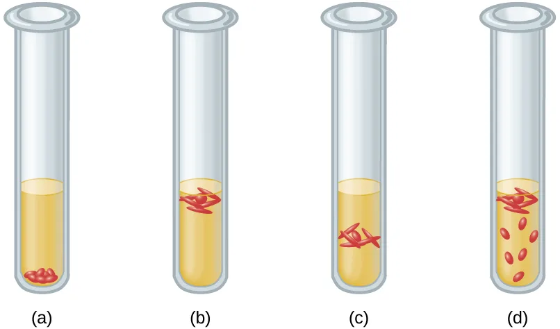
Which type of bacteria is growing in tube (a)?
Cells clustered at the very bottom of the tube, farthest from the oxygen diffusing in at the top, belong to the organism that is killed by oxygen.Which type of bacteria is growing in tube (b)?
Cells growing right at the tube’s opening, where the oxygen concentration is highest, belong to the organism that cannot grow without an abundant supply of oxygen.Which type of bacteria is growing in tube (c)?
Cells growing in a narrow band just below the surface, avoiding both the fully oxygenated top and the oxygen-free bottom, belong to the organism that needs a low, minimum level of oxygen.Which type of bacteria is growing in tube (d)?
Cells scattered throughout the tube but denser near the oxygen-rich top belong to the organism that grows better with oxygen but can also grow without it.Give examples of microorganisms for each category of growth requirements
Pseudomonas aeruginosa is a common pathogen that infects the airways of patients with cystic fibrosis. It does not grow in the absence of oxygen. The bacterium is probably which of the following?
Recall which category of organism cannot grow at all without oxygen.Streptococcus mutans is a major cause of cavities. It resides in the gum pockets, does not have catalase activity, and can be grown outside of an anaerobic chamber. The bacterium is probably which of the following?
Recall which category of organism lacks catalase, does not use oxygen, but is not harmed by its presence and needs no special anaerobic equipment.Why do the instructions for the growth of Neisseria gonorrhoeae recommend a CO₂-enriched atmosphere?
Recall the term for an organism that grows best in a higher-than-atmospheric concentration of CO₂.In terms of oxygen requirements, what type of organism would most likely be responsible for a foodborne illness associated with canned foods?
Show model answer
Did your answer mention:
Why are some obligate anaerobes able to grow in tissues (e.g., gum pockets) that are not completely free of oxygen?
Show model answer
Did your answer mention:
Why should Haemophilus influenzae be grown in a candle jar?
Show model answer
Did your answer mention:
A microbiology instructor prepares cultures for a gram-staining practical laboratory by inoculating growth medium with a gram-positive coccus (nonmotile) and a gram-negative rod (motile). The goal is to demonstrate staining of a mixed culture. The flask is incubated at 35 °C for 24 hours without aeration. A sample is stained and reveals only gram-negative rods. Both cultures are known facultative anaerobes. Give a likely reason for success of the gram-negative rod. Assume that the cultures have comparable intrinsic growth rates.
Show model answer
Did your answer mention:
This section is adapted from Microbiology, Section 9.2: Oxygen Requirements for Microbial Growth by Nina Parker, Mark Schneegurt, Anh-Hue Thi Tu, Philip Lister, Brian M. Forster, and OpenStax, © OpenStax, licensed under CC BY-NC-SA 4.0. Access the original for free at openstax.org. Changes: all six figures re-encoded as WebP and rendered as mediafigures after image and PDF inspection; the two schematic tube figures (OSC_Microbio_09_02_tubO, the body figure, and OSC_Microbio_09_02_tubO_img, the exercise figure) are set kind="diagram", overriding the manifest’s photo guess, since both are drawn artwork rather than photographs; the four photographic figures keep kind="photo"; OSC_Microbio_09_02_bog is eager="true" as the first figure on the page; the body tube figure’s alt is rewritten, tube by tube, from the image, keeping the source’s own oxygen-class identifications since its caption names them; the blood-agar/motility figure’s alt is rewritten because the source alt claims the left plate is labeled “alpha hemolysis,” a label the image does not print — only the right plate carries a printed “beta hemolysis” label — logged as a suspected source-alt defect; the foot, anaerobic-jar, bog, and catalase-test alts are rewritten from the image for detail the source alt omits, without changing what they identify; the three numbered <equation> blocks (Reactions 1–3) are rendered as their own plain-text Unicode paragraphs with their source (1)/(2)/(3) labels, never $…$, since they are chemical reactions rather than arithmetic; the exercise-figure OSC_Microbio_09_02_tubO_img (bare media in the Matching exercise) is rendered once as a mediafigure under the second objective with an author-written caption, since the source prints none, and its alt describes only where the cells sit in each lettered tube, never naming an oxygen class, so the paired multiple-choice items are not answered by the alt; the Case in Point and Clinical Focus boxes are rendered as callouts in source order; the Case in Point’s footnote (a CDC web page) becomes an inline parenthetical citation with its bare access URL dropped; both boxes’ closing questions stay inside their callouts as unanswered plain bullets; the Clinical Focus box’s closing “Jump to the next / go back to the previous Clinical Focus box” links are replaced by a sentence naming where the case continues, Temperature and Microbial Growth, and where it began — the house sentence names How Microbes Grow, since the source’s own “go back to the previous” link points at this section’s own Part 1, which is where the case’s second part starts, not where the whole case began; the two body Check Your Understanding boxes (2 + 2 bullets) are rendered as body items at their note positions, verbatim source stems, all four graded because one sentence of the module fixes each answer; the five source Multiple Choice items and the four-row Matching exercise (rendered as one mediafigure plus four multiple-choice items, one per lettered tube, each offering the source’s five oxygen classes in source list order, source-keyed) are adapted into Practice unchanged; all three unkeyed Short Answer questions stay self-checks whose model answers and rubrics are assembled only from this module’s own sentences — the gum-pockets one from the mixed-infections sentence, the Haemophilus influenzae one from the candle-jar sentences (explicitly noting the module does not itself identify the organism’s oxygen requirements and stopping at what the module gives), and the canned-foods one from the obligate-anaerobe habitat and Clostridioides/Clostridium sentences, since the module never mentions canned foods or a sealed environment and the model answer says so and stops at the general habitat and genus facts it does give; the unkeyed Critical Thinking question (the gram-stain competition scenario) stays a self-check assembled from the module’s facultative-anaerobe definition and the motility difference given in the stem; the third objective’s group keeps its four source Multiple Choice items as its graded floor, so no filler is added there; one author-written textin cloze on the minimum permissive oxygen concentration is added, from the module’s own body sentence, to bring the first objective’s group to the book’s floor; the hydrogen-peroxide textin in the second Check Your Understanding box takes accept="H2O2" (the grader folds it and the Unicode subscript form H₂O₂ to the same normalized string, so only one is listed) since the page prints the formula repeatedly in the surrounding prose; key terms compiled from the module’s eighteen defined terms and the book’s Glossary appendix; four terms (anaerobic jar, anaerobic chamber, minimum permissive oxygen concentration, maximum permissive oxygen concentration) have no appendix entry and are taken from the module’s own defining sentences. No source exercise item is otherwise omitted.