Structure and Function of DNA
By the end of this section, you will be able to:
- Describe the biochemical structure of deoxyribonucleotides
- Identify the base pairs used in the synthesis of deoxyribonucleotides
- Explain why the double helix of DNA is described as antiparallel
In Microbial Metabolism, we discussed the microbial catabolism of three classes of macromolecules: proteins, lipids and carbohydrates. In this chapter, we will discuss the genetic role of a fourth class of molecules: nucleic acids. Like other macromolecules, nucleic acids are composed of monomers, called nucleotides, which are polymerized to form large strands. Each nucleic acid strand contains certain nucleotides that appear in a certain order within the strand, called its base sequence. The base sequence of deoxyribonucleic acid (DNA) is responsible for carrying and retaining the hereditary information in a cell. In Mechanisms of Microbial Genetics, we will discuss in detail the ways in which DNA uses its own base sequence to direct its own synthesis, as well as the synthesis of RNA and proteins, which, in turn, gives rise to products with diverse structure and function. In this section, we will discuss the basic structure and function of DNA.
DNA Nucleotides
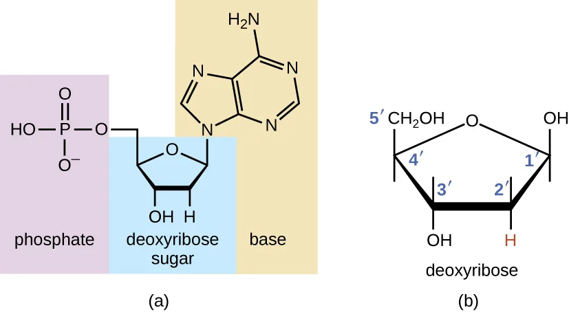
The building blocks of nucleic acids are nucleotides. Nucleotides that compose DNA are called deoxyribonucleotides. The three components of a deoxyribonucleotide are a five-carbon sugar called deoxyribose, a phosphate group, and a nitrogenous base, a nitrogen-containing ring structure that is responsible for complementary base pairing between nucleic acid strands (the figure below). The carbon atoms of the five-carbon deoxyribose are numbered 1′, 2′, 3′, 4′, and 5′ (1′ is read as “one prime”). A nucleoside comprises the five-carbon sugar and nitrogenous base.
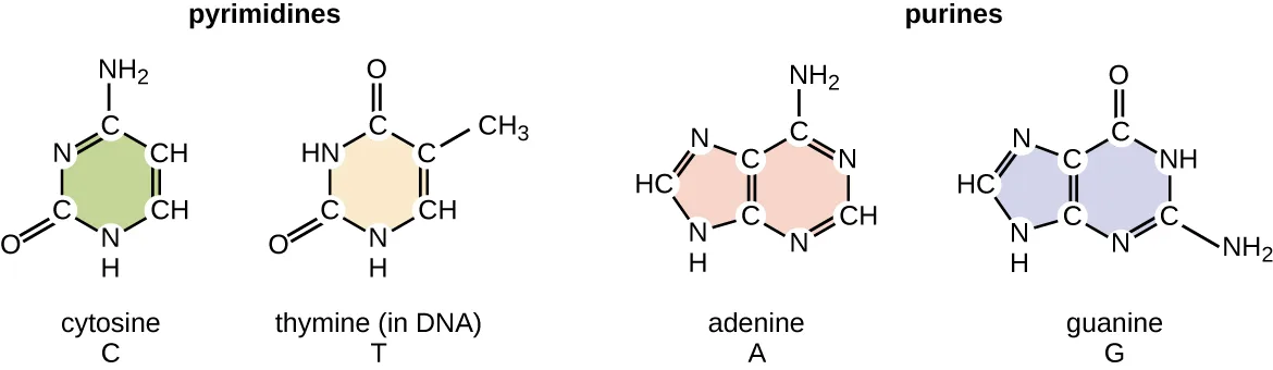
The deoxyribonucleotide is named according to the nitrogenous bases (the figure below). The nitrogenous bases adenine (A) and guanine (G) are the purines; they have a double-ring structure with a six-carbon ring fused to a five-carbon ring. The pyrimidines, cytosine (C) and thymine (T), are smaller nitrogenous bases that have only a six-carbon ring structure.
Individual nucleoside triphosphates combine with each other by covalent bonds known as 5′-3′ phosphodiester bonds, or linkages whereby the phosphate group attached to the 5′ carbon of the sugar of one nucleotide bonds to the hydroxyl group of the 3′ carbon of the sugar of the next nucleotide. Phosphodiester bonding between nucleotides forms the sugar-phosphate backbone, the alternating sugar-phosphate structure composing the framework of a nucleic acid strand (the figure below). During the polymerization process, deoxynucleotide triphosphates (dNTP) are used. To construct the sugar-phosphate backbone, the two terminal phosphates are released from the dNTP as a pyrophosphate. The resulting strand of nucleic acid has a free phosphate group at the 5′ carbon end and a free hydroxyl group at the 3′ carbon end. The two unused phosphate groups from the nucleotide triphosphate are released as pyrophosphate during phosphodiester bond formation. Pyrophosphate is subsequently hydrolyzed, releasing the energy used to drive nucleotide polymerization.
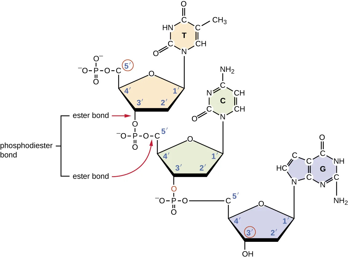
Extended description
Reading top to bottom, three nucleotides are linked by two phosphodiester bonds. The top nucleotide (thymine base) has its 5′-carbon phosphate joined to the 3′ carbon of the sugar below it (cytosine base); that nucleotide’s own 5′-carbon phosphate is in turn joined to the 3′ carbon of the sugar below it (guanine base), whose 3′ carbon is left as a free OH at the bottom of the strand. A bracket labeled ‘phosphodiester bond’ spans each of these two linkages, with red arrows pointing to the two ’ester bond’ labels — one at the phosphate’s join to the upper sugar’s 5′ carbon, one at its join to the lower sugar’s 3′ carbon — that together make up that phosphodiester bond.
Check Your Understanding
What is meant by the 5′ and 3′ ends of a nucleic acid strand?
Recall which of the two terminal groups — phosphate or hydroxyl — the strand carries at each numbered carbon end.Discovering the Double Helix
By the early 1950s, considerable evidence had accumulated indicating that DNA was the genetic material of cells, and now the race was on to discover its three-dimensional structure. Around this time, Austrian biochemist Erwin Chargaff (1905–2002) examined the content of DNA in different species and discovered that adenine, thymine, guanine, and cytosine were not found in equal quantities, and that it varied from species to species, but not between individuals of the same species. He found that the amount of adenine was very close to equaling the amount of thymine, and the amount of cytosine was very close to equaling the amount of guanine, or A = T and G = C. These relationships are also known as Chargaff’s rules. (N. Kresge et al., “Chargaff’s Rules: The Work of Erwin Chargaff,” Journal of Biological Chemistry 280 [2005]: e21.)
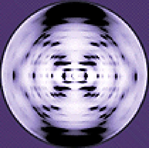
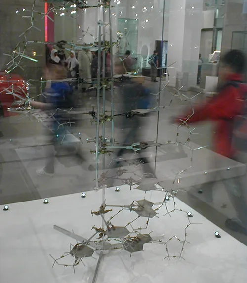
Other scientists were also actively exploring this field during the mid-20th century. In 1952, American scientist Linus Pauling (1901–1994) was the world’s leading structural chemist and odds-on favorite to solve the structure of DNA. Pauling had earlier discovered the structure of protein α helices, using X-ray diffraction, and, based upon X-ray diffraction images of DNA made in his laboratory, he proposed a triple-stranded model of DNA. (L. Pauling, “A Proposed Structure for the Nucleic Acids,” Proceedings of the National Academy of Sciences of the United States of America 39, no. 2 [1953]: 84–97.) At the same time, British researchers Rosalind Franklin (1920–1958) and her graduate student R.G. Gosling were also using X-ray diffraction to understand the structure of DNA (the figure below). It was Franklin’s scientific expertise that resulted in the production of more well-defined X-ray diffraction images of DNA that would clearly show the overall double-helix structure of DNA.
James Watson (1928–), an American scientist, and Francis Crick (1916–2004), a British scientist, were working together in the 1950s to discover DNA’s structure. They used Chargaff’s rules and Franklin and Wilkins’ X-ray diffraction images of DNA fibers to piece together the purine-pyrimidine pairing of the double helical DNA molecule (the figure below). In April 1953, Watson and Crick published their model of the DNA double helix in Nature. (J.D. Watson and F.H.C. Crick, “A Structure for Deoxyribose Nucleic Acid,” Nature 171, no. 4356 [1953]: 737–738.) The same issue additionally included papers by Wilkins and colleagues (M.H.F. Wilkins et al., “Molecular Structure of Deoxypentose Nucleic Acids,” Nature 171, no. 4356 [1953]: 738–740), as well as by Franklin and Gosling (R. Franklin and R.G. Gosling, “Molecular Configuration in Sodium Thymonucleate,” Nature 171, no. 4356 [1953]: 740–741), each describing different aspects of the molecular structure of DNA. In 1962, James Watson, Francis Crick, and Maurice Wilkins were awarded the Nobel Prize in Physiology and Medicine. Unfortunately, by then Franklin had died, and Nobel prizes at the time were not awarded posthumously. Work continued, however, on learning about the structure of DNA. In 1973, Alexander Rich (1924–2015) and colleagues were able to analyze DNA crystals to confirm and further elucidate DNA structure. (R.O. Day et al., “A Crystalline Fragment of the Double Helix: The Structure of the Dinucleoside Phosphate Guanylyl-3′,5′-Cytidine,” Proceedings of the National Academy of Sciences of the United States of America 70, no. 3 [1973]: 849–853.)
Check Your Understanding
Which scientists are given most of the credit for describing the molecular structure of DNA?
This section names several scientists whose work fed into the discovery; identify the pair who published the model and became famous for it.DNA Structure
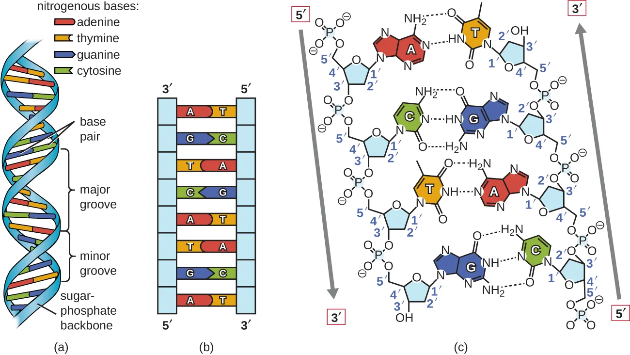
Watson and Crick proposed that DNA is made up of two strands that are twisted around each other to form a right-handed helix. The two DNA strands are antiparallel, such that the 3′ end of one strand faces the 5′ end of the other (the figure below). The 3′ end of each strand has a free hydroxyl group, while the 5′ end of each strand has a free phosphate group. The sugar and phosphate of the polymerized nucleotides form the backbone of the structure, whereas the nitrogenous bases are stacked inside. These nitrogenous bases on the interior of the molecule interact with each other, base pairing.
Analysis of the diffraction patterns of DNA has determined that there are approximately 10 bases per turn in DNA. The asymmetrical spacing of the sugar-phosphate backbones generates major grooves (where the backbone is far apart) and minor grooves (where the backbone is close together) (the figure below). These grooves are locations where proteins can bind to DNA. The binding of these proteins can alter the structure of DNA, regulate replication, or regulate transcription of DNA into RNA.
Extended description
Panel (a) shows the double helix as a twisted ladder: the sugar-phosphate backbones run along the outside as ribbons, and paired bases form the rungs, colored red for adenine, yellow for thymine, blue for guanine, and green for cytosine, with alternating wide major grooves and narrow minor grooves. Panel (b) untwists the same ladder into a flat rung diagram labeled by base letters, with the left strand’s 3′ end at top and 5′ end at bottom while the right strand’s 5′ end is at top and 3′ end at bottom, showing the antiparallel orientation. Panel (c) zooms into four rungs’ worth of chemical structure (an A-T pair, then a C-G pair, then a T-A pair, then a G-C pair, reading top to bottom), showing the sugar-phosphate backbone linking each nucleotide’s 3′ carbon to the next nucleotide’s 5′ phosphate, and dashed lines marking two hydrogen bonds in each A-T pair and three hydrogen bonds in each C-G pair.
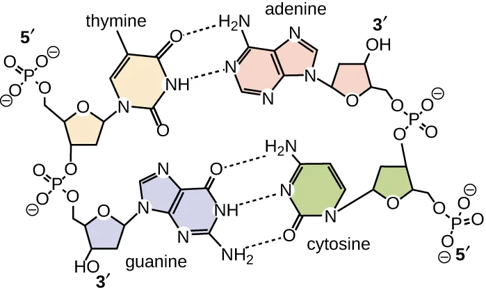
Base pairing takes place between a purine and pyrimidine. In DNA, adenine (A) and thymine (T) are complementary base pairs, and cytosine (C) and guanine (G) are also complementary base pairs, explaining Chargaff’s rules (the figure below). The base pairs are stabilized by hydrogen bonds; adenine and thymine form two hydrogen bonds between them, whereas cytosine and guanine form three hydrogen bonds between them.
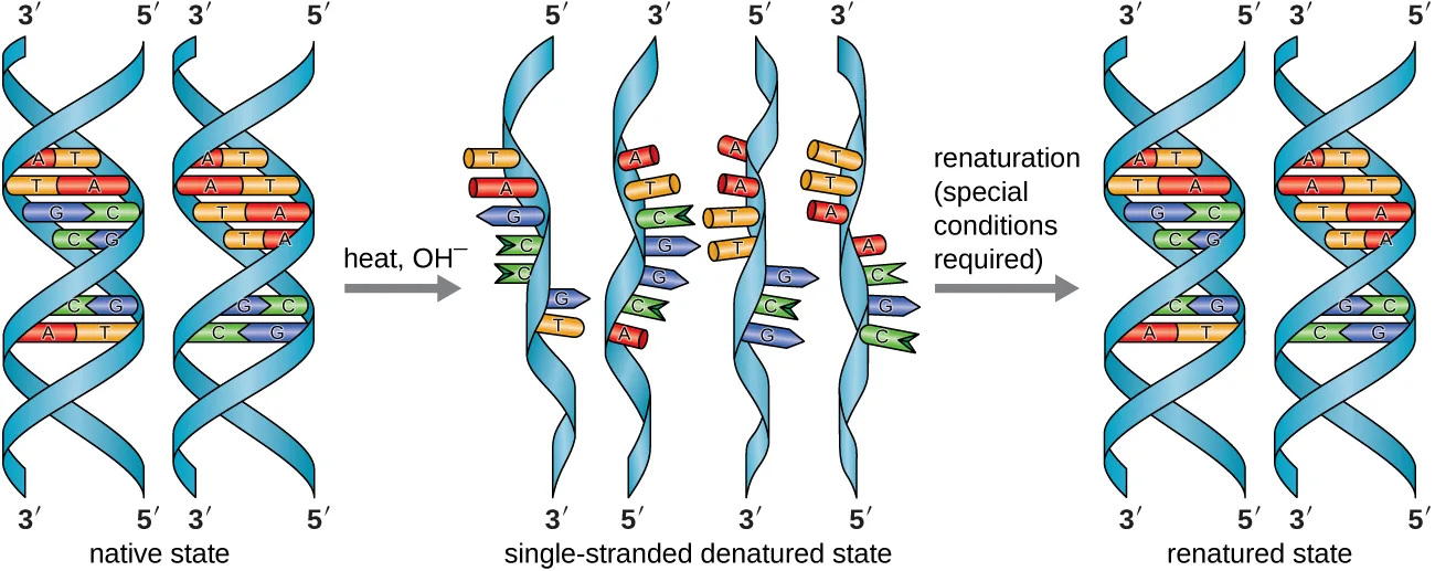
In the laboratory, exposing the two DNA strands of the double helix to high temperatures or to certain chemicals can break the hydrogen bonds between complementary bases, thus separating the strands into two separate single strands of DNA (single-stranded DNA [ssDNA]). This process is called DNA denaturation and is analogous to protein denaturation, as described in Proteins. The ssDNA strands can also be put back together as double-stranded DNA (dsDNA), through reannealing or renaturing by cooling or removing the chemical denaturants, allowing these hydrogen bonds to reform. The ability to artificially manipulate DNA in this way is the basis for several important techniques in biotechnology (the figure below). Because of the additional hydrogen bonding between the C = G base pair, DNA with a high GC content is more difficult to denature than DNA with a lower GC content.
Extended description
Reading left to right: the native state shows two separate double helices, each a twisted ladder with paired rungs (A-T and C-G) between two backbone ribbons, each strand end-labeled 3′ or 5′. An arrow labeled ‘heat, OH⁻’ points to the single-stranded denatured state, where the same strands appear separated into four individual ribbons, still end-labeled 3′ and 5′, with their bases projecting unpaired along each ribbon’s length. A second arrow labeled ‘renaturation (special conditions required)’ points to the renatured state, where the strands have reformed into two double helices with their original base pairs restored.
Link to Learning
View an animation on DNA structure from the DNA Learning Center to learn more.
Check Your Understanding
What are the two complementary base pairs of DNA and how are they bonded together?
Recall the two purine-pyrimidine pairings this section names as complementary, and the different hydrogen-bond counts given for each.DNA Function
DNA stores the information needed to build and control the cell. The transmission of this information from mother to daughter cells is called vertical gene transfer and it occurs through the process of DNA replication. DNA is replicated when a cell makes a duplicate copy of its DNA, then the cell divides, resulting in the correct distribution of one DNA copy to each resulting cell. DNA can also be enzymatically degraded and used as a source of nucleosides and nucleotides for the cell. Unlike other macromolecules, DNA does not serve a structural role in cells.
Check Your Understanding
How does DNA transmit genetic information to offspring?
Name the process by which a cell makes a duplicate copy of its DNA before dividing, so that each resulting cell receives a copy.Eye on Ethics. Paving the Way for Women in Science and Health Professions
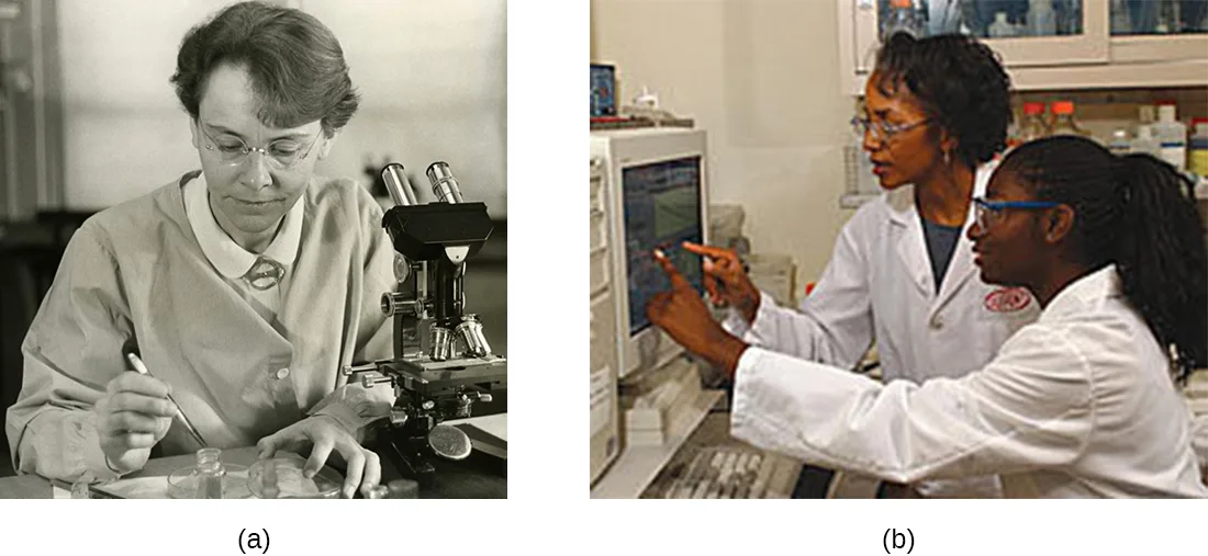
Historically, women have been underrepresented in the sciences and in medicine, and often their pioneering contributions have gone relatively unnoticed. For example, although Rosalind Franklin performed the X-ray diffraction studies demonstrating the double helical structure of DNA, it is Watson and Crick who became famous for this discovery, building on her data. There still remains great controversy over whether their acquisition of her data was appropriate and whether personality conflicts and gender bias contributed to the delayed recognition of her significant contributions. Similarly, Barbara McClintock did pioneering work in maize (corn) genetics from the 1930s through 1950s, discovering transposons (jumping genes), but she was not recognized until much later, receiving a Nobel Prize in Physiology or Medicine in 1983 (the figure below).
Today, women still remain underrepresented in many fields of science and medicine. While more than half of the undergraduate degrees in science are awarded to women, only 46% of doctoral degrees in science are awarded to women. In academia, the number of women at each level of career advancement continues to decrease, with women holding less than one-third of the positions of Ph.D.-level scientists in tenure-track positions, and less than one-quarter of the full professorships at 4-year colleges and universities (N.H. Wolfinger, “For Female Scientists, There’s No Good Time to Have Children,” The Atlantic, July 29, 2013). Even in the health professions, like nearly all other fields, women are often underrepresented in many medical careers and earn significantly less than their male counterparts, as shown in a 2013 study published by the Journal of the American Medical Association (S.A. Seabury et al., “Trends in the Earnings of Male and Female Health Care Professionals in the United States, 1987 to 2010,” Journal of the American Medical Association Internal Medicine 173, no. 18 [2013]: 1748–1750).
Why do such disparities continue to exist and how do we break these cycles? The situation is complex and likely results from the combination of various factors, including how society conditions the behaviors of girls from a young age and supports their interests, both professionally and personally. Some have suggested that women do not belong in the laboratory, including Nobel Prize winner Tim Hunt, whose 2015 public comments suggesting that women are too emotional for science (E. Chung, “Tim Hunt, Sexism and Science: The Real ‘Trouble With Girls’ in Labs,” CBC News Technology and Science, June 12, 2015) were met with widespread condemnation.
Perhaps girls should be supported more from a young age in the areas of science and math (the figure below). Science, technology, engineering, and mathematics (STEM) programs sponsored by the American Association of University Women (AAUW) (American Association of University Women, “Building a STEM Pipeline for Girls and Women”) and National Aeronautics and Space Administration (NASA) (National Aeronautics and Space Administration, “Outreach Programs: Women and Girls Initiative”) are excellent examples of programs that offer such support. Contributions by women in science should be made known more widely to the public, and marketing targeted to young girls should include more images of historically and professionally successful female scientists and medical professionals, encouraging all bright young minds, including girls and women, to pursue careers in science and medicine.
Clinical Focus. Part 2
Based upon his symptoms, Alex’s physician suspects that he is suffering from a foodborne illness that he acquired during his travels. Possibilities include bacterial infection (e.g., enterotoxigenic E. coli, Vibrio cholerae, Campylobacter jejuni, Salmonella), viral infection (rotavirus or norovirus), or protozoan infection (Giardia lamblia, Cryptosporidium parvum, or Entamoeba histolytica).
His physician orders a stool sample to identify possible causative agents (e.g., bacteria, cysts) and to look for the presence of blood because certain types of infectious agents (like C. jejuni, Salmonella, and E. histolytica) are associated with the production of bloody stools.
Alex’s stool sample showed neither blood nor cysts. Following analysis of his stool sample and based upon his recent travel history, the hospital physician suspected that Alex was suffering from traveler’s diarrhea caused by enterotoxigenic E. coli (ETEC), the causative agent of most traveler’s diarrhea. To verify the diagnosis and rule out other possibilities, Alex’s physician ordered a diagnostic lab test of his stool sample to look for DNA sequences encoding specific virulence factors of ETEC. The physician instructed Alex to drink lots of fluids to replace what he was losing and discharged him from the hospital.
ETEC produces several plasmid-encoded virulence factors that make it pathogenic compared with typical E. coli. These include the secreted toxins heat-labile enterotoxin (LT) and heat-stabile enterotoxin (ST), as well as colonization factor (CF). Both LT and ST cause the excretion of chloride ions from intestinal cells to the intestinal lumen, causing a consequent loss of water from intestinal cells, resulting in diarrhea. CF encodes a bacterial protein that aids in allowing the bacterium to adhere to the lining of the small intestine.
- Why did Alex’s physician use genetic analysis instead of either isolation of bacteria from the stool sample or direct Gram stain of the stool sample alone?
The case continues in Structure and Function of Cellular Genomes. The case began in Using Microbiology to Discover the Secrets of Life.
Summary
- Nucleic acids are composed of nucleotides, each of which contains a pentose sugar, a phosphate group, and a nitrogenous base. Deoxyribonucleotides within DNA contain deoxyribose as the pentose sugar.
- DNA contains the pyrimidines cytosine and thymine, and the purines adenine and guanine.
- Nucleotides are linked together by phosphodiester bonds between the 5′ phosphate group of one nucleotide and the 3′ hydroxyl group of another. A nucleic acid strand has a free phosphate group at the 5′ end and a free hydroxyl group at the 3′ end.
- Chargaff discovered that the amount of adenine is approximately equal to the amount of thymine in DNA, and that the amount of the guanine is approximately equal to cytosine. These relationships were later determined to be due to complementary base pairing.
- Watson and Crick, building on the work of Chargaff, Franklin and Gosling, and Wilkins, proposed the double helix model and base pairing for DNA structure.
- DNA is composed of two complementary strands oriented antiparallel to each other with the phosphodiester backbones on the exterior of the molecule. The nitrogenous bases of each strand face each other and complementary bases hydrogen bond to each other, stabilizing the double helix.
- Heat or chemicals can break the hydrogen bonds between complementary bases, denaturing DNA. Cooling or removing chemicals can lead to renaturation or reannealing of DNA by allowing hydrogen bonds to reform between complementary bases.
- DNA stores the instructions needed to build and control the cell. This information is transmitted from parent to offspring through vertical gene transfer.
Key terms
- nucleic acid — class of macromolecules composed of nucleotide monomers polymerized into strands.
- nucleotide — nucleic acid monomer composed of a pentose sugar, a phosphate group, and a nitrogenous base.
- base sequence — identity of the specific nucleotides present in a nucleic acid strand and their order within the strand.
- deoxyribonucleic acid (DNA) — double-stranded nucleic acid composed of deoxyribonucleotides that serves as the genetic material of the cell.
- deoxyribonucleotides — DNA nucleotides containing deoxyribose as the pentose sugar component.
- nitrogenous base — nitrogen-containing ring structure within a nucleotide that is responsible for complementary base pairing between nucleic acid strands.
- adenine — purine nitrogenous base found in nucleotides.
- guanine — purine nitrogenous base found in nucleotides.
- purines — nitrogenous bases containing a double-ring structure with a six-carbon ring fused to a five-carbon ring; includes adenine and guanine.
- pyrimidines — nitrogenous bases containing a single six-carbon ring; includes cytosine and thymine in DNA.
- cytosine — pyrimidine nitrogenous base found in nucleotides.
- thymine — pyrimidine nitrogenous base found only in DNA nucleotides.
- phosphodiester bonds — linkage whereby the phosphate group attached to the 5′ carbon of the sugar of one nucleotide bonds to the hydroxyl group of the 3′ carbon of the sugar of the next nucleotide.
- sugar-phosphate backbone — alternating sugar-phosphate structure composing the framework of a nucleic acid strand that results from phosphodiester bond formation between nucleotides.
- antiparallel — two strands of DNA helix oriented in opposite directions; one strand is oriented in the 5′ to 3′ direction, while the other is oriented in the 3′ to 5′ direction.
- complementary base pairs — base pairing due to hydrogen bonding that occurs between a specific purine and a specific pyrimidine; A bonds with T (in DNA), and C bonds with G.
- vertical gene transfer — transfer of genes from parent to offspring.
Practice
Describe the biochemical structure of deoxyribonucleotides
Which of the following is not found within DNA?
Recall the four kinds of molecules DNA is actually built from and stabilized by, and identify the one that belongs to proteins instead.The end of a nucleic acid strand with a free phosphate group is called the ________.
Recall which numbered carbon of the terminal sugar carries the unattached phosphate group, and how that end of the strand is named.What is the role of phosphodiester bonds within the sugar-phosphate backbone of DNA?
This bond links successive sugars through their 5′ phosphate and 3′ hydroxyl groups; think about what structure that linkage builds, not what holds the two strands’ bases together.Identify the base pairs used in the synthesis of deoxyribonucleotides
If 30% of the bases within a DNA molecule are adenine, what is the percentage of thymine?
Apply the equality between adenine and thymine that Chargaff discovered.Which of the following statements about base pairing in DNA is incorrect?
Recall which two bases actually pair together, and check each statement about hydrogen bonds and helix geometry against that pairing.During denaturation of DNA, which of the following happens?
Consider which of the two bond types in DNA — the ones stacking bases together or the ones lining the backbone — is broken by heat or chemicals.The A-T base pair has more hydrogen bonding than the C-G base pair.
Compare the hydrogen-bond counts given for each base pair.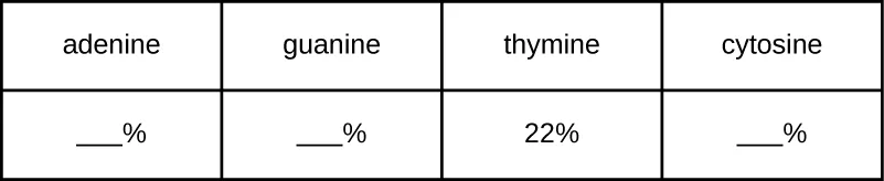
A certain DNA sample is found to have a makeup consisting of 22% thymine. Use Chargaff’s rules to fill in the percentages for the other three nitrogenous bases.
Use A = T and G = C to complete the remaining three percentages so all four sum to 100%.Why is DNA with a high GC content more difficult to denature than that with a low GC content?
Recall which base pair this section says has additional hydrogen bonding, and what that additional bonding does to how easily the strands separate.Explain why the double helix of DNA is described as antiparallel
If a DNA strand contains the sequence 5′-ATTCCGGATCGA-3′, which of the following is the sequence of the complementary strand of DNA?
Remember that the complementary strand is written in the opposite direction from the original, not the same direction.The work of Rosalind Franklin and R.G. Gosling was important in demonstrating the helical nature of DNA.
Recall whose X-ray diffraction images demonstrated DNA’s helical shape.What is meant by the term antiparallel?
Recall which end of each strand faces which end of its partner strand.In considering the structure of the DNA double helix, how would you expect the structure to differ if there was base pairing between two purines? Between two pyrimidines?
Show model answer
Did your answer mention:
This section is adapted from Microbiology, Section 10.2: Structure and Function of DNA by Nina Parker, Mark Schneegurt, Anh-Hue Thi Tu, Philip Lister, Brian M. Forster, and OpenStax, © OpenStax, licensed under CC BY-NC-SA 4.0. Access the original for free at openstax.org. Changes: all ten figures (the module’s nine body figures plus the Chargaff Art Connection exercise image) re-encoded as WebP; the manifest’s photo guess is overridden to kind="diagram" for the Nucleotide, Bases, NAcidStrand, DoubHelix, BasePairs, Denatured, and Chargaff Art Connection figures (all drawn structures or a rendered table, not photographs) and kept as kind="photo" for the X-ray diffraction pattern, the wire-model museum photo, and the two Eye on Ethics photographs; every alt was rewritten from the image itself rather than the source alt’s screen-reader phrasing (“1-prime”, “B)” for a lettered panel), and a longdesc was added for the NAcidStrand, DoubHelix, and Denatured process figures to walk their labelled steps in reading order. The module’s eleven footnotes (six body journal citations — Kresge 2005, Pauling 1953, Watson and Crick 1953, Wilkins et al. 1953, Franklin and Gosling 1953, Day et al. 1973 — and five web-sourced citations inside the Eye on Ethics — Wolfinger/The Atlantic, Seabury/JAMA, Chung/CBC News, AAUW, NASA) are rendered as inline parenthetical citations after the sentences they support, with the Eye on Ethics citations’ bare access URLs and “Accessed” dates dropped. The Pauling footnote’s journal name is corrected from “Proceedings of the National Academy of Science” (the source’s one-word typo, missing the plural) to “Proceedings of the National Academy of Sciences,” logged as an erratum; the “Journal of the American Medical Association Internal Medicine” citation and the “heat-stabile” spelling are transcribed as printed (defensible on the source’s own terms). Primes are normalized to U+2032 (′) throughout, including in the two title primes of the Day et al. citation; the source’s FIB key “5ʹ end” (using the modifier letter prime, U+02B9) is rendered as the textin answer “5′ end” with accept="5 prime end|five prime end", since the grader folds the curly and straight apostrophes onto U+2032 but not U+02B9. The Link to Learning, Eye on Ethics, and Clinical Focus boxes are rendered as callouts in source order; the Clinical Focus box’s “Jump to the next / go back to the previous Clinical Focus box” links are replaced by a sentence naming where the case continues, Structure and Function of Cellular Genomes, and where it began, Using Microbiology to Discover the Secrets of Life; its closing question stays inside the callout as an unanswered plain bullet. All four body Check Your Understanding bullets are graded body items, since each is fixed by one sentence or passage of this section: three multiplechoice (the 5′/3′ ends; the scientists credited for the double-helix model; the hydrogen-bond counts of the two complementary base pairs) and one textin keeping the source’s own stem verbatim (“How does DNA transmit genetic information to offspring?”), answer “DNA replication” with accept="replication", fixed by the sentence “it occurs through the process of DNA replication”; a page-wide grep confirmed no other stem or hint prints “replication” ahead of this item. Of the source’s unkeyed exercises, all three Short Answer questions and one of the two Critical Thinking questions are graded multiplechoice from this module’s own sentences and figure — the phosphodiester-bond role, the antiparallel definition, the GC-content/denaturation reasoning (keyed by the single sentence “Because of the additional hydrogen bonding between the C = G base pair, DNA with a high GC content is more difficult to denature than DNA with a lower GC content,” without relying on the separate two-versus-three hydrogen-bond count given earlier), and the Chargaff percentage table (mediafigure, keyed by the Chargaff’s-rules sentence, distractors built from the module’s own bases in other, rule-violating combinations); the source prints no answer key for any of these five. The remaining Critical Thinking question (purine-purine and pyrimidine-pyrimidine base pairing) stays a selfcheck: it asks the learner to reason about a pairing the module never describes, so its model answer states only what the module gives — the ring-size difference between purines and pyrimidines — and stops there. All 13 of the module’s source exercises (5 Multiple Choice, 2 True/False as two-option multiple choice, 1 Fill in the Blank as textin, 3 Short Answer, and 2 Critical Thinking) are adapted into the Practice block; none is omitted. Key terms are compiled from the module’s 17 defined terms, all 17 definitions taken directly from the book’s Glossary appendix (none sentence-derived). Same-module figure and table cross-references are rendered as describing prose (“the figure below”); cross-references to other modules are absolute site-root links (Microbial Metabolism, Mechanisms of Microbial Genetics, Proteins).