DNA Replication
By the end of this section, you will be able to:
- Explain the meaning of semiconservative DNA replication
- Explain why DNA replication is bidirectional and includes both a leading and lagging strand
- Explain why Okazaki fragments are formed
- Describe the process of DNA replication and the functions of the enzymes involved
- Identify the differences between DNA replication in bacteria and eukaryotes
- Explain the process of rolling circle replication
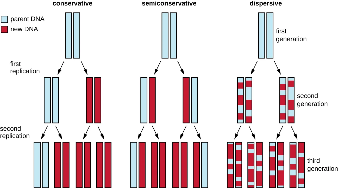
The elucidation of the structure of the double helix by James Watson and Francis Crick in 1953 provided a hint as to how DNA is copied during the process of replication. Separating the strands of the double helix would provide two templates for the synthesis of new complementary strands, but exactly how new DNA molecules were constructed was still unclear. In one model, semiconservative replication, the two strands of the double helix separate during DNA replication, and each strand serves as a template from which the new complementary strand is copied; after replication, each double-stranded DNA includes one parental or “old” strand and one “new” strand. There were two competing models also suggested: conservative and dispersive, which are shown in the figure below.
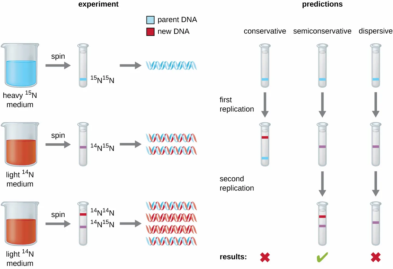
Matthew Meselson (1930–) and Franklin Stahl (1929–) devised an experiment in 1958 to test which of these models correctly represents DNA replication (shown below). They grew E. coli for several generations in a medium containing a “heavy” isotope of nitrogen (¹⁵N) that was incorporated into nitrogenous bases and, eventually, into the DNA. This labeled the parental DNA. The E. coli culture was then shifted into a medium containing ¹⁴N and allowed to grow for one generation. The cells were harvested and the DNA was isolated. The DNA was separated by ultracentrifugation, during which the DNA formed bands according to its density. DNA grown in ¹⁵N would be expected to form a band at a higher density position than that grown in ¹⁴N. Meselson and Stahl noted that after one generation of growth in ¹⁴N, the single band observed was intermediate in position in between DNA of cells grown exclusively in ¹⁵N or ¹⁴N. This suggested either a semiconservative or dispersive mode of replication. Some cells were allowed to grow for one more generation in ¹⁴N and spun again. The DNA harvested from cells grown for two generations in ¹⁴N formed two bands: one DNA band was at the intermediate position between ¹⁵N and ¹⁴N, and the other corresponded to the band of ¹⁴N DNA. These results could only be explained if DNA replicates in a semiconservative manner. If DNA replication was dispersive, a single purple band positioned closer to the red ¹⁴N band would have been observed, as more ¹⁴N was added in a dispersive manner to replace ¹⁵N. Therefore, the other two models were ruled out. As a result of this experiment, we now know that during DNA replication, each of the two strands that make up the double helix serves as a template from which new strands are copied. The new strand will be complementary to the parental or “old” strand. The resulting DNA molecules have the same sequence and are divided equally into the two daughter cells.
Extended description
Left panel, top to bottom: a beaker of heavy ¹⁵N medium is spun, giving a single low (heavy) band labeled ¹⁵N¹⁵N, which resolves into all-blue double helices; a beaker of light ¹⁴N medium is spun, giving a single intermediate band labeled ¹⁴N¹⁵N, which resolves into helices with one blue and one red strand; a second round in ¹⁴N medium gives two bands, a light band labeled ¹⁴N¹⁴N (all-red helices) and an intermediate band labeled ¹⁴N¹⁵N (mixed helices). Right panel: a predictions table with three columns, conservative, semiconservative, and dispersive. All three start with one heavy (blue) band. After the first replication, conservative predicts one heavy and one light band, while semiconservative and dispersive both predict a single intermediate (purple) band. After the second replication, only the semiconservative column is shown continuing: it predicts one light and one intermediate band. Below the tubes, the results row marks conservative with a red X, semiconservative with a green check mark, and dispersive with a red X.
Check Your Understanding
What would have been the conclusion of Meselson and Stahl’s experiment if, after the first generation, they had found two bands of DNA — one heavy and one light — instead of a single intermediate band?
Compare the three columns of the predictions figure after the first round of replication — which one draws two separate bands rather than a single merged band?DNA Replication in Bacteria
DNA replication has been well studied in bacteria primarily because of the small size of the genome and the mutants that are available. E. coli has 4.6 million base pairs (Mbp) in a single circular chromosome and all of it is replicated in approximately 42 minutes, starting from a single origin of replication and proceeding around the circle bidirectionally. This means that approximately 1000 nucleotides are added per second. The process is quite rapid and occurs with few errors.
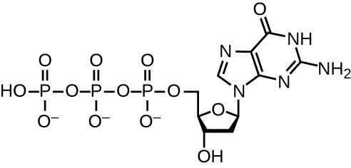
DNA replication uses a large number of proteins and enzymes, listed in the table below. One of the key players is the enzyme DNA polymerase, also known as DNA pol. In bacteria, three main types of DNA polymerases are known: DNA pol I, DNA pol II, and DNA pol III. It is now known that DNA pol III is the enzyme required for DNA synthesis; DNA pol I and DNA pol II are primarily required for repair. DNA pol III adds deoxyribonucleotides each complementary to a nucleotide on the template strand, one by one to the 3′-OH group of the growing DNA chain. The addition of these nucleotides requires energy. This energy is present in the bonds of three phosphate groups attached to each nucleotide (a triphosphate nucleotide), similar to how energy is stored in the phosphate bonds of adenosine triphosphate (ATP), shown below for one such nucleotide. When the bond between the phosphates is broken and diphosphate is released, the energy released allows for the formation of a covalent phosphodiester bond by dehydration synthesis between the incoming nucleotide and the free 3′-OH group on the growing DNA strand.
Initiation
The initiation of replication occurs at specific nucleotide sequence called the origin of replication, where various proteins bind to begin the replication process. E. coli has a single origin of replication (as do most prokaryotes), called oriC, on its one chromosome. The origin of replication is approximately 245 base pairs long and is rich in adenine-thymine (AT) sequences.
Some of the proteins that bind to the origin of replication are important in making single-stranded regions of DNA accessible for replication. Chromosomal DNA is typically wrapped around histones (in eukaryotes and archaea) or histone-like proteins (in bacteria), and is supercoiled, or extensively wrapped and twisted on itself. This packaging makes the information in the DNA molecule inaccessible. However, enzymes called topoisomerases change the shape and supercoiling of the chromosome. For bacterial DNA replication to begin, the supercoiled chromosome is relaxed by topoisomerase II, also called DNA gyrase. An enzyme called helicase then separates the DNA strands by breaking the hydrogen bonds between the nitrogenous base pairs. Recall that AT sequences have fewer hydrogen bonds and, hence, have weaker interactions than guanine-cytosine (GC) sequences. These enzymes require ATP hydrolysis. As the DNA opens up, Y-shaped structures called replication forks are formed. Two replication forks are formed at the origin of replication, allowing for bidirectional replication and formation of a structure that looks like a bubble when viewed with a transmission electron microscope; as a result, this structure is called a replication bubble. The DNA near each replication fork is coated with single-stranded binding proteins to prevent the single-stranded DNA from rewinding into a double helix.
Once single-stranded DNA is accessible at the origin of replication, DNA replication can begin. However, DNA pol III is able to add nucleotides only in the 5′ to 3′ direction (a new DNA strand can be only extended in this direction). This is because DNA polymerase requires a free 3′-OH group to which it can add nucleotides by forming a covalent phosphodiester bond between the 3′-OH end and the 5′ phosphate of the next nucleotide. This also means that it cannot add nucleotides if a free 3′-OH group is not available, which is the case for a single strand of DNA. The problem is solved with the help of an RNA sequence that provides the free 3′-OH end. Because this sequence allows the start of DNA synthesis, it is appropriately called the primer. The primer is five to 10 nucleotides long and complementary to the parental or template DNA. It is synthesized by RNA primase, which is an RNA polymerase. Unlike DNA polymerases, RNA polymerases do not need a free 3′-OH group to synthesize an RNA molecule. Now that the primer provides the free 3′-OH group, DNA polymerase III can now extend this RNA primer, adding DNA nucleotides one by one that are complementary to the template strand, as shown in the three-model diagram above.
Elongation
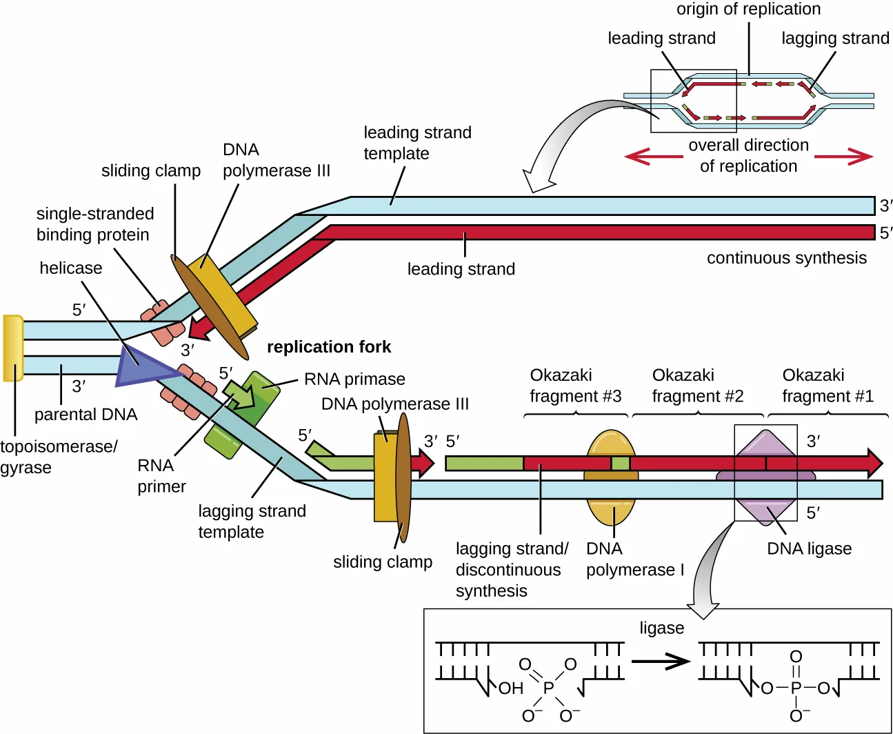
During elongation in DNA replication, the addition of nucleotides occurs at its maximal rate of about 1000 nucleotides per second. DNA polymerase III can only extend in the 5′ to 3′ direction, which poses a problem at the replication fork. The DNA double helix is antiparallel; that is, one strand is oriented in the 5′ to 3′ direction and the other is oriented in the 3′ to 5′ direction (see Structure and Function of DNA). During replication, one strand, which is complementary to the 3′ to 5′ parental DNA strand, is synthesized continuously toward the replication fork because polymerase can add nucleotides in this direction. This continuously synthesized strand is known as the leading strand. The other strand, complementary to the 5′ to 3′ parental DNA, grows away from the replication fork, so the polymerase must move back toward the replication fork to begin adding bases to a new primer, again in the direction away from the replication fork. It does so until it bumps into the previously synthesized strand and then it moves back again, as shown in the replication-fork diagram below. These steps produce small DNA sequence fragments known as Okazaki fragments, each separated by RNA primer. Okazaki fragments are named after the Japanese research team and married couple Reiji and Tsuneko Okazaki, who first discovered them in 1966. The strand with the Okazaki fragments is known as the lagging strand, and its synthesis is said to be discontinuous.
The leading strand can be extended from one primer alone, whereas the lagging strand needs a new primer for each of the short Okazaki fragments. The overall direction of the lagging strand will be 3′ to 5′, and that of the leading strand 5′ to 3′. A protein called the sliding clamp holds the DNA polymerase in place as it continues to add nucleotides. The sliding clamp is a ring-shaped protein that binds to the DNA and holds the polymerase in place. Beyond its role in initiation, topoisomerase also prevents the overwinding of the DNA double helix ahead of the replication fork as the DNA is opening up; it does so by causing temporary nicks in the DNA helix and then resealing it. As synthesis proceeds, the RNA primers are replaced by DNA. The primers are removed by the exonuclease activity of DNA polymerase I, and the gaps are filled in. The nicks that remain between the newly synthesized DNA (that replaced the RNA primer) and the previously synthesized DNA are sealed by the enzyme DNA ligase that catalyzes the formation of covalent phosphodiester linkage between the 3′-OH end of one DNA fragment and the 5′ phosphate end of the other fragment, stabilizing the sugar-phosphate backbone of the DNA molecule.
Extended description
Inset (top right): a replication bubble with the origin of replication at its center; on the top strand a solid arrow points left from the origin (leading strand) and short arrows point left on the other side (lagging strand); on the bottom strand a solid arrow points right from the origin (leading strand) and short arrows point right on the other side (lagging strand); flanking arrows read ‘overall direction of replication’, pointing outward both ways. Main diagram (left half of the bubble, left to right): parental DNA enters from the left, still double-stranded and labeled 5′ (top) and 3′ (bottom); topoisomerase/gyrase sits at the far left; helicase, drawn as a wedge, splits the strands; single-stranded binding proteins coat both single strands just past helicase. On the upper (leading-strand template) branch, DNA polymerase III and a sliding clamp sit where the template reads 3′ (right) to 5′ (left), synthesizing the leading strand continuously toward the right, ending 5′ at the right and 3′ at the fork; a label reads ‘continuous synthesis’. On the lower (lagging-strand template) branch, RNA primase attaches a green RNA primer near the fork; further right, DNA polymerase III and a sliding clamp elongate a primed segment 5′ to 3′ (left to right); further right still, three labeled segments — Okazaki fragment #3, #2, and #1, read right to left in order of synthesis — each begin with a green RNA primer joined to red new DNA; DNA polymerase I sits on Okazaki fragment #2’s primer, replacing it with DNA; DNA ligase sits between Okazaki fragments #1 and #2, sealing the nick. A boxed close-up below shows ligase joining a fragment’s free 3′-OH to the next fragment’s 5′ phosphate, releasing a phosphate group.
Termination
Once the complete chromosome has been replicated, termination of DNA replication must occur. Although much is known about initiation of replication, less is known about the termination process. Following replication, the resulting complete circular genomes of prokaryotes are concatenated, meaning that the circular DNA chromosomes are interlocked and must be separated from each other. This is accomplished through the activity of bacterial topoisomerase IV, which introduces double-stranded breaks into DNA molecules, allowing them to separate from each other; the enzyme then reseals the circular chromosomes. The resolution of concatemers is an issue unique to prokaryotic DNA replication because of their circular chromosomes. Because both bacterial DNA gyrase and topoisomerase IV are distinct from their eukaryotic counterparts, these enzymes serve as targets for a class of antimicrobial drugs called quinolones.
| Enzyme or Factor | Function |
|---|---|
| DNA pol I | Exonuclease activity removes RNA primer and replaces it with newly synthesized DNA |
| DNA pol III | Main enzyme that adds nucleotides in the 5′ to 3′ direction |
| Helicase | Opens the DNA helix by breaking hydrogen bonds between the nitrogenous bases |
| Ligase | Seals the gaps between the Okazaki fragments on the lagging strand to create one continuous DNA strand |
| Primase | Synthesizes RNA primers needed to start replication |
| Single-stranded binding proteins | Bind to single-stranded DNA to prevent hydrogen bonding between DNA strands, reforming double-stranded DNA |
| Sliding clamp | Helps hold DNA pol III in place when nucleotides are being added |
| Topoisomerase II (DNA gyrase) | Relaxes supercoiled chromosome to make DNA more accessible for the initiation of replication; helps relieve the stress on DNA when unwinding, by causing breaks and then resealing the DNA |
| Topoisomerase IV | Introduces double-stranded breaks into concatenated chromosomes to release them from each other, and then reseals the DNA |
(Source note: the source’s table says topoisomerase IV introduces a “single-stranded break”; the module’s own Termination paragraph above says it “introduces double-stranded breaks,” and type II topoisomerases act through transient double-strand breaks (Levine, Hiasa, and Marians, Biochimica et Biophysica Acta 1400 (1998): 29–43), so the table row follows the paragraph.)
The Molecular Machinery Involved in Bacterial DNA Replication
Check Your Understanding
The enzyme that breaks the hydrogen bonds holding the two strands of DNA together so that replication can occur is ________.
Name the enzyme that separates the DNA strands right after the supercoiled chromosome is relaxed.Is it the lagging strand or the leading strand that is synthesized in the direction toward the opening of the replication fork?
Recall which strand’s polymerase moves continuously forward, following the fork as it opens, rather than looping back for each new fragment.The enzyme responsible for removing the RNA primers in newly replicated bacterial DNA is ________.
Name the polymerase whose exonuclease activity removes primers and fills the resulting gaps, distinct from the polymerase that does most of the elongating.DNA Replication in Eukaryotes
Eukaryotic genomes are much more complex and larger than prokaryotic genomes and are typically composed of multiple linear chromosomes, compared in the table below. The human genome, for example, has 3 billion base pairs per haploid set of chromosomes, and 6 billion base pairs are inserted during replication. There are multiple origins of replication on each eukaryotic chromosome, shown below; the human genome has 30,000 to 50,000 origins of replication. The rate of replication is approximately 100 nucleotides per second—10 times slower than prokaryotic replication.
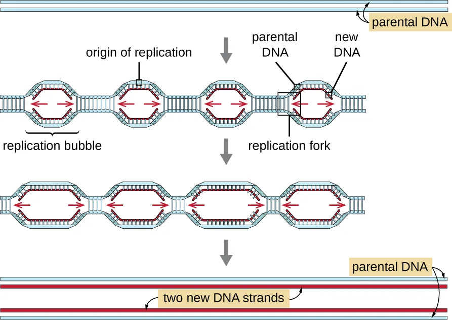
The essential steps of replication in eukaryotes are the same as in prokaryotes. Before replication can start, the DNA has to be made available as a template. Eukaryotic DNA is highly supercoiled and packaged, which is facilitated by many proteins, including histones (see Structure and Function of Cellular Genomes). At the origin of replication, a prereplication complex composed of several proteins, including helicase, forms and recruits other enzymes involved in the initiation of replication, including topoisomerase to relax supercoiling, single-stranded binding protein, RNA primase, and DNA polymerase. Following initiation of replication, in a process similar to that found in prokaryotes, elongation is facilitated by eukaryotic DNA polymerases. The leading strand is continuously synthesized by the eukaryotic polymerase enzyme pol ε, while the lagging strand is synthesized by pol δ. (Source note: the source assigns pol δ to the leading strand and pol ε to the lagging strand. Genetic and biochemical evidence places pol ε on the leading strand and pol δ on the lagging strand (Pursell et al., Science 317 (2007): 127–130; Johansson and Dixon, Cold Spring Harbor Perspectives in Biology 5 (2013): a012799), so this page swaps the two.) A sliding clamp protein holds the DNA polymerase in place so that it does not fall off the DNA. The enzyme ribonuclease H (RNase H), instead of a DNA polymerase as in bacteria, removes the RNA primer, which is then replaced with DNA nucleotides. The gaps that remain are sealed by DNA ligase.
Because eukaryotic chromosomes are linear, one might expect that their replication would be more straightforward. As in prokaryotes, the eukaryotic DNA polymerase can add nucleotides only in the 5′ to 3′ direction. In the leading strand, synthesis continues until it reaches either the end of the chromosome or another replication fork progressing in the opposite direction. On the lagging strand, DNA is synthesized in short stretches, each of which is initiated by a separate primer. When the replication fork reaches the end of the linear chromosome, there is no place to make a primer for the DNA fragment to be copied at the end of the chromosome. These ends thus remain unpaired and, over time, they may get progressively shorter as cells continue to divide.
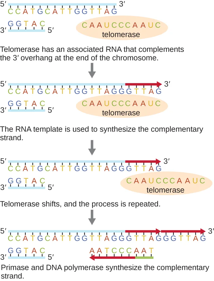
The ends of the linear chromosomes are known as telomeres and consist of noncoding repetitive sequences. The telomeres protect coding sequences from being lost as cells continue to divide. In humans, a six base-pair sequence, TTAGGG, is repeated 100 to 1000 times to form the telomere. The discovery of the enzyme telomerase, shown below, clarified our understanding of how chromosome ends are maintained. Telomerase contains a catalytic part and a built-in RNA template. It attaches to the end of the chromosome, and complementary bases to the RNA template are added on the 3′ end of the DNA strand. Once the 3′ end of the lagging strand template is sufficiently elongated, DNA polymerase can add the nucleotides complementary to the ends of the chromosomes. In this way, the ends of the chromosomes are replicated. In humans, telomerase is typically active in germ cells and adult stem cells; it is not active in adult somatic cells and may be associated with the aging of these cells. Eukaryotic microbes including fungi and protozoans also produce telomerase to maintain chromosomal integrity. For her discovery of telomerase and its action, Elizabeth Blackburn (1948–) received the Nobel Prize for Medicine or Physiology in 2009.
Extended description
Four stacked panels, each showing a long top DNA strand (5′ left, 3′ right) reading 5′-CCATGCATTGGTTAG-3′ and a much shorter complementary bottom strand reading 3′-GGTAC-5′ beneath its left end, leaving a single-stranded 3′ overhang on the top strand. Panel 1: an oval labeled telomerase, containing the RNA sequence CAAUCCCAAUC, base-pairs with the overhang, its RNA extending past the strand’s 3′ end. Panel 2: using that RNA as template, the top strand is extended (shown in a second color) to 5′-CCATGCATTGGTTAGGGTTAG-3′, telomerase still attached at the new 3′ end. Panel 3: telomerase has shifted rightward to the new 3′ end, ready to repeat the extension. Panel 4: the top strand carries two such extensions; a green RNA primer has bound near its new 3′ end, and a red arrow shows a new complementary strand being synthesized from that primer back toward the original short strand.
| Property | Bacteria | Eukaryotes |
|---|---|---|
| Genome structure | Single circular chromosome | Multiple linear chromosomes |
| Number of origins per chromosome | Single | Multiple |
| Rate of replication | 1000 nucleotides per second | 100 nucleotides per second |
| Telomerase | Not present | Present |
| RNA primer removal | DNA pol I | RNase H |
| Strand elongation | DNA pol III | pol δ, pol ε |
Comparison of Bacterial and Eukaryotic Replication
Link to Learning
This animation compares the process of prokaryotic and eukaryotic DNA replication.
Check Your Understanding
How does the origin of replication differ between eukaryotes and prokaryotes?
Compare the Number of origins per chromosome row of the comparison table above.What polymerase enzymes are responsible for DNA synthesis during eukaryotic replication?
Name the two eukaryotic polymerases that elongate the leading and lagging strands — different names from the bacterial enzyme that does both.What is found at the ends of the chromosomes in eukaryotes and why?
Show model answer
Did your answer mention:
DNA Replication of Extrachromosomal Elements: Plasmids and Viruses
To copy their nucleic acids, plasmids and viruses frequently use variations on the pattern of DNA replication described for prokaryote genomes. For more information on the wide range of viral replication strategies, see The Viral Life Cycle.
Rolling Circle Replication
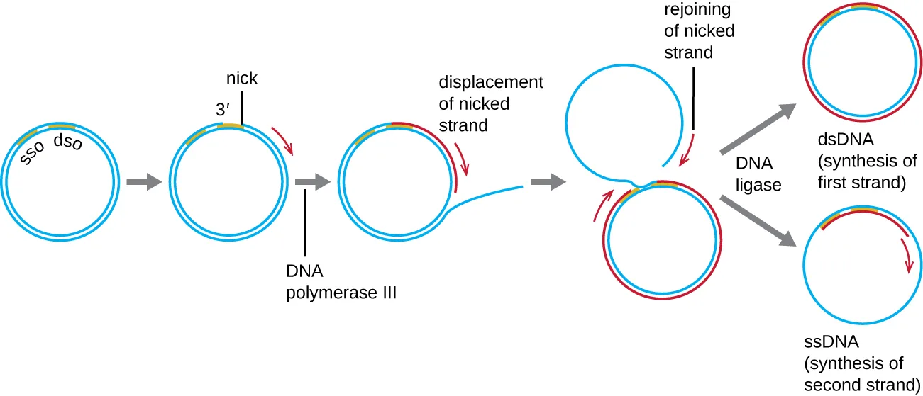
Whereas many bacterial plasmids (see Unique Characteristics of Prokaryotic Cells) replicate by a process similar to that used to copy the bacterial chromosome, other plasmids, several bacteriophages, and some viruses of eukaryotes use rolling circle replication, shown below. The circular nature of plasmids and the circularization of some viral genomes on infection make this possible. Rolling circle replication begins with the enzymatic nicking of one strand of the double-stranded circular molecule at the double-stranded origin (dso) site. In bacteria, DNA polymerase III binds to the 3′-OH group of the nicked strand and begins to unidirectionally replicate the DNA using the un-nicked strand as a template, displacing the nicked strand as it does so. Completion of DNA replication at the site of the original nick results in full displacement of the nicked strand, which may then recircularize into a single-stranded DNA molecule. RNA primase then synthesizes a primer to initiate DNA replication at the single-stranded origin (sso) site of the single-stranded DNA (ssDNA) molecule, resulting in a double-stranded DNA (dsDNA) molecule identical to the other circular DNA molecule.
Extended description
Five stages, left to right. (1) A double-stranded circle with a short sso segment next to a short dso segment. (2) A nick appears at the 3′ end of the dso, with an arrow showing the direction of unwinding. (3) DNA polymerase III, labeled beneath the circle, copies the un-nicked strand clockwise around the circle while displacing the nicked strand, which now trails off the circle as a growing single-stranded loop. (4) The displaced loop has grown until it is nearly as large as the original circle, still attached at the nick site, labeled ‘displacement of nicked strand’; an arrow labeled ‘rejoining of nicked strand’ shows its two ends coming together. (5) Two outcomes are shown side by side after DNA ligase acts: a complete double-stranded circle labeled ‘dsDNA (synthesis of first strand)’, and a single-stranded circle labeled ‘ssDNA (synthesis of second strand)’, with an arrow around it showing where the second strand is subsequently synthesized.
Check Your Understanding
Is there a lagging strand in rolling circle replication? Why or why not?
Show model answer
Did your answer mention:
Summary
- The DNA replication process is semiconservative, which results in two DNA molecules, each having one parental strand of DNA and one newly synthesized strand.
- In bacteria, the initiation of replication occurs at the origin of replication, where supercoiled DNA is unwound by DNA gyrase, made single-stranded by helicase, and bound by single-stranded binding protein to maintain its single-stranded state. Primase synthesizes a short RNA primer, providing a free 3′-OH group to which DNA polymerase III can add DNA nucleotides.
- During elongation, the leading strand of DNA is synthesized continuously from a single primer. The lagging strand is synthesized discontinuously in short Okazaki fragments, each requiring its own primer. The RNA primers are removed and replaced with DNA nucleotides by bacterial DNA polymerase I, and DNA ligase seals the gaps between these fragments.
- Termination of replication in bacteria involves the resolution of circular DNA concatemers by topoisomerase IV to release the two copies of the circular chromosome.
- Eukaryotes typically have multiple linear chromosomes, each with multiple origins of replication. Overall, replication in eukaryotes is similar to that in prokaryotes.
- The linear nature of eukaryotic chromosomes necessitates telomeres to protect genes near the end of the chromosomes. Telomerase extends telomeres, preventing their degradation, in some cell types.
- Rolling circle replication is a type of rapid unidirectional DNA synthesis of a circular DNA molecule used for the replication of some plasmids.
Key terms
- replication — process by which DNA is copied.
- semiconservative replication — model of DNA replication in which the two strands of the double helix separate during DNA replication, and each strand serves as a template from which the new complementary strand is copied, so that after replication each double-stranded DNA includes one parental strand and one new strand.
- DNA polymerase — class of enzymes that adds nucleotides to the free 3′-OH group of a growing DNA chain that are complementary to the template strand.
- initiation of replication — the stage of DNA replication that occurs at a specific nucleotide sequence called the origin of replication, where various proteins bind to begin the replication process.
- origin of replication — specific nucleotide sequence where replication begins.
- supercoiled — extensive wrapping and twisting of a DNA molecule, allowing the DNA to fit within a small space.
- topoisomerase II — enzyme responsible for facilitating topological transitions of DNA, relaxing it from its supercoiled state.
- DNA gyrase — bacterial topoisomerase that relaxes the supercoiled chromosome to make DNA more accessible for the initiation of replication.
- helicase — enzyme that unwinds DNA by breaking the hydrogen bonds between the nitrogenous base pairs, using ATP.
- replication forks — Y-shaped structure that forms during the process of replication as DNA unwinds and opens up to separate the DNA strands.
- replication bubble — circular structure formed when the DNA strands are separated for replication.
- single-stranded binding proteins — protein that coats the single strands of DNA near each replication fork to prevent the single-stranded DNA from rewinding into a double helix.
- primer — short complementary sequence of five to 10 RNA nucleotides synthesized on the template strand by primase that provides a free 3′-OH group to which DNA polymerase can add DNA nucleotides.
- primase — RNA polymerase enzyme that synthesizes the RNA primer required to initiate DNA synthesis.
- elongation in DNA replication — stage of DNA replication during which DNA polymerase adds nucleotides, complementary to the parental strand, to the 3′ end of a growing DNA strand.
- leading strand — strand of DNA made continuously in the 5′ to 3′ direction by DNA polymerase.
- Okazaki fragments — short fragment of DNA made during lagging strand synthesis.
- lagging strand — strand of DNA made discontinuously by DNA polymerase.
- exonuclease — enzymatic activity that removes RNA primers in DNA introduced by primase.
- DNA ligase — enzyme that catalyzes the formation of a covalent phosphodiester linkage between the 3′-OH end of one DNA fragment and the 5′ phosphate end of another DNA fragment.
- termination of DNA replication — stage of replication during which DNA replication is halted once the chromosome has been fully replicated.
- telomere — repetitive, noncoding sequence found at the end of a linear eukaryotic chromosome that protects the genes near the end of the chromosome from deletion as the DNA molecule is repeatedly replicated.
- telomerase — enzyme that attaches to the end of a linear chromosome and adds nucleotides to the 3′ end of one of the DNA strands, maintaining the telomere sequence, thus preventing loss of DNA from the end of the chromosome.
- rolling circle replication — type of rapid unidirectional DNA synthesis of a circular DNA molecule.
Practice
Explain the meaning of semiconservative DNA replication
The DNA replication process is ________, which results in two DNA molecules, each having one parental strand of DNA and one newly synthesized strand.
Name the model this section’s summary opens with — one parental strand paired with one new strand in each resulting molecule.In the semiconservative model of DNA replication, what does each resulting double-stranded DNA molecule contain?
Recall the phrase used to describe each hybrid molecule after replication.Below is a DNA sequence. Envision that this is a section of a DNA molecule that has separated in preparation for replication, so you are only seeing one DNA strand. Construct the complementary DNA sequence (indicating 5′ and 3′ ends). DNA sequence: 3′-TACTGACTGACGATC-5′
Show model answer
Pairing each base of the template with its complement (A↔T, G↔C) and keeping the new strand antiparallel to the template — so the new strand’s 5′ end aligns under the template’s 3′ end — gives:
3′-T A C T G A C T G A C G A T C-5′ (template)
5′-A T G A C T G A C T G C T A G-3′ (new complementary strand)
The complementary DNA sequence is 5′-ATGACTGACTGCTAG-3′.
Did your answer mention:
Review the diagram comparing the three proposed models of DNA replication and the diagram of the Meselson–Stahl experiment above. Why was it important that Meselson and Stahl continue their experiment to at least two rounds of replication after isotopic labeling of the starting DNA with ¹⁵N, instead of stopping the experiment after only one round of replication?
Show model answer
Did your answer mention:
Explain why DNA replication is bidirectional and includes both a leading and lagging strand
E. coli’s entire circular chromosome is replicated in approximately 42 minutes, starting from a single origin of replication and proceeding around the circle ________.
Name the term for replication proceeding in both directions away from the origin at once.The strand of DNA made continuously in the 5′ to 3′ direction by DNA polymerase is called the ________.
Name the strand that grows continuously toward the replication fork, needing only one primer.Which of the following would be synthesized using 5′-CAGTTCGGA-3′ as a template?
Pair each base with its complement and remember the new strand is antiparallel, so its 3′ end lines up under the template’s 5′ end.Explain why Okazaki fragments are formed
More primers are used in lagging strand synthesis than in leading strand synthesis.
Recall the section’s description of how the leading strand and the lagging strand are each primed.Why are Okazaki fragments formed during lagging strand synthesis?
Recall which direction DNA polymerase can add nucleotides in, and what that means for the strand growing away from the fork.Short DNA sequence fragments produced during discontinuous lagging strand synthesis, each separated by an RNA primer, are called ________ fragments.
Name the Japanese research team and married couple this fragment type is named after.Describe the process of DNA replication and the functions of the enzymes involved
Which of the following is the enzyme that replaces the RNA nucleotides in a primer with DNA nucleotides?
Recall which polymerase’s exonuclease activity removes RNA primers and fills the resulting gap with DNA.The enzyme responsible for relaxing supercoiled DNA to allow for the initiation of replication is called ________.
Name the topoisomerase mentioned by both of its names in the Initiation subsection.Which of the following is not involved in the initiation of replication?
Three of these four are named among the initiation-stage players; the fourth seals gaps later, during elongation.Why is primase required for DNA replication?
Recall what DNA polymerase requires before it can add its first nucleotide to a template strand.What is the role of single-stranded binding protein in DNA replication?
Recall what would happen to the separated strands near the fork if nothing kept them apart.If deoxyribonucleotides that lack the 3′-OH groups are added during the replication process, what do you expect will occur?
Show model answer
Did your answer mention:
Sort each function below under the enzyme of bacterial DNA replication it describes.
DNA polymerase I
DNA polymerase III
Helicase
DNA ligase
Identify the differences between DNA replication in bacteria and eukaryotes
Which of the following enzymes involved in DNA replication is unique to eukaryotes?
Recall which enzyme’s role — extending the ends of linear chromosomes — has no counterpart in circular bacterial chromosomes.Eukaryotes typically have multiple ________ chromosomes, each with multiple origins of replication.
Recall the genome-structure row of the comparison table above.Sort each replication property below under bacteria or eukaryotes.
Bacteria
Eukaryotes
Explain the process of rolling circle replication
Unidirectional replication of a circular DNA molecule like a plasmid that involves nicking one DNA strand and displacing it while synthesizing a new strand is called ________.
Name the process this section’s final subsection is titled after.In rolling circle replication, what happens to the nicked strand as DNA polymerase III replicates the un-nicked strand?
Recall what becomes of the strand that was nicked once the polymerase begins copying the other strand.Rolling circle replication is a type of rapid ________ DNA synthesis of a circular DNA molecule used for the replication of some plasmids.
Recall the one direction the polymerase moves around the circle, unlike bidirectional chromosome replication.This section is adapted from Microbiology, Section 11.2: DNA Replication by Nina Parker, Mark Schneegurt, Anh-Hue Thi Tu, Philip Lister, Brian M. Forster, and OpenStax, © OpenStax, licensed under CC BY-NC-SA 4.0. Access the original for free at openstax.org. Changes: all seven source figures re-encoded as WebP and rendered as mediafigures, kind="diagram" throughout after image inspection; the three-models figure’s and the Meselson–Stahl figure’s source alts (716 and 740 characters) were condensed to fit the 600-character cap since their captions already carry the explanation, and the replication-fork figure’s source alt (703 characters) was likewise condensed with the full walk-through moved to longdesc; longdescs were added for the replication-fork, Meselson–Stahl, telomerase, and rolling-circle diagrams, reading the artwork’s own labels, arrows, and 5′/3′ ends (the telomerase longdesc quotes the actual base sequences drawn); same-module figure and table cross-references are rendered as describing prose (“shown below,” “shown above”) rather than print numbers; the Critical Thinking item “Review [the three-models figure] and [the Meselson–Stahl figure]…” replaces both figure links with describing phrases naming what each diagram shows; both same-module cross-references to other authored sections are absolute site-root links, and the reference to The Viral Life Cycle likewise; both CALS tables (The Molecular Machinery Involved in Bacterial DNA Replication; Comparison of Bacterial and Eukaryotic Replication) are transcribed as Markdown from the CNXML cells, checked against the PDF page, never from the summary attribute, with the table’s own spanning title kept as an italic line beneath each table rather than as a table row; the Molecular Machinery table gets a sortbins in Practice built from four of its nine enzymes (DNA polymerase I, DNA polymerase III, helicase, DNA ligase) as bins, two distinguishing phrases each (one from the table, one from the body paragraph describing the same enzyme) — the remaining five enzymes (primase, single-stranded binding proteins, sliding clamp, topoisomerase II/DNA gyrase, topoisomerase IV) are left out of the bin set to keep the item within the sortbins bin cap; the Comparison table gets its own two-bin sortbins sorting all six of its rows; the Link to Learning keeps its URL, described in the source’s own sentence; the eight body Check Your Understanding bullets are rendered at their note positions — the first (Meselson–Stahl two-band hypothetical) is graded as a multiple choice keyed from the Meselson–Stahl figure’s own artwork, whose predictions panel draws the conservative column’s first-replication tube with two separate bands (a heavy and a light band) rather than the single intermediate band drawn for the semiconservative and dispersive columns (a figure-keyed conversion, since the caption paragraph does not state this prediction outright); the source CNXML sentence fs-id1167662443949 is garbled (“the red 1414 would have been observed, as more 14 was added … to replace 15”, missing every “N”), so both its body-paragraph and self-check-model-answer transcriptions on this page read “¹⁴N” and “¹⁵N” as the only sensible repair — logged as a suspected source defect; two of the Termination-section bullets (helicase; DNA polymerase I) are textin term recalls and the third (“lagging or leading strand”) is a two-option multiple choice built from the question’s own alternatives; two of the Eukaryotes-section bullets (origin-of-replication difference; polymerase enzymes) are multiple choice, keyed from the comparison table and the body paragraph respectively, and the third (“what is at the ends of chromosomes and why”) stays a self-check because its honest answer joins a definition with a reason from two different sentences; the Rolling-Circle-section bullet stays a self-check because “is there a lagging strand — why or why not” needs an inference the module does not state outright, assembled from the module’s own description of unidirectional, single-primer synthesis; of the section’s twelve source exercises, all four Multiple Choice, the one True/False, and both Fill in the Blank items are unchanged (the fill-in blank naming “DNA gyrase or topoisomerase II” keeps DNA gyrase as the graded answer with accept="topoisomerase II|gyrase" per the parent’s naming); one source Multiple Choice option (“3′-AGGCTTGAC-4′”) is corrected to “3′-AGGCTTGAC-5′,” a one-character source typo (a nonexistent “4′” end), disclosed here and logged as an erratum, with no change to the keyed answer; two of the three unkeyed Short Answer questions (primase; single-stranded binding protein) are graded as multiple choice from the module’s own single defining sentence for each, with distractors drawn from the sibling enzymes’ own stated roles, and the third (constructing the complementary strand of a given sequence) is a fully worked self-check with every base paired and both strands’ 5′/3′ ends shown; both unkeyed Critical Thinking questions stay self-checks, one assembled from the Meselson–Stahl paragraph’s own account of why a second round was needed, the other from the module’s own statement of what a free 3′-OH group is needed for; key terms compiled from the module’s 24 defined terms and the book’s Glossary appendix, with two definitions (“semiconservative replication,” “initiation of replication”) taken from the module’s own defining sentence because neither has its own appendix entry; three Practice items (the “semiconservative” cloze, with accept="semi-conservative" since a hyphen between two letters does not fold; the “bidirectionally” cloze; and the “linear” cloze) are built from single ## Summary sentences, and two (“leading strand,” “Okazaki”) are term-recall clozes built from a single body sentence naming the term verbatim, added as fillers to bring every objective group to the book’s practice floor. No source exercise item is omitted. Two claim corrections, each with a visible Source note: the eukaryotic leading and lagging strands are assigned to pol ε and pol δ respectively (the source has them the other way round), and the Molecular Machinery table’s topoisomerase IV row says “double-stranded breaks” as the module’s own Termination paragraph does (the source table says “single-stranded break”).