Protein Synthesis (Translation)
By the end of this section, you will be able to:
- Describe the genetic code and explain why it is considered almost universal
- Explain the process of translation and the functions of the molecular machinery of translation
- Compare translation in eukaryotes and prokaryotes
The synthesis of proteins consumes more of a cell’s energy than any other metabolic process. In turn, proteins account for more mass than any other macromolecule of living organisms. They perform virtually every function of a cell, serving as both functional (e.g., enzymes) and structural elements. The process of translation, or protein synthesis, the second part of gene expression, involves the decoding by a ribosome of an mRNA message into a polypeptide product.
The Genetic Code
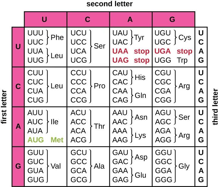
Translation of the mRNA template converts nucleotide-based genetic information into the “language” of amino acids to create a protein product. A protein sequence consists of 20 commonly occurring amino acids. Each amino acid is defined within the mRNA by a triplet of nucleotides called a codon. The relationship between an mRNA codon and its corresponding amino acid is called the genetic code.
The three-nucleotide code means that there is a total of 64 possible combinations (, with four different nucleotides possible at each of the three different positions within the codon). This number is greater than the number of amino acids and a given amino acid is encoded by more than one codon (see the genetic code table below). This redundancy in the genetic code is called degeneracy. Typically, whereas the first two positions in a codon are important for determining which amino acid will be incorporated into a growing polypeptide, the third position, called the wobble position, is less critical. In some cases, if the nucleotide in the third position is changed, the same amino acid is still incorporated.
Whereas 61 of the 64 possible triplets code for amino acids, three of the 64 codons do not code for an amino acid; they terminate protein synthesis, releasing the polypeptide from the translation machinery. These are called stop codons or nonsense codons. Another codon, AUG, also has a special function. In addition to specifying the amino acid methionine, it also typically serves as the start codon to initiate translation. The reading frame, the way nucleotides in mRNA are grouped into codons, for translation is set by the AUG start codon near the 5′ end of the mRNA. Each set of three nucleotides following this start codon is a codon in the mRNA message.
The genetic code is nearly universal. With a few exceptions, virtually all species use the same genetic code for protein synthesis, which is powerful evidence that all extant life on earth shares a common origin. However, unusual amino acids such as selenocysteine and pyrrolysine have been observed in archaea and bacteria. In the case of selenocysteine, the codon used is UGA (normally a stop codon). However, UGA can encode for selenocysteine using a stem-loop structure (known as the selenocysteine insertion sequence, or SECIS element), which is found at the 3′ untranslated region of the mRNA. Pyrrolysine uses a different stop codon, UAG. The incorporation of pyrrolysine requires the pylS gene and a unique transfer RNA (tRNA) with a CUA anticodon.
Extended description
The table is a 4×4 grid of blocks, one block for each combination of the codon’s first and second letters. Reading down the far-left column gives the first letter of the codon (U, C, A, or G); reading across the top row gives the second letter (U, C, A, or G); and reading down the third-letter column at the right edge of each row block gives the third letter (U, C, A, or G) for whichever line inside that block is being read. A codon is found by combining the row, column, and sub-row letters in that order, then reading the amino acid abbreviation — or, in the two red cells of that block, the word ‘stop’ — printed beside the group of codons that share it. The start codon, AUG, is picked out in green inside the third row block.
Check Your Understanding
How many bases are in each codon?
Recall the length of the nucleotide sequence that specifies one amino acid.Using the genetic code table above, what amino acid is coded for by the codon AAU?
Find the block for first letter A and second letter A, then read down to the sub-row for third letter U.What happens when a stop codon is reached?
Show model answer
Did your answer mention:
The Protein Synthesis Machinery
In addition to the mRNA template, many molecules and macromolecules contribute to the process of translation. The composition of each component varies across taxa; for instance, ribosomes may consist of different numbers of ribosomal RNAs (rRNAs) and polypeptides depending on the organism. However, the general structures and functions of the protein synthesis machinery are comparable from bacteria to human cells. Translation requires the input of an mRNA template, ribosomes, tRNAs, and various enzymatic factors.
Ribosomes
A ribosome is a complex macromolecule composed of catalytic rRNAs (called ribozymes) and structural rRNAs, as well as many distinct polypeptides. Mature rRNAs make up approximately 50% of each ribosome. Prokaryotes have 70S ribosomes, whereas eukaryotes have 80S ribosomes in the cytoplasm and rough endoplasmic reticulum, and 70S ribosomes in mitochondria and chloroplasts. Ribosomes dissociate into large and small subunits when they are not synthesizing proteins and reassociate during the initiation of translation. In E. coli, the small subunit is described as 30S (which contains the 16S rRNA subunit), and the large subunit is 50S (which contains the 5S and 23S rRNA subunits), for a total of 70S (Svedberg units are not additive). Eukaryote ribosomes have a small 40S subunit (which contains the 18S rRNA subunit) and a large 60S subunit (which contains the 5S, 5.8S and 28S rRNA subunits), for a total of 80S. The small subunit is responsible for binding the mRNA template, whereas the large subunit binds tRNAs (discussed in the next subsection).
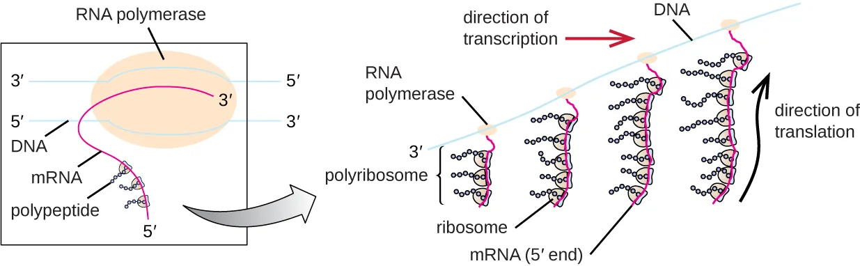
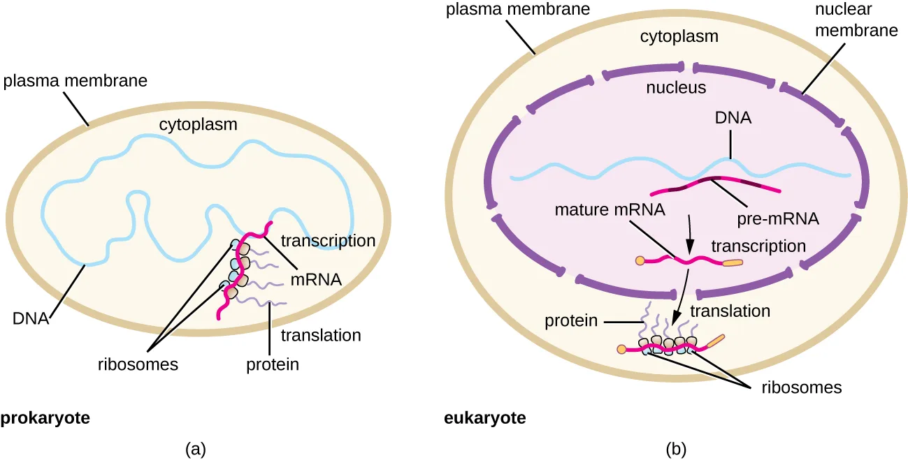
Each mRNA molecule is simultaneously translated by many ribosomes, all synthesizing protein in the same direction: reading the mRNA from 5′ to 3′ and synthesizing the polypeptide from the N terminus to the C terminus. The complete structure containing an mRNA with multiple associated ribosomes is called a polyribosome (or polysome). In both bacteria and archaea, before transcriptional termination occurs, each protein-encoding transcript is already being used to begin synthesis of numerous copies of the encoded polypeptide(s) because the processes of transcription and translation can occur concurrently, forming polyribosomes (see the figure below). The reason why transcription and translation can occur simultaneously is because both of these processes occur in the same 5′ to 3′ direction, they both occur in the cytoplasm of the cell, and because the RNA transcript is not processed once it is transcribed. This allows a prokaryotic cell to respond to an environmental signal requiring new proteins very quickly. In contrast, in eukaryotic cells, simultaneous transcription and translation is not possible. Although polyribosomes also form in eukaryotes, they cannot do so until RNA synthesis is complete and the RNA molecule has been modified and transported out of the nucleus.
Extended description
At the left, a boxed close-up shows RNA polymerase, drawn as a large oval, sitting on double-stranded DNA (labeled 3′ and 5′ on the upper strand, 5′ and 3′ on the lower) and spinning out a single magenta RNA strand — labeled mRNA, with its free 5′ end already carrying one ribosome that is translating a short polypeptide. An arrow points from this boxed panel to a wider view at the right: RNA polymerase molecules run left to right along the DNA in the direction of transcription; behind each one, progressively longer magenta mRNA strands trail off in the direction of translation, each strand studded with a row of ribosomes — more ribosomes on the longer, older strands — forming a polyribosome.
Transfer RNAs
Transfer RNAs (tRNAs) are structural RNA molecules and, depending on the species, many different types of tRNAs exist in the cytoplasm. Bacterial species typically have between 60 and 90 types. Serving as adaptors, each tRNA type binds to a specific codon on the mRNA template and adds the corresponding amino acid to the polypeptide chain. Therefore, tRNAs are the molecules that actually “translate” the language of RNA into the language of proteins. As the adaptor molecules of translation, it is surprising that tRNAs can fit so much specificity into such a small package. The tRNA molecule interacts with three factors: aminoacyl tRNA synthetases, ribosomes, and mRNA.
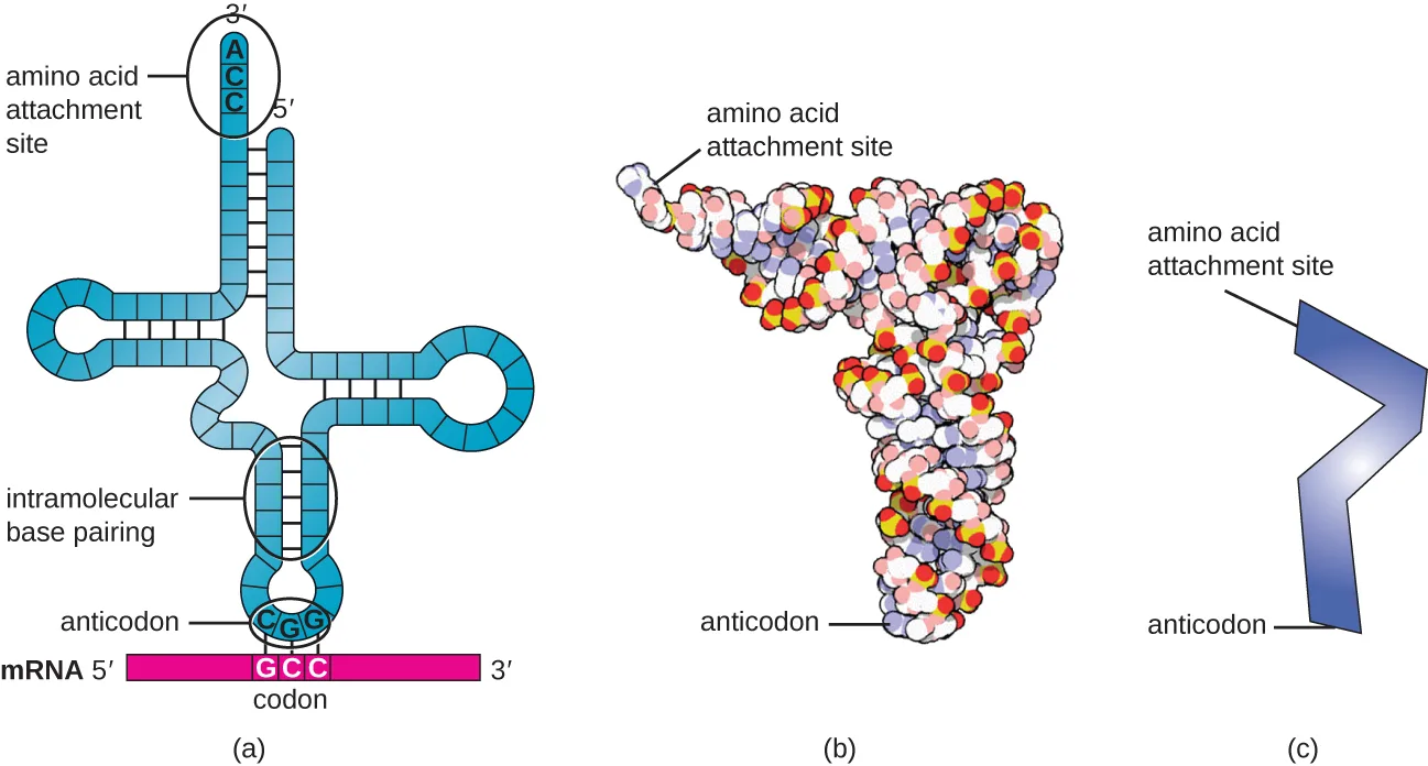
Mature tRNAs take on a three-dimensional structure when complementary bases exposed in the single-stranded RNA molecule hydrogen bond with each other (see the figure below). This shape positions the amino-acid binding site, called the CCA amino acid binding end, which is a cytosine-cytosine-adenine sequence at the 3′ end of the tRNA, and the anticodon at the other end. The anticodon is a three-nucleotide sequence that bonds with an mRNA codon through complementary base pairing.
An amino acid is added to the end of a tRNA molecule through the process of tRNA “charging,” during which each tRNA molecule is linked to its correct or cognate amino acid by a group of enzymes called aminoacyl tRNA synthetases. At least one type of aminoacyl tRNA synthetase exists for each of the 20 amino acids. During this process, the amino acid is first activated by the addition of adenosine monophosphate (AMP) and then transferred to the tRNA, making it a charged tRNA, and AMP is released.
Extended description
(a) The folded strand’s 3′ end sits at the top of the amino-acid-attachment arm and reads A-C-C; the 5′ end is also at the top, on the neighboring arm. Following the strand down from the fold, the base of the cross carries a three-letter anticodon, shown paired by short dashed lines to a complementary three-letter codon printed on the mRNA strand just below, which is itself labeled 5′ at the left and 3′ at the right. (b) The same molecule rendered as a space-filling model, narrow at the amino-acid-attachment end and widening toward the anticodon end. (c) The same molecule reduced to a single zigzag line, labeled at the same two ends.
Check Your Understanding
Describe the structure and composition of the prokaryotic ribosome.
Show model answer
Did your answer mention:
In what direction is the mRNA template read?
All ribosomes translating a given mRNA move along it in the same direction, reading toward its 3′ end.Describe the structure and function of a tRNA.
Show model answer
Did your answer mention:
The Mechanism of Protein Synthesis
Translation is similar in prokaryotes and eukaryotes. Here we will explore how translation occurs in E. coli, a representative prokaryote, and specify any differences between bacterial and eukaryotic translation.
Initiation
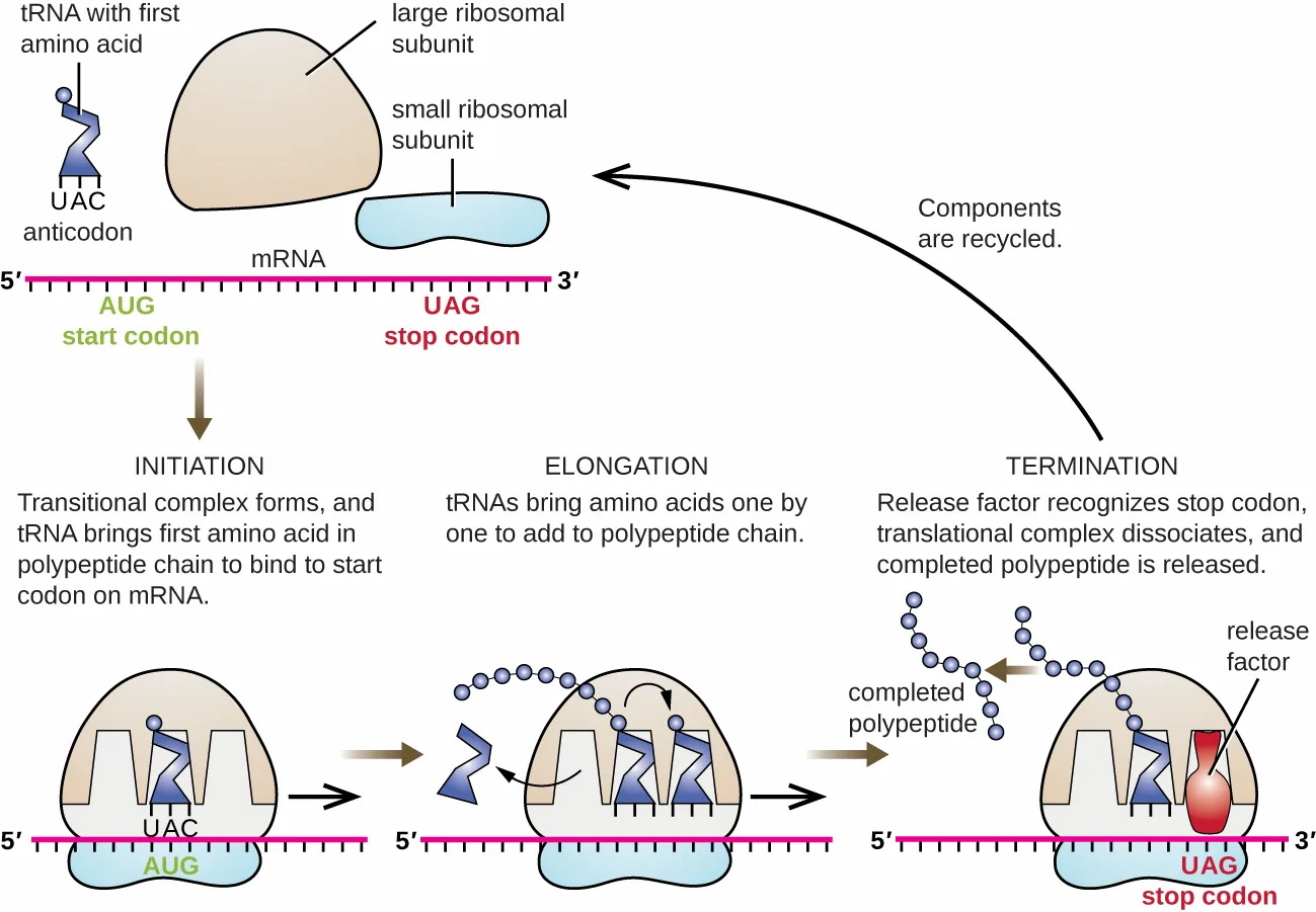
The initiation of protein synthesis begins with the formation of an initiation complex. In E. coli, this complex involves the small 30S ribosome, the mRNA template, three initiation factors that help the ribosome assemble correctly, guanosine triphosphate (GTP) that acts as an energy source, and a special initiator tRNA carrying N-formyl-methionine (fMet-tRNAfMet) (see the figure below). The initiator tRNA interacts with the start codon AUG of the mRNA and carries a formylated methionine (fMet). Because of its involvement in initiation, fMet is inserted at the beginning (N terminus) of every polypeptide chain synthesized by E. coli. In E. coli mRNA, a leader sequence upstream of the first AUG codon, called the Shine-Dalgarno sequence (also known as the ribosomal binding site AGGAGG), interacts through complementary base pairing with the rRNA molecules that compose the ribosome. This interaction anchors the 30S ribosomal subunit at the correct location on the mRNA template. At this point, the 50S ribosomal subunit then binds to the initiation complex, forming an intact ribosome.
In eukaryotes, initiation complex formation is similar, with the following differences:
- The initiator tRNA is a different specialized tRNA carrying methionine, called Met-tRNAi
- Instead of binding to the mRNA at the Shine-Dalgarno sequence, the eukaryotic initiation complex recognizes the 5′ cap of the eukaryotic mRNA, then tracks along the mRNA in the 5′ to 3′ direction until the AUG start codon is recognized. At this point, the 60S subunit binds to the complex of Met-tRNAi, mRNA, and the 40S subunit.
Extended description
At the upper left, a tRNA carrying the first amino acid and bearing the anticodon UAC approaches the mRNA at its start codon, AUG, together with the small ribosomal subunit (drawn as a flat oval) and the large ribosomal subunit (drawn as a dome); the same mRNA strand shows a stop codon, UAG, further along toward its 3′ end. Below, three panels run left to right. In INITIATION, the initiation complex has formed: the tRNA sits in the middle (P) of the ribosome’s three sites, which run E, P, A from left to right at the start codon. In ELONGATION, tRNAs add amino acids one by one: a tRNA carrying a long chain of linked circles occupies the P site, a tRNA carrying a single circle occupies the A site, and an empty tRNA is leaving the E site. In TERMINATION, a release factor (drawn in red) has bound the stop codon UAG, now aligned with the A site; the completed polypeptide chain and the release factor separate from the ribosome. A curved arrow labeled ‘Components are recycled’ returns the dissociated subunits to the upper-left starting point.
Elongation
In prokaryotes and eukaryotes, the basics of elongation of translation are the same. In E. coli, the binding of the 50S ribosomal subunit to produce the intact ribosome forms three functionally important ribosomal sites: The A (aminoacyl) site binds incoming charged aminoacyl tRNAs. The P (peptidyl) site binds charged tRNAs carrying amino acids that have formed peptide bonds with the growing polypeptide chain but have not yet dissociated from their corresponding tRNA. The E (exit) site releases dissociated tRNAs so that they can be recharged with free amino acids. There is one notable exception to this assembly line of tRNAs: During initiation complex formation, bacterial fMet-tRNAfMet or eukaryotic Met-tRNAi enters the P site directly without first entering the A site, providing a free A site ready to accept the tRNA corresponding to the first codon after the AUG.
Elongation proceeds with single-codon movements of the ribosome each called a translocation event. During each translocation event, the charged tRNAs enter at the A site, then shift to the P site, and then finally to the E site for removal. Ribosomal movements, or steps, are induced by conformational changes that advance the ribosome by three bases in the 3′ direction. Peptide bonds form between the amino group of the amino acid attached to the A-site tRNA and the carboxyl group of the amino acid attached to the P-site tRNA. The formation of each peptide bond is catalyzed by peptidyl transferase, an RNA-based ribozyme that is integrated into the 50S ribosomal subunit. The amino acid bound to the P-site tRNA is also linked to the growing polypeptide chain. As the ribosome steps across the mRNA, the former P-site tRNA enters the E site, detaches from the amino acid, and is expelled. Several of the steps during elongation, including binding of a charged aminoacyl tRNA to the A site and translocation, require energy derived from GTP hydrolysis, which is catalyzed by specific elongation factors. Amazingly, the E. coli translation apparatus takes only 0.05 seconds to add each amino acid, meaning that a 200 amino-acid protein can be translated in just 10 seconds.
Termination
The termination of translation occurs when a nonsense codon (UAA, UAG, or UGA) is encountered for which there is no complementary tRNA. On aligning with the A site, these nonsense codons are recognized by release factors in prokaryotes and eukaryotes that result in the P-site amino acid detaching from its tRNA, releasing the newly made polypeptide. The small and large ribosomal subunits dissociate from the mRNA and from each other; they are recruited almost immediately into another translation initiation complex.
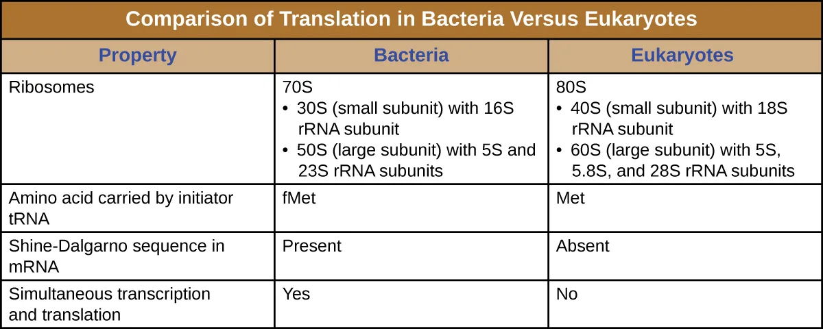
In summary, there are several key features that distinguish prokaryotic gene expression from that seen in eukaryotes. These are illustrated in the figure below and listed in the table below.
Extended description
(a) The prokaryotic cell is bounded by a single plasma membrane; a wavy DNA strand loops through the cytoplasm and gives rise to an mRNA strand still being synthesized, which is already studded with ribosomes translating it into a protein chain — transcription and translation shown occurring together in the same open space. (b) The eukaryotic cell has an outer plasma membrane and, within it, a separate nucleus bounded by its own nuclear membrane; inside the nucleus a DNA strand gives rise to a pre-mRNA strand that is processed into a mature mRNA, which then crosses into the cytoplasm, where ribosomes translate it into a protein chain — transcription and translation shown as separated in space.
| Property | Bacteria | Eukaryotes |
|---|---|---|
| Ribosomes | 70S | 80S |
| Small subunit | 30S (16S rRNA subunit) | 40S (18S rRNA subunit) |
| Large subunit | 50S (5S and 23S rRNA subunits) | 60S (5S, 5.8S, and 28S rRNA subunits) |
| Amino acid carried by initiator tRNA | fMet | Met |
| Shine-Dalgarno sequence in mRNA | Present | Absent |
| Simultaneous transcription and translation | Yes | No |
Protein Targeting, Folding, and Modification
During and after translation, polypeptides may need to be modified before they are biologically active. Post-translational modifications include:
- removal of translated signal sequences—short tails of amino acids that aid in directing a protein to a specific cellular compartment
- proper “folding” of the polypeptide and association of multiple polypeptide subunits, often facilitated by chaperone proteins, into a distinct three-dimensional structure
- proteolytic processing of an inactive polypeptide to release an active protein component, and
- various chemical modifications (e.g., phosphorylation, methylation, or glycosylation) of individual amino acids.
Check Your Understanding
What are the components of the initiation complex for translation in prokaryotes?
Show model answer
Did your answer mention:
What are two differences between initiation of prokaryotic and eukaryotic translation? Sort each phrase under the type of initiation it describes.
Prokaryotic initiation
Eukaryotic initiation
Check Your Understanding
What occurs at each of the three active sites of the ribosome?
Show model answer
Did your answer mention:
What causes termination of translation?
Show model answer
Did your answer mention:
Summary
- In translation, polypeptides are synthesized using mRNA sequences and cellular machinery, including tRNAs that match mRNA codons to specific amino acids and ribosomes composed of RNA and proteins that catalyze the reaction.
- The genetic code is degenerate in that several mRNA codons code for the same amino acids. The genetic code is almost universal among living organisms.
- Prokaryotic (70S) and cytoplasmic eukaryotic (80S) ribosomes are each composed of a large subunit and a small subunit of differing sizes between the two groups. Each subunit is composed of rRNA and protein. Organelle ribosomes in eukaryotic cells resemble prokaryotic ribosomes.
- Some 60 to 90 species of tRNA exist in bacteria. Each tRNA has a three-nucleotide anticodon as well as a binding site for a cognate amino acid. All tRNAs with a specific anticodon will carry the same amino acid.
- Initiation of translation occurs when the small ribosomal subunit binds with initiation factors and an initiator tRNA at the start codon of an mRNA, followed by the binding to the initiation complex of the large ribosomal subunit.
- In prokaryotic cells, the start codon codes for N-formyl-methionine carried by a special initiator tRNA. In eukaryotic cells, the start codon codes for methionine carried by a special initiator tRNA. In addition, whereas ribosomal binding of the mRNA in prokaryotes is facilitated by the Shine-Dalgarno sequence within the mRNA, eukaryotic ribosomes bind to the 5′ cap of the mRNA.
- During the elongation stage of translation, a charged tRNA binds to mRNA in the A site of the ribosome; a peptide bond is catalyzed between the two adjacent amino acids, breaking the bond between the first amino acid and its tRNA; the ribosome moves one codon along the mRNA; and the first tRNA is moved from the P site of the ribosome to the E site and leaves the ribosomal complex.
- Termination of translation occurs when the ribosome encounters a stop codon, which does not code for a tRNA. Release factors cause the polypeptide to be released, and the ribosomal complex dissociates.
- In prokaryotes, transcription and translation may be coupled, with translation of an mRNA molecule beginning as soon as transcription allows enough mRNA exposure for the binding of a ribosome, prior to transcription termination. Transcription and translation are not coupled in eukaryotes because transcription occurs in the nucleus, whereas translation occurs in the cytoplasm or in association with the rough endoplasmic reticulum.
- Polypeptides often require one or more post-translational modifications to become biologically active.
Key terms
- translation — process of protein synthesis whereby a ribosome decodes an mRNA message into a polypeptide product.
- protein synthesis — process of protein synthesis whereby a ribosome decodes an mRNA message into a polypeptide product.
- codon — three-nucleotide sequence within mRNA that specifies a particular amino acid to be incorporated into the polypeptide being synthesized.
- genetic code — correspondence between mRNA nucleotide codons and the translated amino acids.
- degeneracy — redundancy in the genetic code because a given amino acid is encoded by more than one nucleotide triplet codon.
- wobble position — third position of a codon that, when changed, typically results in the incorporation of the same amino acid because of the degeneracy of the genetic code.
- stop codons — one of three codons for which there is no tRNA with a complementary anticodon; a signal within the mRNA for termination of translation.
- nonsense codons — codons for which there is no tRNA with a complementary anticodon; a signal within the mRNA for termination of translation.
- start codon — AUG codon, specifying methionine, which is typically the codon that initiates translation.
- reading frame — way nucleotides in mRNA are grouped into codons.
- initiation of translation — stage of translation during which an initiation complex composed of the small ribosomal subunit, the mRNA template, initiation factors, GTP, and a special initiator tRNA forms, and the large ribosomal subunit then binds to the initiation complex.
- polyribosome — structure including an mRNA molecule that is being translated by multiple ribosomes concurrently.
- polysome — structure including an mRNA molecule that is being translated by multiple ribosomes concurrently.
- CCA amino acid binding end — region of a mature tRNA that binds to an amino acid.
- anticodon — three-nucleotide sequence of a mature tRNA that interacts with an mRNA codon through complementary base pairing.
- cognate amino acid — amino acid added to a specific tRNA molecule that correctly corresponds to the tRNA’s anticodon and, hence, the mRNA’s codon, reflecting the genetic code.
- aminoacyl tRNA synthetase — enzyme that binds to a tRNA molecule and catalyzes the addition of the correct amino acid to the tRNA.
- charged tRNA — activated tRNA molecule carrying its cognate amino acid.
- initiation of protein synthesis — beginning of translation with the formation of an initiation complex composed of the small ribosomal subunit, the mRNA template, initiation factors, GTP, and a special initiator tRNA.
- initiation factors — proteins that participate in ribosome assembly during initiation.
- elongation of translation — stage of translation during which amino acids are added one by one to the C-terminus of the growing polypeptide.
- A (aminoacyl) site — functional site of an intact ribosome that binds incoming charged aminoacyl tRNAs.
- P (peptidyl) site — functional site of an intact ribosome that binds charged tRNAs carrying amino acids that have formed peptide bonds with the growing polypeptide chain but have not yet dissociated from their corresponding tRNA.
- E (exit) site — functional site of an intact ribosome that releases dissociated uncharged tRNAs so that they can be recharged with free amino acids.
- peptidyl transferase — RNA-based ribozyme that is part of the 50S ribosomal subunit and catalyzes formation of the peptide bond between the amino acid bound to a tRNA and the growing polypeptide chain.
- termination of translation — stage of translation during which a nonsense codon aligns with the A site, signaling release factors to release of the polypeptide, leading to the dissociation of the small and large ribosomal subunits from the mRNA and from each other.
Practice
Describe the genetic code and explain why it is considered almost universal
Which of the following is the name of the three-base sequence in the mRNA that binds to a tRNA molecule?
This sequence sits on the mRNA, not on the tRNA.Each codon within the genetic code encodes a different amino acid.
Recall how many codons exist compared with how many amino acids are commonly incorporated into proteins.The third position within a codon, in which changes often result in the incorporation of the same amino acid into the growing polypeptide, is called the ________.
This position is less critical than the first two for determining the amino acid.What is meant by the genetic code being nearly universal?
Show model answer
Did your answer mention:
Prior to the elucidation of the genetic code, prominent scientists, including Francis Crick, had predicted that each mRNA codon, coding for one of the 20 amino acids, needed to be at least three nucleotides long. Why is it not possible for codons to be any shorter?
Show model answer
Did your answer mention:
Below is an antisense DNA sequence. Translate the mRNA molecule synthesized using the genetic code, recording the resulting amino acid sequence, indicating the N and C termini. Antisense DNA strand: 3′-TACTGACTGACGATC-5′
Show model answer
5′-AUGACUGACUGCUAG-3′. Reading this from the 5′ end in codons: AUG, ACU, GAC, UGC, UAG. Using the genetic code, AUG codes for methionine (the start codon), ACU codes for threonine, GAC codes for aspartate, UGC codes for cysteine, and UAG is a stop codon. The resulting polypeptide, from the N terminus to the C terminus, is methionine–threonine–aspartate–cysteine; translation terminates at the UAG stop codon, so no amino acid is added for it.Did your answer mention:
Explain the process of translation and the functions of the molecular machinery of translation
Which component is the last to join the initiation complex during the initiation of translation?
The small subunit, mRNA, and initiator tRNA assemble first; one component completes the intact ribosome.During elongation in translation, to which ribosomal site does an incoming charged tRNA molecule bind?
This is the first of the three functional sites a charged tRNA passes through.Which of the following is the amino acid that appears at the N-terminus of all newly translated prokaryotic and eukaryotic polypeptides?
This is the amino acid carried by the initiator tRNA that recognizes the start codon.When the ribosome reaches a nonsense codon, which of the following occurs?
No tRNA carries an anticodon complementary to a nonsense codon.The enzyme that adds an amino acid to a tRNA molecule is called ________.
At least one type of this enzyme exists for each of the 20 amino acids.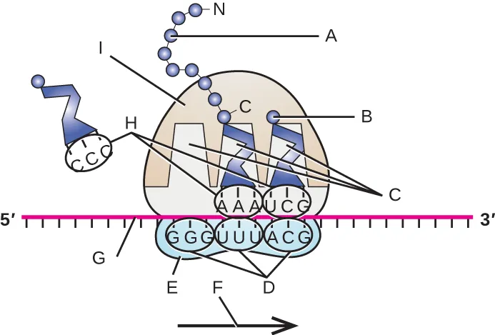
Using the figure above, match each lettered leader line to the part of translation it points to.
Show model answer
Did your answer mention:
Compare translation in eukaryotes and prokaryotes
Sort each property below under whether it describes translation in bacteria or in eukaryotes, using the comparison table above.
Bacteria
Eukaryotes
Transcription and translation are not coupled in eukaryotes because transcription occurs in the ________, whereas translation occurs in the cytoplasm.
This is the membrane-bound compartment that separates the two processes in eukaryotic cells.Eukaryotic ribosomes located in the mitochondria and chloroplasts are ________, the same size class as prokaryotic ribosomes.
Organelle ribosomes in eukaryotic cells resemble prokaryotic ribosomes, not the 80S ribosomes of the surrounding cytoplasm.This section is adapted from Microbiology, Section 11.4: Protein Synthesis (Translation) by Nina Parker, Mark Schneegurt, Anh-Hue Thi Tu, Philip Lister, Brian M. Forster, and OpenStax, © OpenStax, licensed under CC BY-NC-SA 4.0. Access the original for free at openstax.org. Changes: figures re-encoded as WebP; alts rewritten from the vendored images rather than copied from the source (the source alts for the genetic-code table, the co-transcriptional-translation diagram, the tRNA panels, the initiation/elongation/termination diagram, and the prokaryote/eukaryote diagram carry typos and are supplemented here with a longdesc walking each figure’s steps or labels; the genetic-code table’s longdesc explains how to read the table rather than listing all 64 codons); the Comparison of Translation in Bacteria Versus Eukaryotes figure, which prints no source caption, is transcribed as a Markdown table (its own sortbins under the third objective) and kept as a figure with no caption line; the ten body Check Your Understanding bullets (three boxes of three, three, and four) are rendered as body items at each note’s position — 4 graded (textin, multiplechoice, sortbins) from this module’s own sentences, table, and genetic-code figure, and 6 left as self-checks whose model answers are drawn from this section’s text; of the source’s four unkeyed Short Answer and two unkeyed Critical Thinking questions, 1 is graded from this module’s own text and table (the prokaryote/eukaryote Short Answer, converted to the Bacteria/Eukaryotes sortbins fed by the comparison table), 4 stay self-checks with model answers assembled from this section’s own text (the “nearly universal” question, including its selenocysteine/pyrrolysine exceptions; the antisense-strand translation, fully worked from the genetic-code table above; why codons cannot be shorter than three nucleotides, worked from the module’s own // arithmetic; and the figure-labelling Critical Thinking item, whose model answer is the letter-to-part mapping read from the image, including the ribosome’s E, P, and A sites read off the leader line lettered C in the order they fall along the mRNA’s 5′→3′ direction), and 1 (the “why does translation terminate at a stop codon” Short Answer) is dropped rather than converted or kept as a Practice self-check because it duplicates two body Check Your Understanding bullets already on the page (the first box’s “what happens when a stop codon is reached?” and the third box’s “what causes termination of translation?”) — the source prints no answer key for any of the five that are used; key terms compiled from the module’s 26 defined terms and the book’s Glossary appendix, with 1 taken from the defining sentence (initiation of protein synthesis — no appendix entry matches its sense; protein synthesis and aminoacyl tRNA synthetase both have appendix entries, under the headwords “translation (protein synthesis)” and “aminoacyl-tRNA synthetase” respectively); the True/False item rendered as a two-option multiple choice; two filler textin items built from this section’s own Summary and body sentences to fill the third objective’s Practice group, one with an accept for its unhyphenated Svedberg spelling (70 S); primes normalized to U+2032 (′) throughout, matching the source’s own mixed use of the right single quote and the prime character.