How Asexual Prokaryotes Achieve Genetic Diversity
By the end of this section, you will be able to:
- Compare the processes of transformation, transduction, and conjugation
- Explain how asexual gene transfer results in prokaryotic genetic diversity
- Explain the structure and consequences for bacterial genetic diversity of transposons
Typically, when we consider genetic transfer, we think of vertical gene transfer, the transmission of genetic information from generation to generation. Vertical gene transfer is by far the main mode of transmission of genetic information in all cells. In sexually reproducing organisms, crossing-over events and independent assortment of individual chromosomes during meiosis contribute to genetic diversity in the population. Genetic diversity is also introduced during sexual reproduction, when the genetic information from two parents, each with different complements of genetic information, are combined, producing new combinations of parental genotypes in the diploid offspring. The occurrence of mutations also contributes to genetic diversity in a population. Genetic diversity of offspring is useful in changing or inconsistent environments and may be one reason for the evolutionary success of sexual reproduction.
When prokaryotes and eukaryotes reproduce asexually, they transfer a nearly identical copy of their genetic material to their offspring through vertical gene transfer. Although asexual reproduction produces more offspring more quickly, any benefits of diversity among those offspring are lost. How then do organisms whose dominant reproductive mode is asexual create genetic diversity? In prokaryotes, horizontal gene transfer (HGT), the introduction of genetic material from one organism to another organism within the same generation, is an important way to introduce genetic diversity. HGT allows even distantly related species to share genes, influencing their phenotypes. It is thought that HGT is more prevalent in prokaryotes but that only a small fraction of the prokaryotic genome may be transferred by this type of transfer at any one time. As the phenomenon is investigated more thoroughly, it may be revealed to be even more common. Many scientists believe that HGT and mutation are significant sources of genetic variation, the raw material for the process of natural selection, in prokaryotes. Although HGT is more common among evolutionarily related organisms, it may occur between any two species that live together in a natural community.
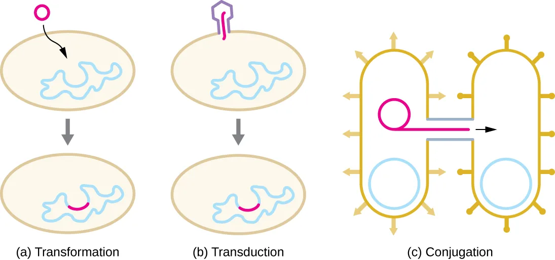
HGT in prokaryotes is known to occur by the three primary mechanisms illustrated below:
- Transformation: naked DNA is taken up from the environment
- Transduction: genes are transferred between cells in a virus (see The Viral Life Cycle)
- Conjugation: use of a hollow tube called a conjugation pilus to transfer genes between cells
Extended description
(a) Transformation: two beige oval cells stacked with a downward arrow between them; the top cell has a magenta circle of free DNA outside it; the bottom cell’s chromosome now carries a magenta arc where the DNA has recombined in. (b) Transduction: the same before/after layout; the top cell has a purple bacteriophage particle attached to its surface; the bottom cell’s chromosome carries the same kind of magenta arc, delivered by the phage instead of taken up directly. (c) Conjugation: unlike (a) and (b), a single static scene, not a before/after pair. Two gold/tan, rod-shaped cells, each ringed with several short radiating pili, are connected by a thin bridge. Each cell has a plain, uncolored circular chromosome. The left cell also holds a small magenta plasmid circle; a magenta line with an arrowhead runs from that plasmid through the bridge into the right cell, showing the plasmid copy being transferred.
Check Your Understanding
What are three ways sexual reproduction introduces genetic variation into offspring?
Show model answer
Did your answer mention:
What is a benefit of asexual reproduction?
Recall what the module says is traded off against the diversity that is lost in asexual reproduction.What are the three mechanisms of horizontal gene transfer in prokaryotes?
Show model answer
Did your answer mention:
Transformation
Frederick Griffith was the first to demonstrate the process of transformation. In 1928, he showed that live, nonpathogenic Streptococcus pneumoniae bacteria could be transformed into pathogenic bacteria through exposure to a heat-killed pathogenic strain. He concluded that some sort of agent, which he called the “transforming principle,” had been passed from the dead pathogenic bacteria to the live, nonpathogenic bacteria. In 1944, Oswald Avery (1877–1955), Colin MacLeod (1909–1972), and Maclyn McCarty (1911–2005) demonstrated that the transforming principle was DNA (see Using Microbiology to Discover the Secrets of Life).
In transformation, the prokaryote takes up naked DNA found in its environment and that is derived from other cells that have lysed on death and released their contents, including their genome, into the environment. Many bacteria are naturally competent, meaning that they actively bind to environmental DNA, transport it across their cell envelopes into their cytoplasm, and make it single stranded. Typically, double-stranded foreign DNA within cells is destroyed by nucleases as a defense against viral infection. However, these nucleases are usually ineffective against single-stranded DNA, so this single-stranded DNA within the cell has the opportunity to recombine into the bacterial genome. A molecule of DNA that contains fragments of DNA from different organisms is called recombinant DNA. (Recombinant DNA will be discussed in more detail in Microbes and the Tools of Genetic Engineering.) If the bacterium incorporates the new DNA into its own genome through recombination, the bacterial cell may gain new phenotypic properties. For example, if a nonpathogenic bacterium takes up DNA for a toxin gene from a pathogen and then incorporates it into its chromosome, it, too, may become pathogenic. Plasmid DNA may also be taken up by competent bacteria and confer new properties to the cell. Overall, transformation in nature is a relatively inefficient process because environmental DNA levels are low because of the activity of nucleases that are also released during cellular lysis. Additionally, genetic recombination is inefficient at incorporating new DNA sequences into the genome.
In nature, bacterial transformation is an important mechanism for the acquisition of genetic elements encoding virulence factors and antibiotic resistance. Genes encoding resistance to antimicrobial compounds have been shown to be widespread in nature, even in environments not influenced by humans. These genes, which allow microbes living in mixed communities to compete for limited resources, can be transferred within a population by transformation, as well as by the other processes of HGT. In the laboratory, we can exploit the natural process of bacterial transformation for genetic engineering to make a wide variety of medicinal products, as discussed in Microbes and the Tools of Genetic Engineering.
Check Your Understanding
Why does a bacterial cell make environmental DNA brought into the cell into a single-stranded form?
Show model answer
Did your answer mention:
Transduction
Viruses that infect bacteria (bacteriophages) may also move short pieces of chromosomal DNA from one bacterium to another in a process called transduction (see the transduction diagram in The Viral Life Cycle). Recall that in generalized transduction, any piece of chromosomal DNA may be transferred to a new host cell by accidental packaging of chromosomal DNA into a phage head during phage assembly. By contrast, specialized transduction results from the imprecise excision of a lysogenic prophage from the bacterial chromosome such that it carries with it a piece of the bacterial chromosome from either side of the phage’s integration site to a new host cell. As a result, the host may acquire new properties. This process is called lysogenic conversion. Of medical significance, a lysogenic phage may carry with it a virulence gene to its new host. Once inserted into the new host’s chromosome, the new host may gain pathogenicity. Several pathogenic bacteria, including Corynebacterium diphtheriae (the causative agent of diphtheria) and Clostridium botulinum (the causative agent of botulism), are virulent because of the introduction of toxin-encoding genes by lysogenic bacteriophages, affirming the clinical relevance of transduction in the exchange of genes involved in infectious disease. Archaea have their own viruses that translocate genetic material from one individual to another.
Check Your Understanding
What is the agent of transduction of prokaryotic cells?
Name the viruses that infect bacteria, introduced at the start of this section.In specialized transduction, where does the transducing piece of DNA come from?
Recall which process packages DNA from a fixed location beside the prophage, rather than a random piece of the chromosome.Case in Point. The Clinical Consequences of Transduction
Paul, a 23-year-old relief worker from Atlanta, traveled to Haiti in 2011 to provide aid following the 2010 earthquake. After working there for several weeks, he suddenly began experiencing abdominal distress, including severe cramping, nausea, vomiting, and watery diarrhea. He also began to experience intense muscle cramping. At a local clinic, the physician suspected that Paul’s symptoms were caused by cholera because there had been a cholera outbreak after the earthquake. Because cholera is transmitted by the fecal-oral route, breaches in sanitation infrastructure, such as often occur following natural disasters, may precipitate outbreaks. The physician confirmed the presumptive diagnosis using a cholera dipstick test. He then prescribed Paul a single dose of doxycycline, as well as oral rehydration salts, instructing him to drink significant amounts of clean water.
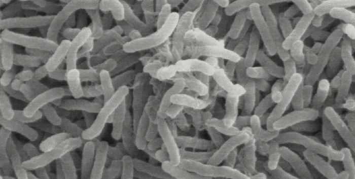
Cholera is caused by the gram-negative curved rod Vibrio cholerae, shown below. Its symptoms largely result from the production of the cholera toxin (CT), which ultimately activates a chloride transporter to pump chloride ions out of the epithelial cells into the gut lumen. Water then follows the chloride ions, causing the prolific watery diarrhea characteristic of cholera. The gene encoding the cholera toxin is incorporated into the bacterial chromosome of V. cholerae through infection of the bacterium with the lysogenic filamentous CTX phage, which carries the CT gene and introduces it into the chromosome on integration of the prophage. Thus, pathogenic strains of V. cholerae result from horizontal gene transfer by specialized transduction.
- Why are outbreaks of cholera more common as a result of a natural disaster?
- Why is muscle cramping a common symptom of cholera? Why is treatment with oral rehydration salts so important for the treatment of cholera?
- In areas stricken by cholera, what are some strategies that people could use to prevent disease transmission?
Conjugation
In conjugation, DNA is directly transferred from one prokaryote to another by means of a conjugation pilus, which brings the organisms into contact with one another. In E. coli, the genes encoding the ability to conjugate are located on a bacterial plasmid called the F plasmid, also known as the fertility factor, and the conjugation pilus is called the F pilus. The F-plasmid genes encode both the proteins composing the F pilus and those involved in rolling circle replication of the plasmid. Cells containing the F plasmid, capable of forming an F pilus, are called F⁺ cells or donor cells, and those lacking an F plasmid are called F⁻ cells or recipient cells.
Conjugation of the F Plasmid
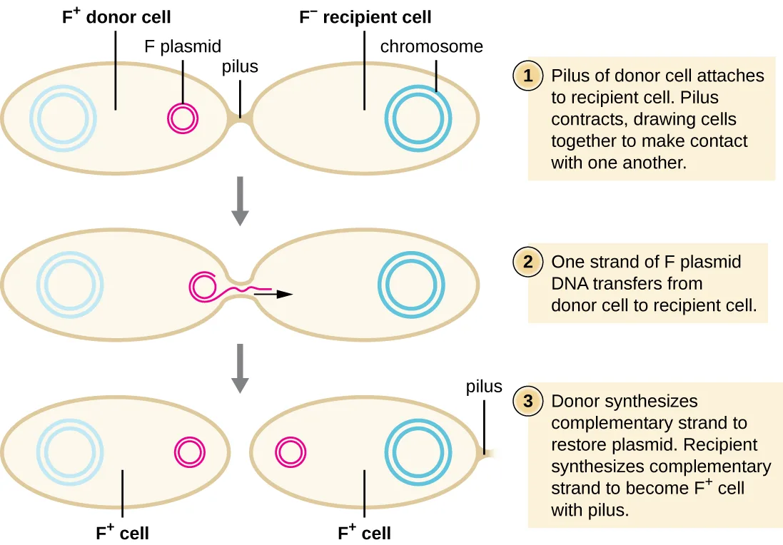
During typical conjugation in E. coli, the F pilus of an F⁺ cell comes into contact with an F⁻ cell and retracts, bringing the two cell envelopes into contact. Then a cytoplasmic bridge forms between the two cells at the site of the conjugation pilus. As rolling circle replication of the F plasmid occurs in the F⁺ cell, a single-stranded copy of the F plasmid is transferred through the cytoplasmic bridge to the F⁻ cell, which then synthesizes the complementary strand, making it double stranded. The F⁻ cell now becomes an F⁺ cell capable of making its own conjugation pilus. Eventually, in a mixed bacterial population containing both F⁺ and F⁻ cells, all cells will become F⁺ cells. Genes on the E. coli F plasmid also encode proteins preventing conjugation between F⁺ cells.
Extended description
Stage 1: an F+ donor cell, containing a small circular F plasmid and a larger circular chromosome, extends a pilus that attaches to an F− recipient cell, containing only a chromosome; the pilus then contracts, pulling the two cells into direct contact. Stage 2: a cytoplasmic bridge has formed where the pilus was; a single strand of the F-plasmid DNA, drawn as a wavy line with an arrowhead, moves from the donor cell through the bridge into the recipient cell. Stage 3: the bridge has closed; the donor cell synthesizes a complementary strand to restore its double-stranded F plasmid, and the recipient cell synthesizes a complementary strand to its transferred strand, producing its own double-stranded F plasmid and growing its own pilus — both cells are now labeled F+ cell.
Conjugation of F′ and Hfr Cells
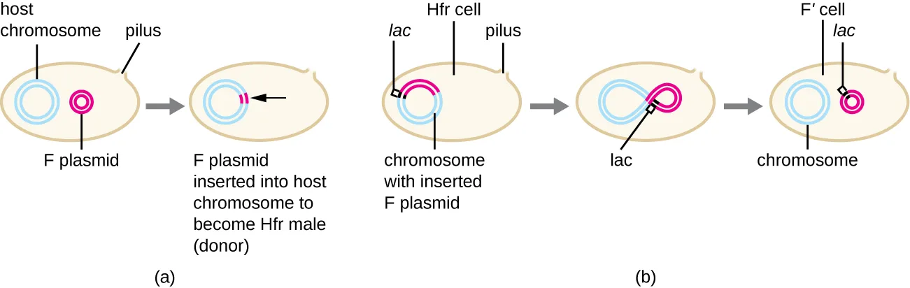
Although typical conjugation in E. coli results in the transfer of the F-plasmid DNA only, conjugation may also transfer chromosomal DNA. This is because the F plasmid occasionally integrates into the bacterial chromosome through recombination between the plasmid and the chromosome, forming an Hfr cell. “Hfr” refers to the high frequency of recombination seen when recipient F⁻ cells receive genetic information from Hfr cells through conjugation. Similar to the imprecise excision of a prophage during specialized transduction, the integrated F plasmid may also be imprecisely excised from the chromosome, producing an F′ plasmid that carries with it some chromosomal DNA adjacent to the integration site. On conjugation, this DNA is introduced to the recipient cell and may be either maintained as part of the F′ plasmid or be recombined into the recipient cell’s bacterial chromosome.
Extended description
Panel (a), left to right: a cell containing a host chromosome (blue loop), a separate F plasmid (small pink loop), and a pilus; an arrow leads to a cell where the F plasmid has integrated into the host chromosome to form one loop, labeled the Hfr cell (donor). Panel (b), left to right: an Hfr cell whose chromosome carries the integrated F plasmid, with the lac gene marked at the plasmid–chromosome junction; an arrow leads to the same DNA folded into a figure-eight shape as the F plasmid begins to excise imprecisely, with lac now positioned at the pinch point; a second arrow leads to the outcome, an F′ cell containing two separate circles — an F′ plasmid that has picked up the lac gene, and the chromosome it came from.
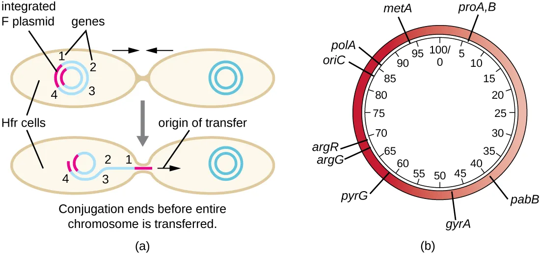
Hfr cells may also treat the bacterial chromosome like an enormous F plasmid and attempt to transfer a copy of it to a recipient F⁻ cell. Because the bacterial chromosome is so large, transfer of the entire chromosome takes a long time. However, contact between bacterial cells during conjugation is transient, so it is unusual for the entire chromosome to be transferred. Host chromosomal DNA near the integration site of the F plasmid, displaced by the unidirectional process of rolling circle replication, is more likely to be transferred and recombined into a recipient cell’s chromosome than host genes farther away. Thus, the relative location of bacterial genes on the Hfr cell’s genome can be mapped based on when they are transferred through conjugation. As a result, prior to the age of widespread bacterial genome sequencing, distances on prokaryotic genome maps were often measured in minutes.
Extended description
Panel (a), top to bottom: an Hfr cell with an integrated F plasmid and four numbered chromosomal genes (1 nearest the integration site, then 2, 3, and 4 farthest away) begins conjugating with an F− recipient cell through a pilus; below, the connection has broken before transfer finishes, so genes 1 and 2 lie closest to the origin of transfer at the break point while genes 3 and 4 remain farther away in the donor cell — the recipient cell’s own chromosome is drawn plain, with no genes shown as having entered it yet. Panel (b): a circular genomic map of E. coli, marked in minutes from 0 at the top around to 100 at the same point, shaded from light near 0 minutes to dark red near 100 minutes; genes are positioned around the circle by how many minutes of conjugational transfer it takes to reach them, including proA,B and pabB (early, lightly shaded) and pyrG, argG, argR, oriC, polA, and metA (later, more darkly shaded), with gyrA near the bottom.
Consequences and Applications of Conjugation
Plasmids are an important type of extrachromosomal DNA element in bacteria and, in those cells that harbor them, are considered to be part of the bacterial genome. From a clinical perspective, plasmids often code for genes involved in virulence. For example, genes encoding proteins that make a bacterial cell resistant to a particular antibiotic are encoded on R plasmids. R plasmids, in addition to their genes for antimicrobial resistance, contain genes that control conjugation and transfer of the plasmid. R plasmids are able to transfer between cells of the same species and between cells of different species. Single R plasmids commonly contain multiple genes conferring resistance to multiple antibiotics.
Genes required for the production of various toxins and molecules important for colonization during infection may also be found encoded on plasmids. For example, verotoxin-producing strains of E. coli (VTEC) appear to have acquired the genes encoding the Shiga toxin from its gram-negative relative Shigella dysenteriae through the acquisition of a large plasmid encoding this toxin. VTEC causes severe diarrheal disease that may result in hemolytic uremic syndrome (HUS), which may be lead to kidney failure and death.
In nonclinical settings, bacterial genes that encode metabolic enzymes needed to degrade specialized atypical compounds like polycyclic aromatic hydrocarbons (PAHs) are also frequently encoded on plasmids. Additionally, certain plasmids have the ability to move from bacterial cells to other cell types, like those of plants and animals, through mechanisms distinct from conjugation. Such mechanisms and their use in genetic engineering are covered in Modern Applications of Microbial Genetics.
Link to Learning
Watch this animation to learn more about the process of conjugation.
Check Your Understanding
What type of replication occurs during conjugation?
Name the replication mode of the F plasmid identified early in the Conjugation section.What occurs to produce an Hfr E. coli cell?
Distinguish integration of the F plasmid into the chromosome from its later excision, which produces a different kind of cell.What types of traits are encoded on plasmids?
Show model answer
Did your answer mention:
Transposition
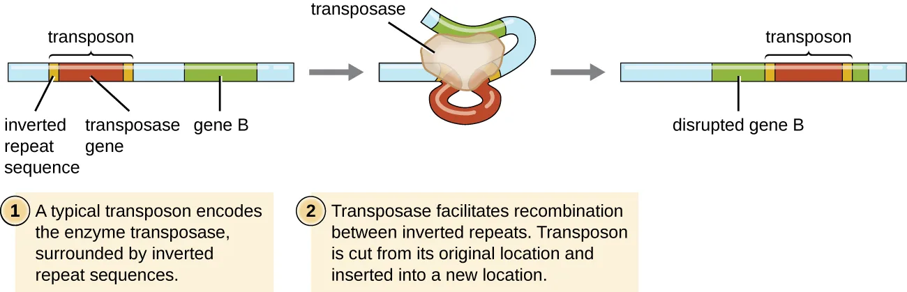
Genetic elements called transposons (transposable elements), or “jumping genes,” are molecules of DNA that include special inverted repeat sequences at their ends and a gene encoding the enzyme transposase. Transposons allow the entire sequence to independently excise from one location in a DNA molecule and integrate into the DNA elsewhere through a process called transposition. Transposons were originally discovered in maize (corn) by American geneticist Barbara McClintock (1902–1992) in the 1940s. Transposons have since been found in all types of organisms, both prokaryotes and eukaryotes. Thus, unlike the three previous mechanisms discussed, transposition is not prokaryote-specific. Most transposons are nonreplicative, meaning they move in a “cut-and-paste” fashion. Some may be replicative, however, retaining their location in the DNA while making a copy to be inserted elsewhere (“copy and paste”). Because transposons can move within a DNA molecule, from one DNA molecule to another, or even from one cell to another, they have the ability to introduce genetic diversity. Movement within the same DNA molecule can alter phenotype by inactivating or activating a gene.
Transposons may carry with them additional genes, moving these genes from one location to another with them. For example, bacterial transposons can relocate antibiotic resistance genes, moving them from chromosomes to plasmids. This mechanism has been shown to be responsible for the colocalization of multiple antibiotic resistance genes on a single R plasmid in Shigella strains causing bacterial dysentery. Such an R plasmid can then be easily transferred among a bacterial population through the process of conjugation.
Extended description
Panel 1, labeled 1: a light-blue chromosome segment carries a transposon — a transposase gene (red) flanked on both sides by short inverted repeat sequences (yellow) — positioned beside a separate green segment labeled gene B. Panel 2, labeled 2: a tan, loop-shaped transposase enzyme has bound the transposon, bending the DNA so its two inverted repeats meet, letting the enzyme cut the transposon out of its original location. Panel 3, unlabeled: the excised transposon has inserted itself into the middle of gene B, splitting the green gene into two segments on either side of the inserted transposase gene and its flanking repeats, labeled disrupted gene B.
Check Your Understanding
What are two ways a transposon can affect the phenotype of a cell it moves to?
Recall what happens when a transposon’s movement disrupts, or lands near, a gene within the same DNA molecule.The table below summarizes the processes discussed in this section.
| Term | Definition |
|---|---|
| Conjugation | Transfer of DNA through direct contact using a conjugation pilus |
| Transduction | Mechanism of horizontal gene transfer in bacteria in which genes are transferred through viral infection |
| Transformation | Mechanism of horizontal gene transfer in which naked environmental DNA is taken up by a bacterial cell |
| Transposition | Process whereby DNA independently excises from one location in a DNA molecule and integrates elsewhere |
Clinical Focus. Part 3
Despite continued antibiotic treatment, Mark’s infection continued to progress rapidly. The infected region continued to expand, and he had to be put on a ventilator to help him breathe. Mark’s physician ordered surgical removal of the infected tissue. Following an initial surgery, Mark’s wound was monitored daily to ensure that the infection did not return, but it continued to spread.
After two additional rounds of surgery, the infection finally seemed to be contained. A few days later, Mark was removed from the ventilator and was able to breathe on his own. However, he had lost a great deal of skin and soft tissue on his lower leg.
- Why does the removal of infected tissue stem the infection?
- What are some likely complications of this method of treatment?
The case continues in Gene Regulation: Operon Theory. The case began in The Functions of Genetic Material.
Summary
- Horizontal gene transfer is an important way for asexually reproducing organisms like prokaryotes to acquire new traits.
- There are three mechanisms of horizontal gene transfer typically used by bacteria: transformation, transduction, and conjugation.
- Transformation allows for competent cells to take up naked DNA, released from other cells on their death, into their cytoplasm, where it may recombine with the host genome.
- In generalized transduction, any piece of chromosomal DNA may be transferred by accidental packaging of the degraded host chromosome into a phage head. In specialized transduction, only chromosomal DNA adjacent to the integration site of a lysogenic phage may be transferred as a result of imprecise excision of the prophage.
- Conjugation is mediated by the F plasmid, which encodes a conjugation pilus that brings an F plasmid-containing F⁺ cell into contact with an F⁻ cell.
- The rare integration of the F plasmid into the bacterial chromosome, generating an Hfr cell, allows for transfer of chromosomal DNA from the donor to the recipient. Additionally, imprecise excision of the F plasmid from the chromosome may generate an F′ plasmid that may be transferred to a recipient by conjugation.
- Conjugation transfer of R plasmids is an important mechanism for the spread of antibiotic resistance in bacterial communities.
- Transposons are molecules of DNA with inverted repeats at their ends that also encode the enzyme transposase, allowing for their movement from one location in DNA to another. Although found in both prokaryotes and eukaryotes, transposons are clinically relevant in bacterial pathogens for the movement of virulence factors, including antibiotic resistance genes.
Key terms
- vertical gene transfer — transfer of genes from parent to offspring.
- horizontal gene transfer (HGT) — introduction of genetic material from one organism to another organism within the same generation.
- transformation — mechanism of horizontal gene transfer in bacteria in which naked environmental DNA is taken up by a bacterial cell.
- transduction — mechanism of horizontal gene transfer in bacteria in which genes are transferred through viral infection.
- conjugation — mechanism of horizontal gene transfer in bacteria in which DNA is directly transferred from one bacterial cell to another by a conjugation pilus.
- conjugation pilus — hollow tube composed of protein encoded by the conjugation plasmid that brings two bacterial cells into contact with each other for the process of conjugation.
- F plasmid (fertility factor) — bacterial plasmid in E. coli containing genes encoding the ability to conjugate, including genes encoding the formation of the conjugation pilus.
- F pilus — specialized type of pilus that aids in DNA transfer between cells; conjugation pilus of E. coli.
- F⁺ cells or donor cells — E. coli cells containing the F plasmid, capable of forming a conjugation pilus.
- F⁻ cells or recipient cells — E. coli cells lacking an F plasmid.
- Hfr cell — E. coli cell in which an F plasmid has integrated into the host cell’s chromosome.
- F′ plasmid — integrated F plasmid imprecisely excised from the chromosome; carries with it some chromosomal DNA adjacent to the integration site.
- R plasmids — plasmids containing genes encoding proteins that make a bacterial cell resistant to one or more antibiotics.
- transposons — molecules of DNA that can independently excise from one location in a DNA molecule and integrate into the DNA elsewhere.
- transposition — process whereby a DNA sequence known as a transposon independently excises from one location in a DNA molecule and integrates elsewhere.
Practice
Compare the processes of transformation, transduction, and conjugation
Which is the mechanism by which improper excision of a prophage from a bacterial chromosome results in packaging of bacterial genes near the integration site into a phage head?
This is the process the section names for a prophage that carries chromosomal DNA from beside its own integration site.Which of the following refers to the uptake of naked DNA from the surrounding environment?
Recall which of the three HGT mechanisms does not involve a virus or a pilus at all.The F plasmid is involved in which of the following processes?
Recall which mechanism depends on the conjugation pilus encoded by the F plasmid.Which of the following refers to the mechanism of horizontal gene transfer naturally responsible for the spread of antibiotic resistance genes within a bacterial population?
Recall which mechanism the section names for the transfer of R plasmids between cells.Briefly describe two ways in which chromosomal DNA from a donor cell may be transferred to a recipient cell during the process of conjugation.
Show model answer
Did your answer mention:
Explain how asexual gene transfer results in prokaryotic genetic diversity
________ is a group of mechanisms that allow for the introduction of genetic material from one organism to another organism within the same generation.
Name the term this whole section defines in its second paragraph.Asexually reproducing organisms lack mechanisms for generating genetic diversity within a population.
Recall whether this section identifies mechanisms, beyond mutation, that let asexually reproducing organisms generate genetic diversity.Sort each phrase under the mechanism of genetic diversity in prokaryotes it describes.
Conjugation
Transduction
Transformation
Transposition
Explain the structure and consequences for bacterial genetic diversity of transposons
A small DNA molecule that has the ability to independently excise from one location in a larger DNA molecule and integrate into the DNA elsewhere is called a ________.
Name the genetic element with inverted repeats at its ends and its own gene for the enzyme transposase, introduced at the start of the Transposition section.Transposons are molecules of DNA with inverted repeats at their ends that also encode the enzyme ________, allowing for their movement from one location in DNA to another.
Name the enzyme, encoded by the transposon itself, that lets it excise and reinsert elsewhere (see Summary).Describe what happens when a nonsense mutation is introduced into the gene encoding transposase within a transposon.
Show model answer
Did your answer mention:
This section is adapted from Microbiology, Section 11.6: How Asexual Prokaryotes Achieve Genetic Diversity by Nina Parker, Mark Schneegurt, Anh-Hue Thi Tu, Philip Lister, Brian M. Forster, and OpenStax, © OpenStax, licensed under CC BY-NC-SA 4.0. Access the original for free at openstax.org. Changes: all six source figures are re-encoded as WebP and rendered as mediafigures; the media manifest guesses kind="photo" for all six, which is correct only for the Vibrio cholerae micrograph — the other five (HGT, conjugation, F-to-Hfr, Hfr, and transposition) are explicit kind="diagram" because each is a genuinely drawn, labeled diagram; the HGT figure’s alt is rewritten because the source alt describes the panels as narrated definitions (“Transformation is when DNA enters into a cell…”) rather than what the artwork shows (paired before/after cell diagrams with color-coded DNA), with a longdesc walking each panel; the conjugation figure’s alt is rewritten because the source alt numbers four steps but the artwork itself carries only three numbered call-out boxes (the third box combines the source alt’s steps 3 and 4); the Hfr genomic-map figure’s alt is corrected because the source alt mislabels the mapped object as “a sample plasmid” of “1000bp total,” when the caption and the map itself show it is the bacterial chromosome/genome mapped in minutes of conjugational transfer (0–100), not base pairs — logged as a source-alt defect; the transposition figure’s alt is rewritten because it narrates a third numbered step that the artwork does not draw a call-out box for (only boxes 1 and 2 are printed); all figure superscript F⁺/F⁻ marks in captions and alts are normalized to the Unicode superscript characters. All three body objectives are used as Practice group headings. All ten body Check Your Understanding bullets are rendered as body items at their note positions: six are graded (four multiple choice and two textin) and four are self-checks with model answers and rubrics assembled only from this module’s own sentences, because their honest answers require assembling several sentences or naming a long list; the four graded multiple-choice items each draw their distractors from this module’s own sibling HGT and transposon descriptions, never invented content. Of the module’s nine source exercises, all four Multiple Choice, both Fill in the Blank, and the one True/False item are adapted into Practice (the True/False as a two-option multiple choice); of the two unkeyed Short Answer questions, neither is fixed by a single module sentence, so both are self-checks with model answers assembled strictly from this module’s own text (the conjugation question from the Conjugation of F′ and Hfr Cells discussion, the transposase-mutation question from the transposase and transposon definitions plus the mutation-type reasoning this book teaches for a nonsense mutation). The Consequences and Applications of Conjugation Check Your Understanding item “What occurs to produce an Hfr E. coli cell?” is rendered as a multiple choice whose distractors are this module’s own descriptions of F′-plasmid excision, typical F⁺/F⁻ conjugation, and transposition, rather than invented alternatives. The four-row Summary of Mechanisms of Genetic Diversity in Prokaryotes table is transcribed from the CNXML cells and feeds one Practice sortbins under “Explain how asexual gene transfer results in prokaryotic genetic diversity,” padded with one additional distinguishing phrase per mechanism drawn from this module’s own text (an F⁺/F⁻ requirement for conjugation, phage packaging for transduction, natural competence for transformation, and the eukaryote/prokaryote reach of transposition), because a table row alone is not enough items for the sortbins minimum. One filler Practice item is author-written strictly from this module’s own text: a cloze textin from the Key Concepts and Summary sentence on transposase, filling the Explain the structure and consequences for bacterial genetic diversity of transposons objective to the three-item floor. Feature boxes are rendered as callouts: the Case in Point (with the Vibrio cholerae micrograph) keeps its three closing questions as unanswered plain bullets at their document position, before the figure, exactly as printed; the Link to Learning keeps its animation link; Clinical Focus Part 3 links the case forward to Gene Regulation: Operon Theory and names The Functions of Genetic Material as where the case began, regardless of which earlier Clinical Focus part the source’s own “previous” link points to. Cross-references to the not-yet-authored Microbes and the Tools of Genetic Engineering (the chapter 12 introduction, cited twice) are left as plain italicized text naming the target rather than links. The two <link document="m58809"> references to The Viral Life Cycle (one with visible link text, one an empty reference to its transduction figure) are both rendered as links to that page, the second as a describing phrase since it names no link text of its own. Key terms are compiled from the module’s 18 defined terms and the book’s Glossary appendix, rendered as 15 bullets: donor cell, F⁻ cell, and recipient cell have no distinct appendix entry, so their meanings are taken from this module’s own defining sentences; F plasmid and fertility factor are defined in the same sentence (“also known as”) and share the one appendix entry F plasmid (fertility factor), so they are combined into a single bullet; the F⁺ cell/donor cell and F⁻ cell/recipient cell pairs, each naming one appendix concept under two body terms, are likewise combined into two bullets rather than four. The source’s “may be lead to kidney failure” (grammatical error, subject-verb agreement) is printed as written, without a silent fix, and logged here and in the ledger as a suspected source defect. The source’s F⁺/F⁻ superscript minus sign, printed inconsistently across the module (as −, -, and – in different <sup> elements), is normalized to F⁺/F⁻ everywhere on this page, and the F’ plasmid’s typewriter apostrophe is normalized to F′ throughout.