Microbes and the Tools of Genetic Engineering
By the end of this section, you will be able to:
- Identify tools of molecular genetics that are derived from microorganisms
- Describe the methods used to create recombinant DNA molecules
- Describe methods used to introduce DNA into prokaryotic cells
- List the types of genomic libraries and describe their uses
- Describe the methods used to introduce DNA into eukaryotic cells
Clinical Focus. Part 1
Kayla, a 24-year-old electrical engineer and running enthusiast, just moved from Arizona to New Hampshire to take a new job. On her weekends off, she loves to explore her new surroundings, going for long runs in the pine forests. In July she spent a week hiking through the mountains. In early August, Kayla developed a low fever, headache, and mild muscle aches, and she felt a bit fatigued. Not thinking much of it, she took some ibuprofen to combat her symptoms and vowed to get more rest.
- What types of medical conditions might be responsible for Kayla’s symptoms?
The case continues in Visualizing and Characterizing DNA, RNA, and Protein.
The science of using living systems to benefit humankind is called biotechnology. Technically speaking, the domestication of plants and animals through farming and breeding practices is a type of biotechnology. However, in a contemporary sense, we associate biotechnology with the direct alteration of an organism’s genetics to achieve desirable traits through the process of genetic engineering. Genetic engineering involves the use of recombinant DNA technology, the process by which a DNA sequence is manipulated in vitro, thus creating recombinant DNA molecules that have new combinations of genetic material. The recombinant DNA is then introduced into a host organism. If the DNA that is introduced comes from a different species, the host organism is now considered to be transgenic.
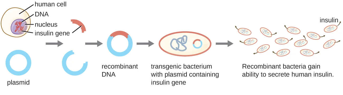
Lydia Villa-Komaroff, working in Walter Gilbert’s lab, demonstrated the groundbreaking power of biotechnology in 1978. The group was the first to show that a transgenic bacterial strain could be used to create human insulin (the diagram below). The insulin gene from humans was inserted into a plasmid. This recombinant DNA plasmid was then inserted into bacteria. As a result, these transgenic microbes are able to produce and secrete human insulin. Many prokaryotes are able to acquire foreign DNA and incorporate functional genes into their own genome through “mating” with other cells (conjugation), viral infection (transduction), and taking up DNA from the environment (transformation). Recall that these mechanisms are examples of horizontal gene transfer—the transfer of genetic material between cells of the same generation.
Extended description
Five stages, left to right. A human cell with its nucleus is shown holding the insulin gene on its DNA; an arrow shows the insulin gene being removed. A plain circular plasmid sits below the cell. An arrow leads to the insulin gene combined with the cut plasmid, forming a recombinant DNA plasmid. An arrow leads to an oval transgenic bacterium containing the recombinant plasmid inside it. A final arrow leads to several small oval bacteria, each still holding the plasmid, secreting dots labeled insulin into the surrounding solution.
Molecular Cloning
Herbert Boyer and Stanley Cohen first demonstrated the complete molecular cloning process in 1974 when they successfully cloned genes from the African clawed frog (Xenopus laevis) into a bacterial plasmid that was then introduced into the bacterial host Escherichia coli.
(Source note: the source dates this experiment to 1973. The 1973 paper by Cohen, Chang, Boyer, and Helling (PNAS 70 (1973): 3240–3244) joined bacterial plasmid DNA only; the Xenopus laevis ribosomal DNA was cloned into E. coli the following year (Morrow et al., PNAS 71 (1974): 1743–1747), so the page prints 1974.) Molecular cloning is a set of methods used to construct recombinant DNA and incorporate it into a host organism; it makes use of a number of molecular tools that are derived from microorganisms.
Restriction Enzymes and Ligases
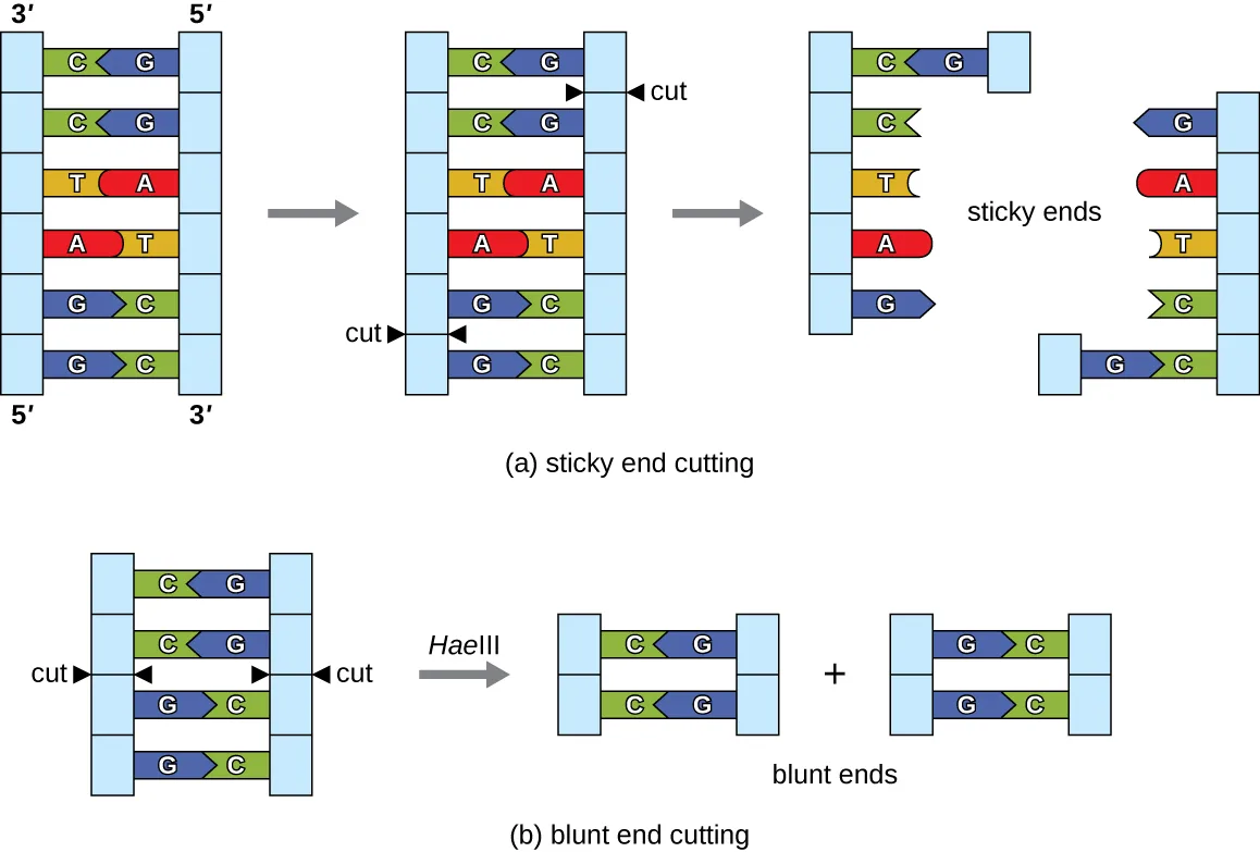
In recombinant DNA technology, DNA molecules are manipulated using naturally occurring enzymes derived mainly from bacteria and viruses. The creation of recombinant DNA molecules is possible due to the use of naturally occurring restriction endonucleases (restriction enzymes), bacterial enzymes produced as a protection mechanism to cut and destroy foreign cytoplasmic DNA that is most commonly a result of bacteriophage infection. Stewart Linn and Werner Arber discovered restriction enzymes in their 1960s studies of how E. coli limits bacteriophage replication on infection. Today, we use restriction enzymes extensively for cutting DNA fragments that can then be spliced into another DNA molecule to form recombinant molecules. Each restriction enzyme cuts DNA at a characteristic recognition site, a specific, usually palindromic, DNA sequence typically between four to six base pairs in length. A palindrome is a sequence of letters that reads the same forward as backward. (The word “level” is an example of a palindrome.) Palindromic DNA sequences contain the same base sequences in the 5′ to 3′ direction on one strand as in the 5′ to 3′ direction on the complementary strand. A restriction enzyme recognizes the DNA palindrome and cuts each backbone at identical positions in the palindrome. Some restriction enzymes cut to produce molecules that have complementary overhangs (sticky ends) while others cut without generating such overhangs, instead producing blunt ends (see the diagram below).
Molecules with complementary sticky ends can easily anneal, or form hydrogen bonds between complementary bases, at their sticky ends. The annealing step allows hybridization of the single-stranded overhangs. Hybridization refers to the joining together of two complementary single strands of DNA. Blunt ends can also attach together, but less efficiently than sticky ends due to the lack of complementary overhangs facilitating the process. In either case, ligation by DNA ligase can then rejoin the two sugar-phosphate backbones of the DNA through covalent bonding, making the molecule a continuous double strand. In 1972, Paul Berg, a Stanford biochemist, was the first to produce a recombinant DNA molecule using this technique, combining the SV40 monkey virus with E. coli bacteriophage lambda to create a hybrid.
Extended description
(a) A six-base-pair double helix reading GGATCC over CCTAGG, top strand 5′ to 3′ left to right; black arrows mark the cut points between the two G’s on each strand. After cutting, the two resulting fragments each carry a single-stranded 5′ GATC overhang — the sticky ends. (b) A four-base-pair double helix reading GGCC over CCGG; black arrows mark cut points directly between the G and the C on each strand. After cutting, both fragments have flush, blunt ends with no overhang.
Plasmids
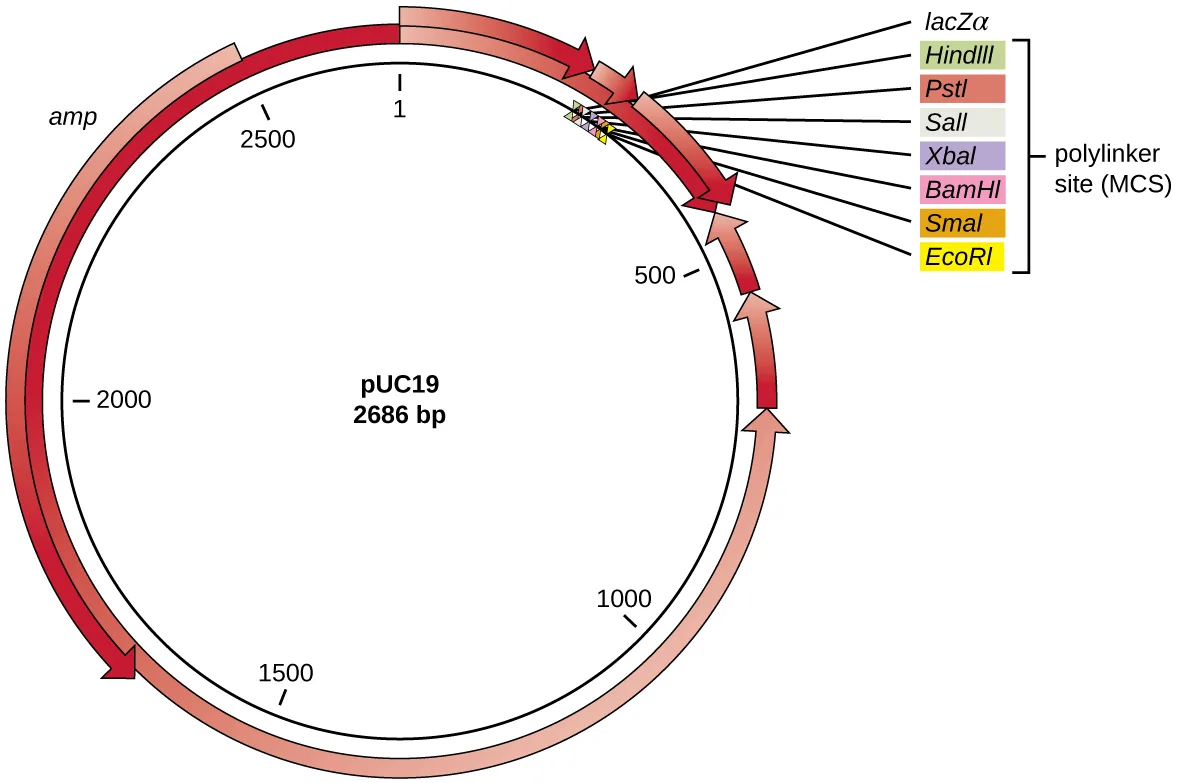
After restriction digestion, genes of interest are commonly inserted into plasmids, small pieces of typically circular, double-stranded DNA that replicate independently of the bacterial chromosome (see Unique Characteristics of Prokaryotic Cells). In recombinant DNA technology, plasmids are often used as vectors, DNA molecules that carry DNA fragments from one organism to another. Plasmids used as vectors can be genetically engineered by researchers and scientific supply companies to have specialized properties, as illustrated by the commonly used plasmid vector pUC19 (see the plasmid map below). Some plasmid vectors contain genes that confer antibiotic resistance; these resistance genes allow researchers to easily find plasmid-containing colonies by plating them on media containing the corresponding antibiotic. The antibiotic kills all host cells that do not harbor the desired plasmid vector, but those that contain the vector are able to survive and grow.
Plasmid vectors used for cloning typically have a polylinker site, or multiple cloning site (MCS). A polylinker site is a short sequence containing multiple unique restriction enzyme recognition sites that are used for inserting DNA into the plasmid after restriction digestion of both the DNA and the plasmid. Having these multiple restriction enzyme recognition sites within the polylinker site makes the plasmid vector versatile, so it can be used for many different cloning experiments involving different restriction enzymes.
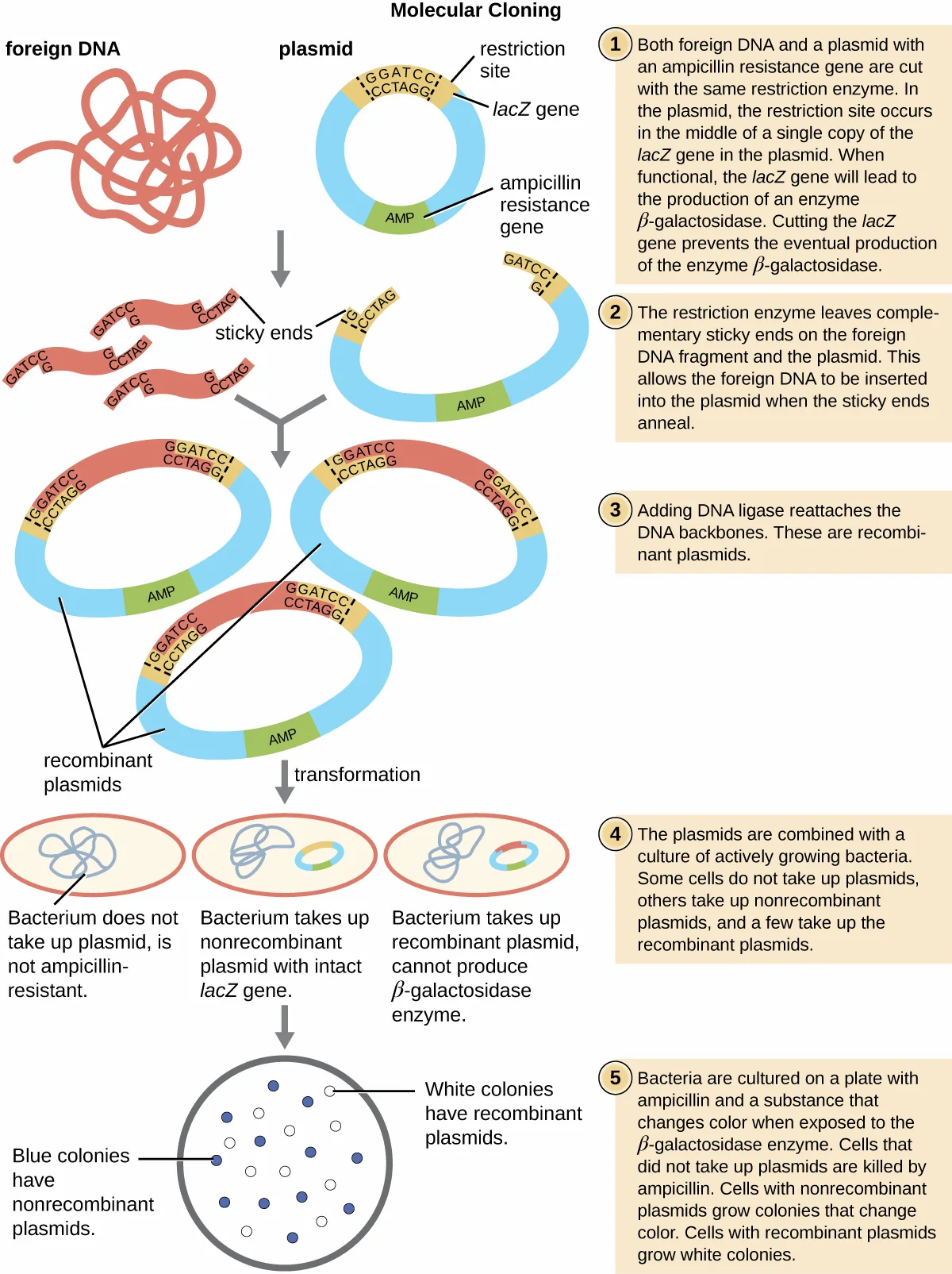
This polylinker site is often found within a reporter gene, another gene sequence artificially engineered into the plasmid that encodes a protein that allows for visualization of DNA insertion. The reporter gene allows a researcher to distinguish host cells that contain recombinant plasmids with cloned DNA fragments from host cells that only contain the non-recombinant plasmid vector. The most common reporter gene used in plasmid vectors is the bacterial lacZ gene encoding beta-galactosidase, an enzyme that naturally degrades lactose but can also degrade a colorless synthetic analog X-gal, thereby producing blue colonies on X-gal–containing media. The lacZ reporter gene is disabled when the recombinant DNA is spliced into the plasmid. Because the LacZ protein is not produced when the gene is disabled, X-gal is not degraded and white colonies are produced, which can then be isolated. This blue-white screening method is described later and shown in the flowchart below. In addition to these features, some plasmids come pre-digested and with an enzyme linked to the linearized plasmid to aid in ligation after the insertion of foreign DNA fragments.
Molecular Cloning using Transformation
The most commonly used mechanism for introducing engineered plasmids into a bacterial cell is transformation, a process in which bacteria take up free DNA from their surroundings. In nature, free DNA typically comes from other lysed bacterial cells; in the laboratory, free DNA in the form of recombinant plasmids is introduced to the cell’s surroundings.
Some bacteria, such as Bacillus spp., are naturally competent, meaning they are able to take up foreign DNA. However, not all bacteria are naturally competent. In most cases, bacteria must be made artificially competent in the laboratory by increasing the permeability of the cell membrane. This can be achieved through chemical treatments that neutralize charges on the cell membrane or by exposing the bacteria to an electric field that creates microscopic pores in the cell membrane. These methods yield chemically competent or electrocompetent bacteria, respectively.
Following the transformation protocol, bacterial cells are plated onto an antibiotic-containing medium to inhibit the growth of the many host cells that were not transformed by the plasmid conferring antibiotic resistance. A technique called blue-white screening is then used for lacZ-encoding plasmid vectors such as pUC19. Blue colonies have a functional beta-galactosidase enzyme because the lacZ gene is uninterrupted, with no foreign DNA inserted into the polylinker site. These colonies typically result from the digested, linearized plasmid religating to itself. However, white colonies lack a functional beta-galactosidase enzyme, indicating the insertion of foreign DNA within the polylinker site of the plasmid vector, thus disrupting the lacZ gene. Thus, white colonies resulting from this blue-white screening contain plasmids with an insert and can be further screened to characterize the foreign DNA. To be sure the correct DNA was incorporated into the plasmid, the DNA insert can then be sequenced.
Link to Learning
View an animation of molecular cloning from the DNA Learning Center.
Check Your Understanding
In blue-white screening, what does a blue colony mean and why is it blue?
Recall which colonies keep an uninterrupted lacZ gene and why that matters for the color reaction.Molecular Cloning Using Conjugation or Transduction
The bacterial process of conjugation (see How Asexual Prokaryotes Achieve Genetic Diversity) can also be manipulated for molecular cloning. F plasmids, or fertility plasmids, are transferred between bacterial cells through the process of conjugation. Recombinant DNA can be transferred by conjugation when bacterial cells containing a recombinant F plasmid are mixed with compatible bacterial cells lacking the plasmid. F plasmids encode a surface structure called an F pilus that facilitates contact between a cell containing an F plasmid and one without an F plasmid. On contact, a cytoplasmic bridge forms between the two cells and the F-plasmid-containing cell replicates its plasmid, transferring a copy of the recombinant F plasmid to the recipient cell. Once it has received the recombinant F plasmid, the recipient cell can produce its own F pilus and facilitate transfer of the recombinant F plasmid to an additional cell. The use of conjugation to transfer recombinant F plasmids to recipient cells is another effective way to introduce recombinant DNA molecules into host cells.
Alternatively, bacteriophages can be used to introduce recombinant DNA into host bacterial cells through a manipulation of the transduction process (see How Asexual Prokaryotes Achieve Genetic Diversity). In the laboratory, DNA fragments of interest can be engineered into phagemids, which are plasmids that have phage sequences that allow them to be packaged into bacteriophages. Bacterial cells can then be infected with these bacteriophages so that the recombinant phagemids can be introduced into the bacterial cells. Depending on the type of phage, the recombinant DNA may be integrated into the host bacterial genome (lysogeny), or it may exist as a plasmid in the host’s cytoplasm.
Extended description
Step 1: both foreign DNA and a plasmid with an ampicillin-resistance gene are cut with the same restriction enzyme; in the plasmid, the restriction site sits within a single copy of the lacZ gene, whose intact product, beta-galactosidase, is lost once the gene is cut. Step 2: the restriction enzyme leaves complementary sticky ends on the foreign DNA fragment and the plasmid, letting the foreign DNA anneal into the plasmid. Step 3: DNA ligase reattaches the DNA backbones, forming recombinant plasmids. Step 4: the plasmids are combined with a culture of growing bacteria; some bacteria take up no plasmid, others take up nonrecombinant plasmid with the lacZ gene intact, and a few take up recombinant plasmid with the lacZ gene disrupted. Step 5: the bacteria are cultured on a plate with ampicillin and a substance that changes color when acted on by beta-galactosidase; ampicillin kills bacteria that took up no plasmid, nonrecombinant-plasmid bacteria form blue colonies, and recombinant-plasmid bacteria form white colonies.
Check Your Understanding
What is the original function of a restriction enzyme?
Recall why bacteria naturally produce these enzymes, before researchers ever put them to use in the laboratory.What two processes are exploited to get recombinant DNA into a bacterial host cell?
Show model answer
Did your answer mention:
Distinguish the uses of an antibiotic resistance gene and a reporter gene in a plasmid vector.
Show model answer
Did your answer mention:
Creating a Genomic Library
Molecular cloning may also be used to generate a genomic library. The library is a complete (or nearly complete) copy of an organism’s genome contained as recombinant DNA plasmids engineered into unique clones of bacteria. Having such a library allows a researcher to create large quantities of each fragment by growing the bacterial host for that fragment. These fragments can be used to determine the sequence of the DNA and the function of any genes present.
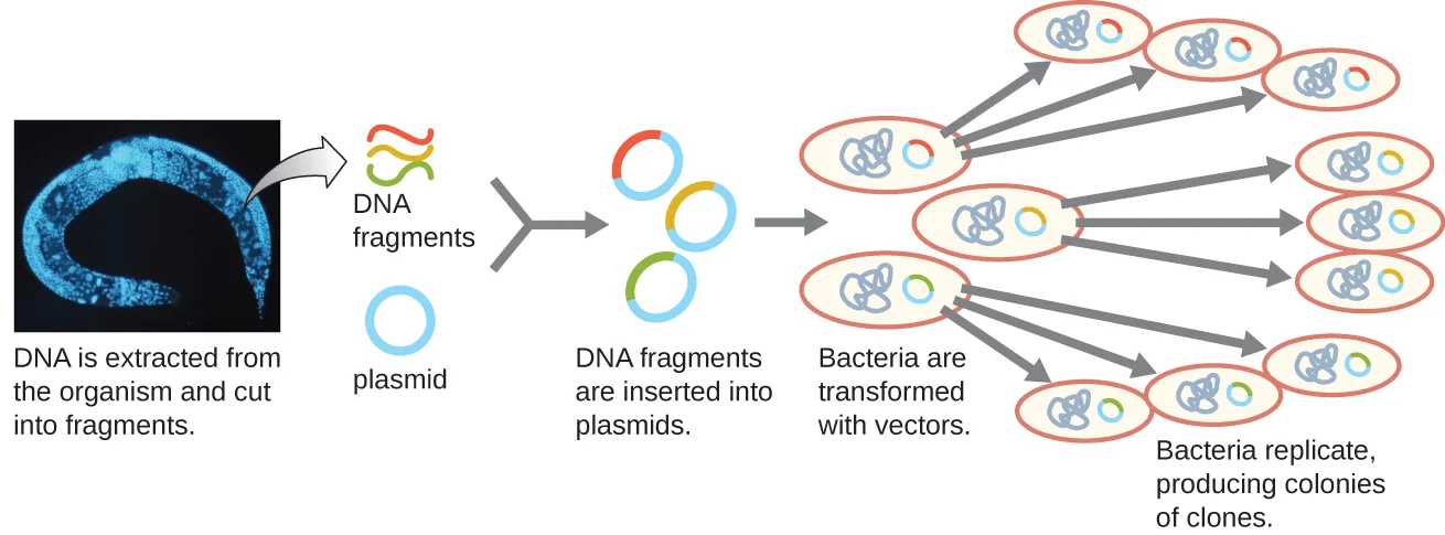
One method for generating a genomic library is to ligate individual restriction enzyme-digested genomic fragments into plasmid vectors cut with the same restriction enzyme (see the diagram below). After transformation into a bacterial host, each transformed bacterial cell takes up a single recombinant plasmid and grows into a colony of cells. All of the cells in this colony are identical clones and carry the same recombinant plasmid. The resulting library is a collection of colonies, each of which contains a fragment of the original organism’s genome, that are each separate and distinct and can each be used for further study. This makes it possible for researchers to screen these different clones to discover the one containing a gene of interest from the original organism’s genome.
Extended description
The figure begins with a blue-tinted micrograph of the source organism’s tissue, labeled ‘DNA is extracted from the organism and cut into fragments.’ An arrow leads to several short, colored DNA fragments and a plain circular plasmid, labeled ‘DNA fragments’ and ‘plasmid.’ A further arrow leads to circular recombinant plasmids, each holding one colored fragment, labeled ‘DNA fragments are inserted into plasmids.’ Arrows lead to several ovals of bacteria, each transformed with one recombinant plasmid, labeled ‘Bacteria are transformed with vectors.’ Final arrows fan out to clusters of identical bacterial colonies, each fanning out from one transformed cell, labeled ‘Bacteria replicate, producing colonies of clones.’
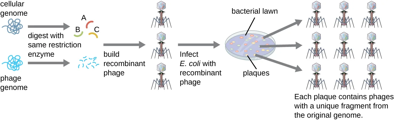
To construct a genomic library using larger fragments of genomic DNA, an E. coli bacteriophage, such as lambda, can be used as a host (see the diagram below). Genomic DNA can be sheared or enzymatically digested and ligated into a pre-digested bacteriophage lambda DNA vector. Then, these recombinant phage DNA molecules can be packaged into phage particles and used to infect E. coli host cells on a plate. During infection within each cell, each recombinant phage will make many copies of itself and lyse the E. coli lawn, forming a plaque. Thus, each plaque from a phage library represents a unique recombinant phage containing a distinct genomic DNA fragment. Plaques can then be screened further to look for genes of interest. One advantage to producing a library using phages instead of plasmids is that a phage particle holds a much larger insert of foreign DNA compared with a plasmid vector, thus requiring a much smaller number of cultures to fully represent the entire genome of the original organism.
Extended description
Left to right: a tangled loop labeled ‘cellular genome’ and a phage particle labeled ‘phage genome’ are each cut, labeled ‘digest with same restriction enzyme,’ producing short colored fragments (labeled A, B, C) from the cellular genome and cut phage DNA. An arrow labeled ‘build recombinant phage’ leads to several phage particles, each packaging one colored fragment. An arrow labeled ‘Infect E. coli with recombinant phage’ leads to a shaded plate labeled ‘bacterial lawn,’ where clear plaques form; each plaque is labeled as containing phages with a unique fragment from the original genome.
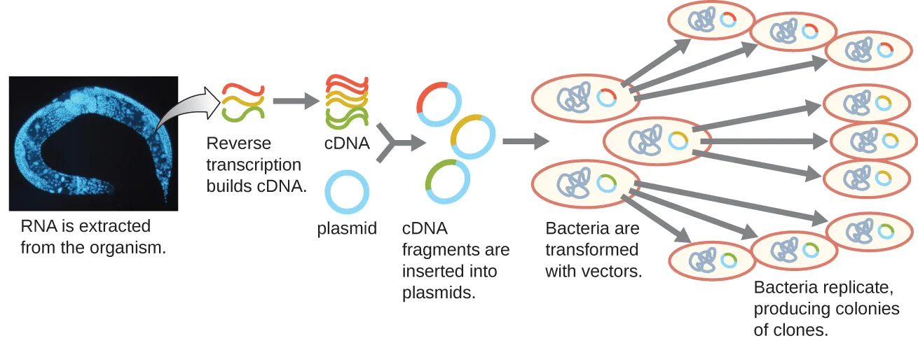
To focus on the expressed genes in an organism or even a tissue, researchers construct libraries using the organism’s messenger RNA (mRNA) rather than its genomic DNA. Whereas all cells in a single organism will have the same genomic DNA, different tissues express different genes, producing different complements of mRNA. For example, all human cells’ genomic DNA contains the gene for insulin, but only cells in the pancreas express mRNA directing the production of insulin. Because mRNA cannot be cloned directly, in the laboratory mRNA must be used as a template by the retroviral enzyme reverse transcriptase to make complementary DNA (cDNA). A cell’s full complement of mRNA can be reverse-transcribed into cDNA molecules, which can be used as a template for DNA polymerase to make double-stranded DNA copies; these fragments can subsequently be ligated into either plasmid vectors or bacteriophage to produce a cDNA library. The benefit of a cDNA library is that it contains DNA from only the expressed genes in the cell. This means that the introns, control sequences such as promoters, and DNA not destined to be translated into proteins are not represented in the library. The focus on translated sequences means that the library cannot be used to study the sequence and structure of the genome in its entirety. The construction of a cDNA genomic library is shown below.
Extended description
The figure begins with a blue-tinted micrograph of the source organism’s tissue, labeled ‘RNA is extracted from the organism.’ An arrow labeled ‘Reverse transcription builds cDNA’ leads to colored double-stranded cDNA fragments and a plain circular plasmid. A further arrow leads to circular recombinant plasmids, each holding one cDNA fragment, labeled ‘cDNA fragments are inserted into plasmids.’ Arrows lead to ovals of bacteria, each transformed with one recombinant plasmid, labeled ‘Bacteria are transformed with vectors,’ and finally to clusters of identical bacterial colonies fanning out from each transformed cell, labeled ‘Bacteria replicate, producing colonies of clones.’
Check Your Understanding
What are the hosts for the genomic libraries described in this section?
Both the plasmid-based and the phage-based methods of building a genomic library ultimately grow their recombinant DNA inside the same kind of cell.What is cDNA?
Recall which molecule reverse transcriptase uses as its template to make this DNA copy.Introducing Recombinant Molecules into Eukaryotic Hosts
The use of bacterial hosts for genetic engineering laid the foundation for recombinant DNA technology; however, researchers have also had great interest in genetically engineering eukaryotic cells, particularly those of plants and animals. The introduction of recombinant DNA molecules into eukaryotic hosts is called transfection. Genetically engineered plants, called transgenic plants, are of significant interest for agricultural and pharmaceutical purposes. The first transgenic plant sold commercially was the Flavr Savr delayed-ripening tomato, which came to market in 1994. Genetically engineered livestock have also been successfully produced, resulting, for example, in pigs with increased nutritional value (Liangxue Lai, Jing X. Kang, Rongfeng Li, Jingdong Wang, William T. Witt, Hwan Yul Yong, Yanhong Hao et al., “Generation of Cloned Transgenic Pigs Rich in Omega-3 Fatty Acids,” Nature Biotechnology 24 no. 4 (2006): 435–436) and goats that secrete pharmaceutical products in their milk (Raylene Ramos Moura, Luciana Magalhães Melo, and Vicente José de Figueirêdo Freitas, “Production of Recombinant Proteins in Milk of Transgenic and Non-Transgenic Goats,” Brazilian Archives of Biology and Technology 54 no. 5 (2011): 927–938).
Electroporation
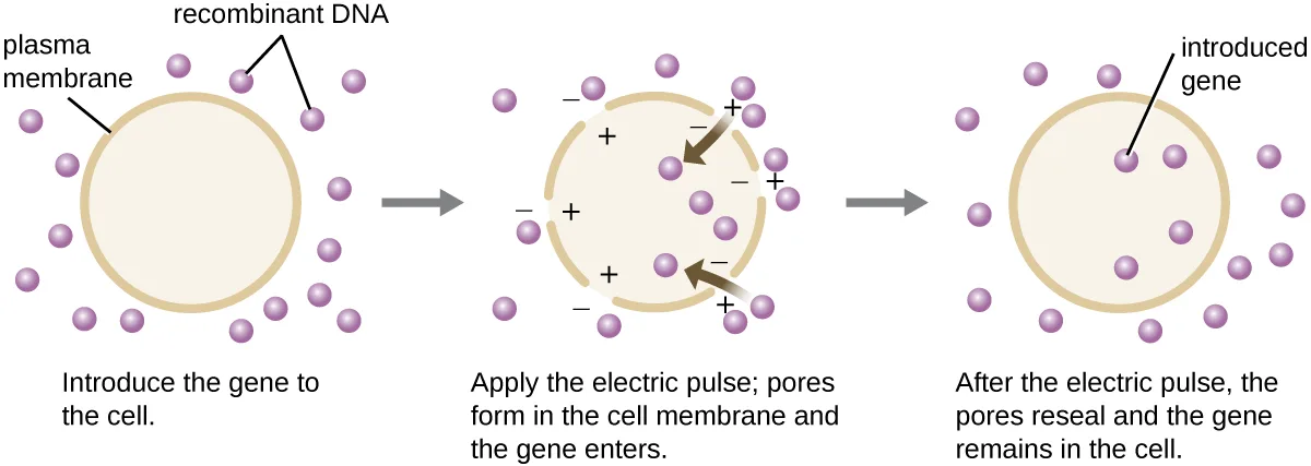
Compared to bacterial cells, eukaryotic cells tend to be less amenable as hosts for recombinant DNA molecules. Because eukaryotes are typically neither competent to take up foreign DNA nor able to maintain plasmids, transfection of eukaryotic hosts is far more challenging and requires more intrusive techniques for success. One method used for transfecting cells in cell culture is called electroporation. A brief electric pulse induces the formation of transient pores in the phospholipid bilayers of cells through which the gene can be introduced. At the same time, the electric pulse generates a short-lived positive charge on one side of the cell’s interior and a negative charge on the opposite side; the charge difference draws negatively charged DNA molecules into the cell (see the diagram below).
Microinjection
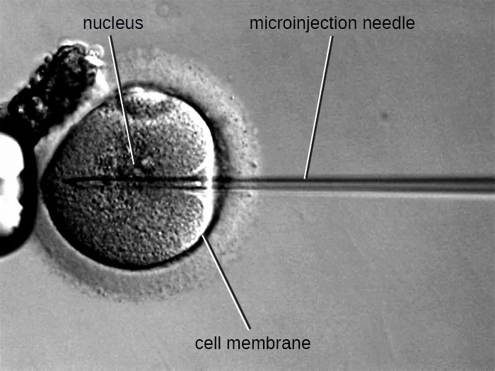
An alternative method of transfection is called microinjection. Because eukaryotic cells are typically larger than those of prokaryotes, DNA fragments can sometimes be directly injected into the cytoplasm using a glass micropipette, as shown below.
Gene Guns
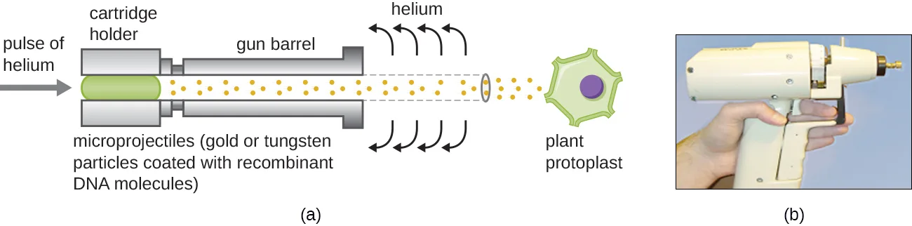
Transfecting plant cells can be even more difficult than animal cells because of their thick cell walls. One approach involves treating plant cells with enzymes to remove their cell walls, producing protoplasts. Then, a gene gun is used to shoot gold or tungsten particles coated with recombinant DNA molecules into the plant protoplasts at high speeds. Recipient protoplast cells can then recover and be used to generate new transgenic plants (see the diagram below).
Shuttle Vectors
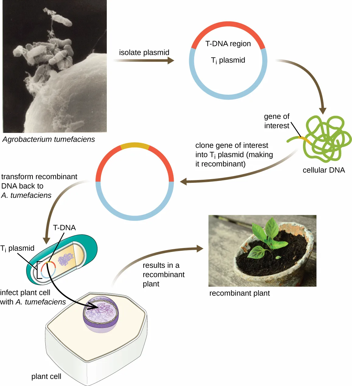
Another method of transfecting plants involves shuttle vectors, plasmids that can move between bacterial and eukaryotic cells. The tumor-inducing (Ti) plasmids originating from the bacterium Agrobacterium tumefaciens are commonly used as shuttle vectors for incorporating genes into plants (see the diagram below). In nature, the Ti plasmids of A. tumefaciens cause plants to develop tumors when they are transferred from bacterial cells to plant cells. Researchers have been able to manipulate these naturally occurring plasmids to remove their tumor-causing genes and insert desirable DNA fragments. The resulting recombinant Ti plasmids can be transferred into the plant genome through the natural transfer of Ti plasmids from the bacterium to the plant host. Once inside the plant host cell, the gene of interest recombines into the plant cell’s genome.
Extended description
The diagram runs in a loop. Top left: a black-and-white micrograph of several rod-shaped Agrobacterium tumefaciens cells. An arrow labeled ‘isolate plasmid’ leads to a circular Ti plasmid with a T-DNA region inside the plasmid ring. A curved arrow labeled ‘clone gene of interest into Ti plasmid (making it recombinant)’ leads from a tangled loop of cellular DNA holding the gene of interest into the plasmid, now redrawn with the gene added within the T-DNA region. An arrow labeled ’transform recombinant DNA back to A. tumefaciens’ leads down to a drawing of an A. tumefaciens cell holding the Ti plasmid and its T-DNA, shown infecting a plant cell at ‘infect plant cell with A. tumefaciens.’ An arrow labeled ‘results in a recombinant plant’ leads right to a color photograph of a small seedling growing in a pot of soil, labeled ‘recombinant plant.’
Viral Vectors
Viral vectors can also be used to transfect eukaryotic cells. In fact, this method is often used in gene therapy to introduce healthy genes into human patients suffering from diseases that result from genetic mutations. Viral genes can be deleted and replaced with the gene to be delivered to the patient (William S.M. Wold and Karoly Toth, “Adenovirus Vectors for Gene Therapy, Vaccination and Cancer Gene Therapy,” Current Gene Therapy 13 no. 6 (2013): 421); the virus then infects the host cell and delivers the foreign DNA into the genome of the targeted cell. Adenoviruses are often used for this purpose because they can be grown to high titer and can infect both nondividing and dividing host cells. However, use of viral vectors for gene therapy can pose some risks for patients, as discussed in Gene Therapy.
Check Your Understanding
What are the methods used to introduce recombinant DNA vectors into animal cells?
Show model answer
Did your answer mention:
Compare and contrast shuttle vectors and viral vectors.
Show model answer
Did your answer mention:
Summary
- Biotechology is the science of utilizing living systems to benefit humankind. In recent years, the ability to directly alter an organism’s genome through genetic engineering has been made possible due to advances in recombinant DNA technology, which allows researchers to create recombinant DNA molecules with new combinations of genetic material.
- Molecular cloning involves methods used to construct recombinant DNA and facilitate their replication in host organisms. These methods include the use of restriction enzymes (to cut both foreign DNA and plasmid vectors), ligation (to paste fragments of DNA together), and the introduction of recombinant DNA into a host organism (often bacteria).
- Blue-white screening allows selection of bacterial transformants that contain recombinant plasmids using the phenotype of a reporter gene that is disabled by insertion of the DNA fragment.
- Genomic libraries can be made by cloning genomic fragments from one organism into plasmid vectors or into bacteriophage.
- cDNA libraries can be generated to represent the mRNA molecules expressed in a cell at a given point.
- Transfection of eukaryotic hosts can be achieved through various methods using electroporation, gene guns, microinjection, shuttle vectors, and viral vectors.
Key terms
- biotechnology — the science of using living systems to benefit humankind.
- genetic engineering — the direct alteration of an organism’s genetics to achieve desirable traits.
- recombinant DNA technology — the process by which DNA from one organism is cut and new pieces of foreign DNA from a second organism are inserted, artificially creating new combinations of genetic material within the organism.
- recombinant DNA molecule — a DNA molecule resulting from the cutting and insertion of DNA from one organism into the DNA of another organism, resulting in a new combination of genetic material.
- transgenic — describing an organism into which foreign DNA from a different species has been introduced.
- molecular cloning — the purposeful fragmentation of DNA followed by attachment to another piece of DNA to produce a recombinant molecule, followed by introduction of this recombinant molecule into an easily manipulated host to allow for the creation of multiple copies of a gene of interest.
- restriction endonucleases (restriction enzymes) — bacterial enzyme that cuts DNA fragments at a unique, often palindromic, recognition site; used in genetic engineering for splicing DNA fragments together into recombinant molecules.
- recognition site — a specific, often palindromic, DNA sequence recognized by a restriction enzyme that is typically four to six base pairs long and reads the same in the 5′ to 3′ direction on one strand as it does in the 5′ to 3′ direction on the complementary strand.
- sticky ends — short, single-stranded complementary overhangs that may be produced when many restriction enzymes cut DNA.
- blunt ends — ends of DNA molecules lacking single-stranded complementary overhangs that are produced when some restriction enzymes cut DNA.
- anneal — formation of hydrogen bonds between the nucleotide base pairs of two single-stranded complementary nucleic acid sequences.
- hybridization — the joining of two complementary single-stranded DNA molecules.
- ligation — repair of the sugar-phosphate backbone of the DNA, making the DNA molecule continuous.
- vectors — DNA molecules that carry DNA fragments from one organism to another.
- polylinker site (multiple cloning site, MCS) — a short sequence containing multiple unique restriction enzyme recognition sites that are used for inserting foreign DNA into the plasmid after restriction digestion of both the foreign DNA and the plasmid.
- reporter gene — genes that encode easily observable characteristics, allowing for their expression to be easily monitored.
- blue-white screening — a technique commonly used for identifying transformed bacterial cells containing recombinant plasmids using lacZ-encoding plasmid vectors.
- phagemids — a plasmid capable of being replicated as a plasmid and also incorporated into a phage head.
- genomic library — a repository of an organism’s entire genome maintained as cloned fragments in the genomes of strains of a host organism.
- clones — a genetically identical cell or individual.
- complementary DNA (cDNA) — a DNA molecule complementary to mRNA that is made through the activity of reverse transcriptase.
- transfection — the introduction of recombinant DNA molecules into eukaryotic hosts.
- electroporation — a genetic engineering technique in which cells are exposed to a short electric pulse, inducing them to take up DNA molecules from their environment.
- microinjection — the direct injection of DNA into the cytoplasm of a eukaryotic cell using a glass micropipette.
- gene gun — an apparatus that shoots gold or tungsten particles coated with recombinant DNA molecules at high speeds into plant protoplasts.
- shuttle vectors — a plasmid that can move between bacterial and eukaryotic cells.
- tumor-inducing (Ti) plasmids — a naturally occurring plasmid of the bacterium Agrobacterium tumefaciens that researchers use as a shuttle vector to introduce a desired DNA fragment into plant cells.
Practice
Identify tools of molecular genetics that are derived from microorganisms
Which of the following is required for repairing the phosphodiester backbone of DNA during molecular cloning?
Recall which enzyme rejoins the two sugar-phosphate backbones after sticky or blunt ends anneal.The enzyme that uses RNA as a template to produce a DNA copy is called:
Name the retroviral enzyme used to build cDNA from mRNA.Is biotechnology always associated with genetic engineering?
Recall the section’s opening distinction between biotechnology in a broad, technical sense and biotechnology in its contemporary, genetic-engineering sense.Describe the methods used to create recombinant DNA molecules
In blue-white screening, what do blue colonies represent?
Recall which colony color results from the lacZ gene being uninterrupted, whether or not the cell took up a plasmid at all.Recombination is a process not usually observed in nature.
Recall that horizontal gene transfer by conjugation, transduction, and transformation, all of which rely on recombination, occur naturally in bacteria.Which is more efficient: blunt-end cloning or sticky-end cloning? Why?
Recall which kind of end lacks a complementary overhang to facilitate the annealing step.Plasmid vectors used for cloning typically have a short sequence containing multiple unique restriction enzyme recognition sites, called a ________ site.
Name the short sequence within a reporter gene where foreign DNA is inserted after restriction digestion.Describe methods used to introduce DNA into prokaryotic cells
All of the following are processes used to introduce DNA molecules into bacterial cells except:
Three of these four terms name mechanisms of horizontal gene transfer this book covers; the fourth is a step of gene expression.It is generally easier to introduce recombinant DNA into prokaryotic cells than into eukaryotic cells.
Recall how this section describes eukaryotic cells’ competence for taking up foreign DNA compared with bacteria’s.Name three elements incorporated into a plasmid vector for efficient cloning.
Show model answer
Did your answer mention:
List the types of genomic libraries and describe their uses
When would a scientist want to generate a cDNA library instead of a genomic library?
Recall what a cDNA library is built from, and how that differs from what a genomic library represents.What is one advantage of generating a genomic library using phages instead of plasmids?
Recall what a single phage particle can carry compared with a single plasmid vector.A repository of an organism’s entire genome maintained as cloned fragments in the genomes of strains of a host organism is called a ________.
Name the term this section defines at the start of the subsection on constructing this kind of collection.Describe the methods used to introduce DNA into eukaryotic cells
The Ti plasmid is used for introducing genes into:
Recall which organism’s cells the Ti plasmid of Agrobacterium tumefaciens naturally transfers its T-DNA into.The process of introducing DNA molecules into eukaryotic cells is called ________.
Name the eukaryotic counterpart to bacterial transformation and conjugation.Plasmids that can move between bacterial and eukaryotic cells are called ________.
Name the class of plasmid that includes the Ti plasmid of Agrobacterium tumefaciens.This section is adapted from Microbiology, Section 12.1: Microbes and the Tools of Genetic Engineering by Nina Parker, Mark Schneegurt, Anh-Hue Thi Tu, Philip Lister, Brian M. Forster, and OpenStax, © OpenStax, licensed under CC BY-NC-SA 4.0. Access the original for free at openstax.org. Changes: all eleven source figures are re-encoded as WebP and rendered as mediafigures; the media manifest guesses kind="photo" for all eleven because every source file is a JPEG, which is correct only for the microinjection micrograph — the other ten (RecombTech, Recuts, pUC19, MolCloning, CloneLibry, PhageLibry, cDNALibry, electropor, GeneGun, and Ti) are explicit kind="diagram" because each is a genuinely drawn, labeled figure, GeneGun and Ti included even though each also carries a photograph panel. The pUC19 alt is corrected for five source typos (“pC19” → pUC19, “plylinker” → polylinker, “HidIII” → HindIII, “PSTI” → PstI, “SaII” → SalI); the MolCloning alt is corrected for three source typos (“DNa” → DNA, “resistang” → resistant, “nto” → not); the Ti alt is corrected for two source typos (“inseted” → inserted, “resuts” → results). Longdesc walk-throughs are added for RecombTech, Recuts, MolCloning, CloneLibry, PhageLibry, cDNALibry, and Ti because each is a multi-step or multi-panel figure whose labelled stages the caption does not name individually. All five body Check Your Understanding boxes are rendered as body items at their note positions: of the eight bullets, four are graded (three multiple choice and one textin) because a single sentence or definition of this module fixes the whole answer, and four are self-checks with model answers and rubrics assembled only from this module’s own sentences, because their honest answers require assembling more than one sentence (the two-process, the antibiotic-resistance-versus-reporter-gene, and the two eukaryotic-transfection self-checks). The graded conversions draw their distractors from this module’s own sibling terms and mechanisms (other transfection methods, other library types, other cloning outcomes), never invented content. Of the module’s thirteen source exercises, all five Multiple Choice, both True/False (as two-option multiple choice), and the one Fill in the Blank are adapted into Practice; of the three unkeyed Short Answer and two unkeyed Critical Thinking questions, two Short Answer and both Critical Thinking questions convert to multiple choice because one sentence of this module fixes their whole answer, and the third Short Answer question (the three plasmid-vector elements) stays a self-check because its honest answer assembles three separate sentences. Two filler textin items (the polylinker/MCS term and the genomic library term) and one filler textin (shuttle vectors) are author-written strictly from this module’s own Key terms to fill the Describe the methods used to create recombinant DNA molecules and Describe the methods used to introduce DNA into eukaryotic cells objectives to the three-item floor. Feature boxes are rendered as callouts: Clinical Focus Part 1 keeps its closing question as an unanswered plain bullet and links forward to Visualizing and Characterizing DNA, RNA, and Protein, where the case continues; the Link to Learning keeps its animation link. The cross-references to Unique Characteristics of Prokaryotic Cells, How Asexual Prokaryotes Achieve Genetic Diversity (twice), and Gene Therapy (twice) are rendered as links, since all land in this run. Key terms are compiled from the module’s 30 distinct defined terms (31 <term> elements; blue-white screening is defined twice and gets one bullet) and the book’s Glossary appendix, rendered as 27 bullets: recombinant DNA and molecule have no distinct appendix entry and are merged into the one bullet recombinant DNA molecule, using the nearest appendix headword’s meaning; restriction enzymes has no distinct appendix entry and is merged into the restriction endonucleases (restriction enzymes) bullet; anneal has no distinct appendix entry and takes the appendix’s annealing definition under the module’s own headword; multiple cloning site (MCS) has no distinct appendix entry and is merged with polylinker site into one bullet, polylinker site (multiple cloning site, MCS); vectors has a doubled appendix entry (an arthropod-vector sense and a DNA-vector sense), and only the DNA-molecule clause is used, matching this module’s sense. The year of the Boyer–Cohen Xenopus cloning experiment is corrected from the source’s 1973 to 1974 with a visible Source note (the 1973 paper joined bacterial plasmids only; the frog-gene clone is Morrow et al. 1974).