Testing the Effectiveness of Antiseptics and Disinfectants
By the end of this section, you will be able to:
- Describe why the phenol coefficient is used
- Compare and contrast the disk-diffusion, use-dilution, and in-use methods for testing the effectiveness of antiseptics, disinfectants, and sterilants
The effectiveness of various chemical disinfectants is reflected in the terms used to describe them. Chemical disinfectants are grouped by the power of their activity, with each category reflecting the types of microbes and viruses its component disinfectants are effective against. High-level germicides have the ability to kill vegetative cells, fungi, viruses, and endospores, leading to sterilization, with extended use. Intermediate-level germicides, as their name suggests, are less effective against endospores and certain viruses, and low-level germicides kill only vegetative cells and certain enveloped viruses, and are ineffective against endospores.
However, several environmental conditions influence the potency of an antimicrobial agent and its effectiveness. For example, length of exposure is particularly important, with longer exposure increasing efficacy. Similarly, the concentration of the chemical agent is also important, with higher concentrations being more effective than lower ones. Temperature, pH, and other factors can also affect the potency of a disinfecting agent.
One method to determine the effectiveness of a chemical agent includes swabbing surfaces before and after use to confirm whether a sterile field was maintained during use. Additional tests are described in the sections that follow. These tests allow for the maintenance of appropriate disinfection protocols in clinical settings, controlling microbial growth to protect patients, health-care workers, and the community.
Phenol Coefficient
The effectiveness of a disinfectant or antiseptic can be determined in a number of ways. Historically, a chemical agent’s effectiveness was often compared with that of phenol, the first chemical agent used by Joseph Lister. In 1903, British chemists Samuel Rideal (1863–1929) and J. T. Ainslie Walker (1868–1930) established a protocol to compare the effectiveness of a variety of chemicals with that of phenol, using as their test organisms Staphylococcus aureus (a gram-positive bacterium) and Salmonella enterica serovar Typhi (a gram-negative bacterium). They exposed the test bacteria to the antimicrobial chemical solutions diluted in water for 7.5 minutes. They then calculated a phenol coefficient for each chemical for each of the two bacteria tested. A phenol coefficient of 1.0 means that the chemical agent has about the same level of effectiveness as phenol. A chemical agent with a phenol coefficient of less than 1.0 is less effective than phenol. An example is formalin, with phenol coefficients of 0.3 (S. aureus) and 0.7 (S. enterica serovar Typhi). A chemical agent with a phenol coefficient greater than 1.0 is more effective than phenol, such as chloramine, with phenol coefficients of 133 and 100, respectively. Although the phenol coefficient was once a useful measure of effectiveness, it is no longer commonly used because the conditions and organisms used were arbitrarily chosen.
Check Your Understanding
What are the differences between the three levels of disinfectant effectiveness? Sort each phrase under the germicide level it describes.
High-level germicides
Intermediate-level germicides
Low-level germicides
Disk-Diffusion Method
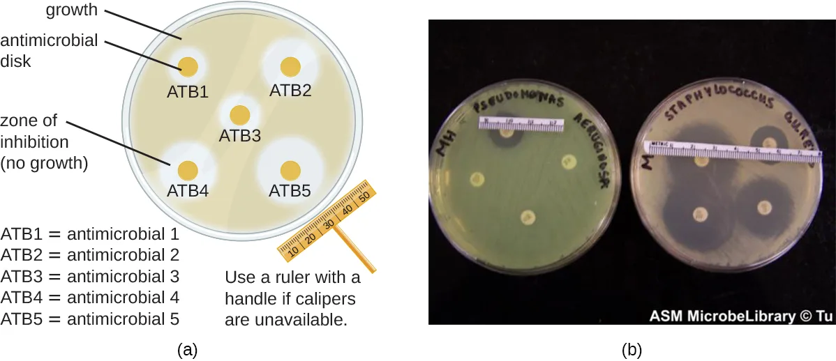
The disk-diffusion method involves applying different chemicals to separate, sterile filter paper disks (shown below). The disks are then placed on an agar plate that has been inoculated with the targeted bacterium and the chemicals diffuse out of the disks into the agar where the bacteria have been inoculated. As the “lawn” of bacteria grows, zones of inhibition of microbial growth are observed as clear areas around the disks. Although there are other factors that contribute to the sizes of zones of inhibition (e.g., whether the agent is water soluble and able to diffuse in the agar), larger zones typically correlate to increased inhibition effectiveness of the chemical agent. The diameter across each zone is measured in millimeters.
Extended description
Panel (a): a circular agar plate with a beige lawn of bacterial growth labeled ‘growth’ near the rim. Five small tan antimicrobial disks are arranged on the plate, labeled ATB1, ATB2, ATB3, ATB4, and ATB5; each disk sits inside a lighter, clear ring labeled ‘zone of inhibition (no growth),’ and the width of the clear ring varies from disk to disk. A caliper-style ruler icon beside the plate carries the instruction ‘Use a ruler with a handle if calipers are unavailable.’ Panel (b): two photographed agar plates side by side, one hand-labeled for a lawn of Pseudomonas aeruginosa and one for a lawn of Staphylococcus aureus, each with a metric measuring strip laid across it and four antimicrobial disks producing clear zones of different sizes.
Check Your Understanding
When comparing the activities of two disinfectants against the same microbe, using the disk-diffusion assay, and assuming both are water soluble and can easily diffuse in the agar, would a more effective disinfectant have a larger zone of inhibition or a smaller one?
Recall what the passage says happens to the size of the zone of inhibition as a chemical disinfectant becomes more effective.Use-Dilution Test
Other methods are also used for measuring the effectiveness of a chemical agent in clinical settings. The use-dilution test is commonly used to determine a chemical’s disinfection effectiveness on an inanimate surface. For this test, a cylinder of stainless steel is dipped in a culture of the targeted microorganism and then dried. The cylinder is then dipped in solutions of disinfectant at various concentrations for a specified amount of time. Finally, the cylinder is transferred to a new test tube containing fresh sterile medium that does not contain disinfectant, and this test tube is incubated. Bacterial survival is demonstrated by the presence of turbidity in the medium, whereas killing of the target organism on the cylinder by the disinfectant will produce no turbidity.
The Association of Official Agricultural Chemists International (AOAC), a nonprofit group that establishes many protocol standards, has determined that a minimum of 59 of 60 replicates must show no growth in such a test to achieve a passing result, and the results must be repeatable from different batches of disinfectant and when performed on different days. Disinfectant manufacturers perform use-dilution tests to validate the efficacy claims for their products, as designated by the EPA.
Check Your Understanding
Is the use-dilution test performed in a clinical setting? Why?
Show model answer
Did your answer mention:
In-Use Test
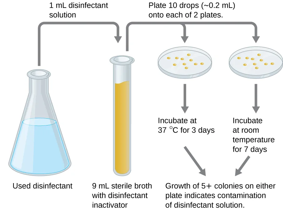
An in-use test can determine whether an actively used solution of disinfectant in a clinical setting is microbially contaminated (shown below). A 1-mL sample of the used disinfectant is diluted into 9 mL of sterile broth medium that also contains a compound to inactivate the disinfectant. Ten drops, totaling approximately 0.2 mL of this mixture, are then inoculated onto each of two agar plates. One plate is incubated at 37 °C for 3 days and the other is incubated at room temperature for 7 days. The plates are monitored for growth of microbial colonies. Growth of five or more colonies on either plate suggests that viable microbial cells existed in the disinfectant solution and that it is contaminated. Such in-use tests monitor the effectiveness of disinfectants in the clinical setting.
Check Your Understanding
What does a positive in-use test indicate?
Recall what the module says a result of five or more colonies growing on either plate means for that disinfectant solution.New Methods to Overcome Specific Challenges
Some pathogens have characteristics that make disinfectant efficacy difficult to prove. Human papilloma virus (HPV), for example, cannot be cultured, so the previously described methods do not work. And since HPV’s protein-only capsid enables it to survive on surfaces for an extended period of time, medical professionals rely on disinfectants to ensure the virus doesn’t spread through facilities or equipment.
New Mexico State University’s Michelle Ozbun, one of the foremost experts on HPV, developed a method to quantify (count) live virus on surfaces outside the body. The typical method for counting viruses like HPV is to grind up infected cells and measure the amount of viral DNA, but this method is imprecise and does not demonstrate the scope of the infection. To test the disinfectant’s live virus was required. Instead of grinding up infected cells, Ozbun applied a stain that only attaches to viral RNA.
Using advanced microscopy, the quantities of infected cells can be determined with great accuracy. For the HPV disinfectant testing, Ozbun and her team applied various disinfectants to three HPV from three sources, and determined that the chemicals reduced the amount of virus by 10,000 times. This technique can be used for disinfectant testing for other protein-capsid viruses (like norovirus and poliovirus) and to produce accurate viral counting for other research or clinical applications.
Clinical Focus. Resolution
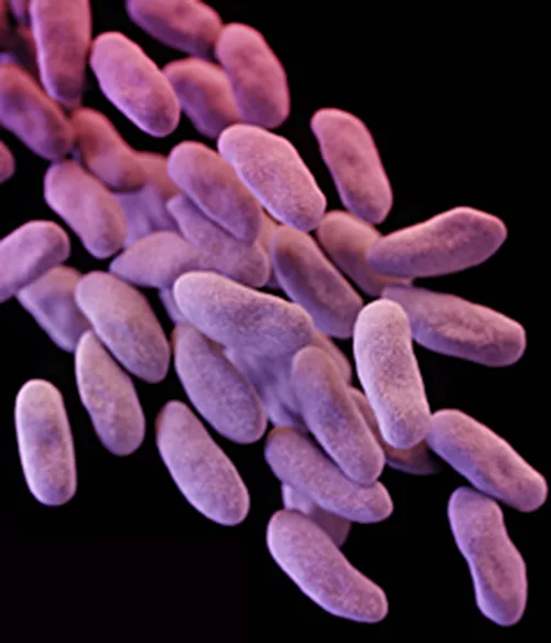
Despite antibiotic treatment, Roberta’s symptoms worsened. She developed pyelonephritis, a severe kidney infection, and was rehospitalized in the intensive care unit (ICU). Her condition continued to deteriorate, and she developed symptoms of septic shock. At this point, her physician ordered a culture from her urine to determine the exact cause of her infection, as well as a drug sensitivity test to determine what antibiotics would be effective against the causative bacterium. The results of this test indicated resistance to a wide range of antibiotics, including the carbapenems, a class of antibiotics that are used as the last resort for many types of bacterial infections. This was an alarming outcome, suggesting that Roberta’s infection was caused by a so-called superbug: a bacterial strain that has developed resistance to the majority of commonly used antibiotics. In this case, the causative agent belonged to the carbapenem-resistant Enterobacteriaceae (CRE), a drug-resistant family of bacteria normally found in the digestive system (shown below). When CRE is introduced to other body systems, as might occur through improperly cleaned surgical instruments, catheters, or endoscopes, aggressive infections can occur.
CRE infections are notoriously difficult to treat, with a 40%–50% fatality rate. To treat her kidney infection and septic shock, Roberta was treated with dialysis, intravenous fluids, and medications to maintain blood pressure and prevent blood clotting. She was also started on aggressive treatment with intravenous administration of a new drug called tigecycline, which has been successful in treating infections caused by drug-resistant bacteria.
After several weeks in the ICU, Roberta recovered from her CRE infection. However, public health officials soon noticed that Roberta’s case was not isolated. Several patients who underwent similar procedures at the same hospital also developed CRE infections, some dying as a result. Ultimately, the source of the infection was traced to the duodenoscopes used in the procedures. Despite the hospital staff meticulously following manufacturer protocols for disinfection, bacteria, including CRE, remained within the instruments and were introduced to patients during procedures.
The case began in Controlling Microbial Growth.
Eye on Ethics. Who Is Responsible?
Carbapenem-resistant Enterobacteriaceae infections due to contaminated endoscopes have become a high-profile problem in recent years. Several CRE outbreaks have been traced to endoscopes, including a case at Ronald Reagan UCLA Medical Center in early 2015 in which 179 patients may have been exposed to a contaminated endoscope. Seven of the patients developed infections, and two later died. Several lawsuits have been filed against Olympus, the manufacturer of the endoscopes. Some claim that Olympus did not obtain FDA approval for design changes that may have led to contamination, and others claim that the manufacturer knowingly withheld information from hospitals concerning defects in the endoscopes.
Lawsuits like these raise difficult-to-answer questions about liability. Invasive procedures are inherently risky, but negative outcomes can be minimized by strict adherence to established protocols. Who is responsible, however, when negative outcomes occur due to flawed protocols or faulty equipment? Can hospitals or health-care workers be held liable if they have strictly followed a flawed procedure? Should manufacturers be held liable—and perhaps be driven out of business—if their lifesaving equipment fails or is found defective? What is the government’s role in ensuring that use and maintenance of medical equipment and protocols are fail-safe?
Protocols for cleaning or sterilizing medical equipment are often developed by government agencies like the FDA, and other groups, like the AOAC, a nonprofit scientific organization that establishes many protocols for standard use globally. These procedures and protocols are then adopted by medical device and equipment manufacturers. Ultimately, the end-users (hospitals and their staff) are responsible for following these procedures and can be held liable if a breach occurs and patients become ill from improperly cleaned equipment.
Unfortunately, protocols are not infallible, and sometimes it takes negative outcomes to reveal their flaws. In 2008, the FDA had approved a disinfection protocol for endoscopes, using glutaraldehyde (at a lower concentration when mixed with phenol), o-phthalaldehyde, hydrogen peroxide, peracetic acid, and a mix of hydrogen peroxide with peracetic acid. However, subsequent CRE outbreaks from endoscope use showed that this protocol alone was inadequate.
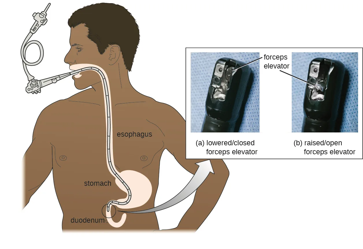
As a result of CRE outbreaks, hospitals, manufacturers, and the FDA are investigating solutions. Many hospitals are instituting more rigorous cleaning procedures than those mandated by the FDA. Manufacturers are looking for ways to redesign duodenoscopes to minimize hard-to-reach crevices where bacteria can escape disinfectants, and the FDA is updating its protocols. In February 2015, the FDA added new recommendations for careful hand cleaning of the duodenoscope elevator mechanism (the location where microbes are most likely to escape disinfection), and issued more careful documentation about quality control of disinfection protocols (shown below).
Extended description
Main drawing: a side view of a person’s head, neck, and torso showing a duodenoscope’s flexible tube entering through the mouth, coiling near a handle, then descending through the esophagus, through the stomach, and into the duodenum, each organ labeled. Inset at right: two close-up photographs of the scope’s tip, each with a pointer line labeled ‘forceps elevator’; the left photo, labeled ‘(a) lowered/closed forceps elevator,’ shows the small metal elevator flap lying flat; the right photo, labeled ‘(b) raised/open forceps elevator,’ shows the same flap tilted upward, opening a gap beside it.
There is no guarantee that new procedures, protocols, or equipment will completely eliminate the risk for infection associated with endoscopes. Yet these devices are used successfully in 500,000–650,000 procedures annually in the United States, many of them lifesaving. At what point do the risks outweigh the benefits of these devices, and who should be held responsible when negative outcomes occur?
Summary
- Chemical disinfectants are grouped by the types of microbes and infectious agents they are effective against. High-level germicides kill vegetative cells, fungi, viruses, and endospores, and can ultimately lead to sterilization. Intermediate-level germicides cannot kill all viruses and are less effective against endospores. Low-level germicides kill vegetative cells and some enveloped viruses, but are ineffective against endospores.
- The effectiveness of a disinfectant is influenced by several factors, including length of exposure, concentration of disinfectant, temperature, and pH.
- Historically, the effectiveness of a chemical disinfectant was compared with that of phenol at killing Staphylococcus aureus and Salmonella enterica serovar Typhi, and a phenol coefficient was calculated.
- The disk-diffusion method is used to test the effectiveness of a chemical disinfectant against a particular microbe.
- The use-dilution test determines the effectiveness of a disinfectant on a surface. In-use tests can determine whether disinfectant solutions are being used correctly in clinical settings.
Key terms
- phenol coefficient — measure of the effectiveness of a chemical agent through comparison with that of phenol on Staphylococcus aureus and Salmonella enterica serovar Typhi.
- disk-diffusion method — a technique for measuring of the effectiveness of one or more antimicrobial agents against a known bacterium; involves measuring the zone(s) of inhibition around the chemical agent(s) in a culture of the bacterium.
- use-dilution test — a technique for determining the effectiveness of a chemical disinfectant on a surface; involves dipping a surface in a culture of the targeted microorganism, disinfecting the surface, and then transferring the surface to a fresh medium to see if bacteria will grow.
- in-use test — a technique for monitoring the correct use of disinfectants in a clinical setting; involves placing used, diluted disinfectant onto an agar plate to see if microbial colonies will grow.
Practice
Describe why the phenol coefficient is used
The effectiveness of chemical disinfectants has historically been compared to that of which of the following?
Name the chemical agent Joseph Lister first used, the one to which every other agent’s effectiveness was once measured.If a chemical disinfectant is more effective than phenol, then its phenol coefficient would be ________ than 1.0.
Recall what a phenol coefficient value above 1.0 versus below 1.0 each indicate about a chemical’s effectiveness relative to phenol.Which of the following refers to a germicide that can kill vegetative cells and certain enveloped viruses but not endospores?
Recall which of the three effectiveness categories this section describes as killing only vegetative cells and some enveloped viruses.A numerical comparison of a chemical agent’s germicidal effectiveness to that of phenol is called a ________.
Name the ratio Rideal and Walker devised in 1903 to standardize disinfectant testing.If used for extended periods of time, ________ germicides may lead to sterility.
Recall which of the three effectiveness categories can eventually sterilize a surface with prolonged use.Why were chemical disinfectants once commonly compared with phenol?
Show model answer
Did your answer mention:
Why is length of exposure to a chemical disinfectant important for its activity?
Recall which direction the module says increasing the length of exposure moves a disinfectant’s efficacy, and compare that with what it says about the other factors in the same paragraph.Compare and contrast the disk-diffusion, use-dilution, and in-use methods for testing the effectiveness of antiseptics, disinfectants, and sterilants
Which type of test is used to determine whether disinfectant solutions actively used in a clinical setting are being used correctly?
Name the test that samples an actively used bottle of disinfectant itself, rather than testing a fresh solution against a microbe on a surface or in a lawn of bacteria.In the disk-diffusion assay, a large zone of inhibition around a disk to which a chemical disinfectant has been applied indicates ________ of the test microbe to the chemical disinfectant.
Name what a wide clear area of no growth around the disk shows about how the microbe responds to the chemical.A test in which a chemical disinfectant is applied to a contaminated stainless-steel cylinder to measure its effectiveness on a surface is called the ________.
Name the test whose passing standard requires at least 59 of 60 replicates to show no growth.Measuring the zones of inhibition produced by chemicals diffusing out of sterile filter-paper disks placed on an inoculated agar plate is called the ________.
Name the method shown by a plate with several small disks, each ringed by a clear area of no growth.What are some advantages of use-dilution and in-use tests compared with the disk-diffusion assay?
Show model answer
Did your answer mention:
This section is adapted from Microbiology, Section 13.4: Testing the Effectiveness of Antiseptics and Disinfectants by Nina Parker, Mark Schneegurt, Anh-Hue Thi Tu, Philip Lister, Brian M. Forster, and OpenStax, © OpenStax, licensed under CC BY-NC-SA 4.0. Access the original for free at openstax.org. Changes: all four source figures are re-encoded as WebP and rendered as mediafigures; the media manifest guesses kind="photo" for all four (JPEG = photo), which is correct only for the CRE micrograph — the disk-diffusion figure (a drawn plate schematic beside a photograph), the in-use-test figure (a fully drawn flow diagram), and the duodenoscope figure (a drawn anatomical illustration beside two photographs of the scope tip) are each explicit kind="diagram" because a genuinely drawn panel is present in each; the disk-diffusion and duodenoscope alts are rewritten from the image with longdesc walk-throughs of their labels, because each is a multi-panel figure whose printed labels (ATB1–ATB5, the ruler instruction; the lowered/raised forceps-elevator captions) the source’s short alt and caption do not fully name; the CRE alt is rewritten from the bare source alt (“Micrograph of oval cells”) to describe the image’s actual appearance; the in-use-test alt omits the diagram’s own baked-in concluding sentence (“growth of 5+ colonies… indicates contamination”), because that exact fact is already stated in the body paragraph immediately above the figure and the alt instead sits directly above a Check Your Understanding item graded on that same fact. All four body Check Your Understanding bullets are rendered at their note positions: the three-level germicide-comparison question is a sortbins (the module’s own three-way compare-and-contrast, without a printed table); the disk-diffusion zone-size question is a multiplechoice over the two alternatives the question itself offers, keyed by the module’s own zone-size sentence; the positive-in-use-test question is a multiplechoice keyed by the module’s own colony-count sentence, its three distractors drawn from this module’s own phenol-coefficient, disk-diffusion, and use-dilution concepts rather than invented; the use-dilution “is it performed in a clinical setting” question stays a selfcheck, because its honest answer draws on three separate sentences (who performs it, for what purpose, and the AOAC’s passing standard) rather than one. Of the module’s nine source exercises, all nine are adapted into Practice: all three Multiple Choice and all three Fill in the Blank with their source keys, and the two unkeyed Short Answer questions and the one unkeyed Critical Thinking question as selfchecks whose model answers and rubric checkpoints are assembled strictly from this module’s own sentences (the phenol-comparison question from the Phenol Coefficient subsection’s opening two sentences; the use-dilution/in-use advantages question from the disk-diffusion, use-dilution, and in-use subsections). The one unkeyed Short Answer question fixed by a single module sentence — “Why is length of exposure to a chemical disinfectant important for its activity?” — is instead graded as a multiplechoice, keyed by the module’s own “longer exposure increasing efficacy” clause, its three distractors built by reversing the direction of this module’s other named factors (concentration, temperature, pH) rather than inventing new claims. The two Practice groups are additionally filled past the book’s floor with three key-term-recall textin items (phenol coefficient; use-dilution test; disk-diffusion method) built from ## Key terms. The Clinical Focus box is rendered as its Resolution part, its figure kept at its document position, and its closing “go back to the previous Clinical Focus box” sentence is replaced with a plain-text sentence naming Controlling Microbial Growth as where the case began, per this run’s chain decision. The cross-reference to Controlling Microbial Growth (Section 13.1) is rendered as a link. The source’s “Human pappiloma virus” and “HVP” are one-word typos for “papilloma” and “HPV,” corrected in place without an inline note; the source’s ungrammatical “To test the disinfectant’s live virus was required” and its “three HPV from three sources” phrasing are transcribed as printed as suspected source defects, not corrected. Key terms are compiled from the module’s four defined terms and the book’s Glossary appendix; all four definitions are taken directly from the Glossary, none sentence-derived — the disk-diffusion method entry keeps the Glossary’s own malformed “a technique for measuring of the effectiveness…” wording verbatim (a suspected Glossary typo, not corrected here); the disk-diffusion method textin accepts the “disc-diffusion method” spelling because the page’s own figure caption (verbatim from the source) calls the same objects “antimicrobial discs."