Testing the Effectiveness of Antimicrobials
By the end of this section, you will be able to:
- Describe how the Kirby-Bauer disk diffusion test determines the susceptibility of a microbe to an antibacterial drug.
- Explain the significance of the minimal inhibitory concentration and the minimal bactericidal concentration relative to the effectiveness of an antimicrobial drug.
Testing the effectiveness of antimicrobial drugs against specific organisms is important in identifying their spectrum of activity and the therapeutic dosage. This type of test, generally described as antimicrobial susceptibility testing (AST), is commonly performed in a clinical laboratory. In this section, we will discuss common methods of testing the effectiveness of antimicrobials.
The Kirby-Bauer Disk Diffusion Test
The Kirby-Bauer disk diffusion test has long been used as a starting point for determining the susceptibility of specific microbes to various antimicrobial drugs. The Kirby-Bauer assay starts with a Mueller-Hinton agar plate on which a confluent lawn is inoculated with a patient’s isolated bacterial pathogen. Filter paper disks impregnated with known amounts of antibacterial drugs to be tested are then placed on the agar plate. As the bacterial inoculum grows, antibiotic diffuses from the circular disk into the agar and interacts with the growing bacteria. Antibacterial activity is observed as a clear circular zone of inhibition around the drug-impregnated disk, similar to the disk-diffusion assay depicted in Testing the Effectiveness of Antiseptics and Disinfectants. The diameter of the zone of inhibition, measured in millimeters and compared to a standardized chart, determines the susceptibility or resistance of the bacterial pathogen to the drug.
There are multiple factors that determine the size of a zone of inhibition in this assay, including drug solubility, rate of drug diffusion through agar, the thickness of the agar medium, and the drug concentration impregnated into the disk. Due to a lack of standardization of these factors, interpretation of the Kirby-Bauer disk diffusion assay provides only limited information on susceptibility and resistance to the drugs tested. The assay cannot distinguish between bacteriostatic and bactericidal activities, and differences in zone sizes cannot be used to compare drug potencies or efficacies. Comparison of zone sizes to a standardized chart will only provide information on the antibacterials to which a bacterial pathogen is susceptible or resistant.
Check Your Understanding
How does one use the information from a Kirby-Bauer assay to predict the therapeutic effectiveness of an antimicrobial drug in a patient?
Show model answer
Did your answer mention:
Micro Connection. Antibiograms: Taking Some of the Guesswork Out of Prescriptions
Unfortunately, infectious diseases don’t take a time-out for lab work. As a result, physicians rarely have the luxury of conducting susceptibility testing before they write a prescription. Instead, they rely primarily on the empirical evidence (i.e., the signs and symptoms of disease) and their professional experience to make an educated guess as to the diagnosis, causative agent(s), and drug most likely to be effective. This approach allows treatment to begin sooner so the patient does not have to wait for lab test results. In many cases, the prescription is effective; however, in an age of increased antimicrobial resistance, it is becoming increasingly more difficult to select the most appropriate empiric therapy. Selecting an inappropriate empiric therapy not only puts the patient at risk but may promote greater resistance to the drug prescribed.
Recently, studies have shown that antibiograms are useful tools in the decision-making process of selecting appropriate empiric therapy. An antibiogram is a compilation of local antibiotic susceptibility data broken down by bacterial pathogen. In a November 2014 study published in the journal Infection Control and Hospital Epidemiology, researchers determined that 85% of the prescriptions ordered in skilled nursing facilities were decided upon empirically, but only 35% of those prescriptions were deemed appropriate when compared with the eventual pathogen identification and susceptibility profile obtained from the clinical laboratory. However, in one nursing facility where use of antibiograms was implemented to direct selection of empiric therapy, appropriateness of empiric therapy increased from 32% before antibiogram implementation to 45% after implementation of antibiograms (J.P. Furuno et al. “Using Antibiograms to Improve Antibiotic Prescribing in Skilled Nursing Facilities.” Infection Control and Hospital Epidemiology 35 no. Suppl S3 (2014):S56–61). Although these data are preliminary, they do suggest that health-care facilities can reduce the number of inappropriate prescriptions by using antibiograms to select empiric therapy, thus benefiting patients and minimizing opportunities for antimicrobial resistance to develop.
Link to Learning
Visit this website to view an interactive antibiogram provided by Stanford University.
Dilution Tests
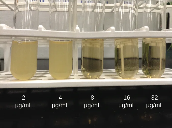
As discussed, the limitations of the Kirby-Bauer disk diffusion test do not allow for a direct comparison of antibacterial potencies to guide selection of the best therapeutic choice. However, antibacterial dilution tests can be used to determine a particular drug’s minimal inhibitory concentration (MIC), the lowest concentration of drug that inhibits visible bacterial growth, and minimal bactericidal concentration (MBC), the lowest drug concentration that kills ≥99.9% of the starting inoculum. Determining these concentrations helps identify the correct drug for a particular pathogen. For the macrobroth dilution assay, a dilution series of the drug in broth is made in test tubes and the same number of cells of a test bacterial strain is added to each tube (see the figure below). The MIC is determined by examining the tubes to find the lowest drug concentration that inhibits visible growth; this is observed as turbidity (cloudiness) in the broth. Tubes with no visible growth are then inoculated onto agar media without antibiotic to determine the MBC. Generally, serum levels of an antibacterial should be at least three to five times above the MIC for treatment of an infection.
The MIC assay can also be performed using 96-well microdilution trays, which allow for the use of small volumes and automated dispensing devices, as well as the testing of multiple antimicrobials and/or microorganisms in one tray (see the figure below). MICs are interpreted as the lowest concentration that inhibits visible growth, the same as for the macrobroth dilution in test tubes. Growth may also be interpreted visually or by using a spectrophotometer or similar device to detect turbidity or a color change if an appropriate biochemical substrate that changes color in the presence of bacterial growth is also included in each well.
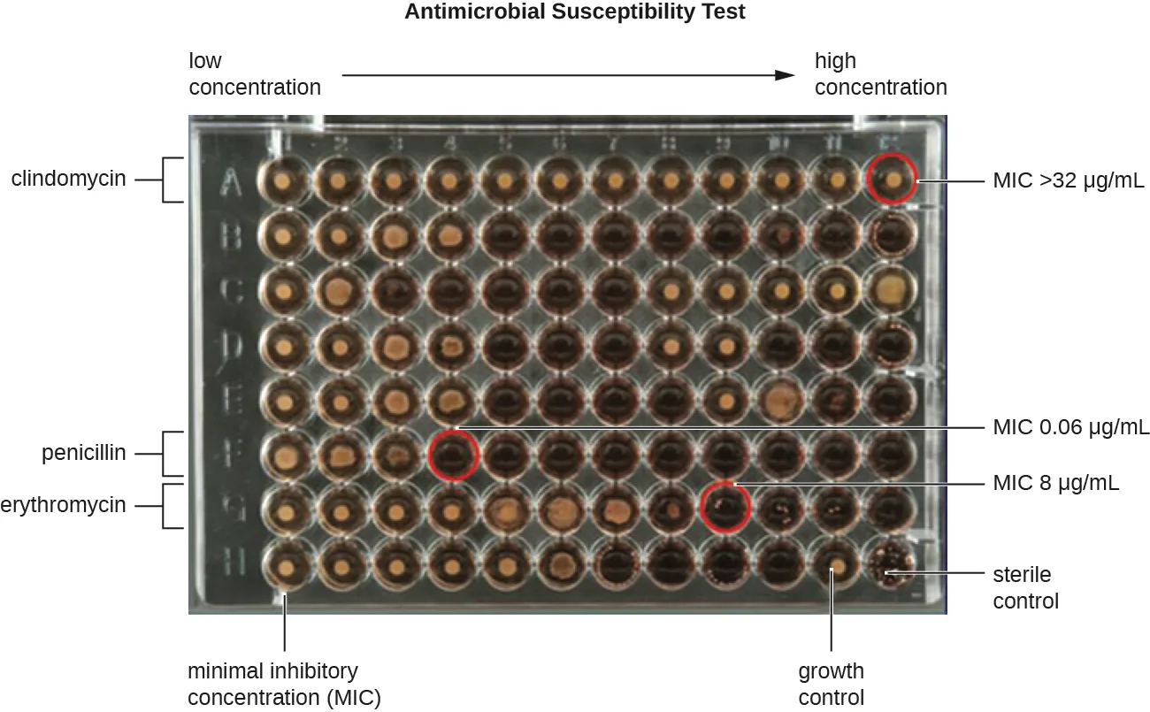
Extended description
The plate has 8 rows, A through H, and 12 columns running left to right beneath an arrow labeled low concentration at the left and high concentration at the right. A bracket beside row A is labeled clindamycin; the well at the far right (highest-concentration) end of row A is circled in red and labeled MIC >32 µg/mL. A bracket beside row F is labeled penicillin; a well toward the left of row F is circled in red and labeled MIC 0.06 µg/mL. A bracket beside row G is labeled erythromycin; a well further right in row G is circled in red and labeled MIC 8 µg/mL. Row H, at the bottom, is labeled minimal inhibitory concentration (MIC) at its left end, growth control further right, and sterile control at its far right end.
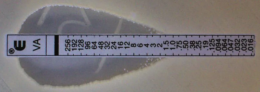
The Etest is an alternative method used to determine MIC, and is a combination of the Kirby-Bauer disk diffusion test and dilution methods. Similar to the Kirby-Bauer assay, a confluent lawn of a bacterial isolate is inoculated onto the surface of an agar plate. Rather than using circular disks impregnated with one concentration of drug, however, commercially available plastic strips that contain a gradient of an antibacterial are placed on the surface of the inoculated agar plate (see the figure below). As the bacterial inoculum grows, antibiotic diffuses from the plastic strips into the agar and interacts with the bacterial cells. Because the rate of drug diffusion is directly related to concentration, an elliptical zone of inhibition is observed with the Etest drug gradient, rather than a circular zone of inhibition observed with the Kirby-Bauer assay. To interpret the results, the intersection of the elliptical zone with the gradient on the drug-containing strip indicates the MIC. Because multiple strips containing different antimicrobials can be placed on the same plate, the MIC of multiple antimicrobials can be determined concurrently and directly compared. However, unlike the macrobroth and microbroth dilution methods, the MBC cannot be determined with the Etest.
Check Your Understanding
Which correctly distinguishes the minimal inhibitory concentration (MIC) from the minimal bactericidal concentration (MBC)?
Reread the sentence that introduces both terms together: one is about inhibiting growth, the other about killing nearly all of the starting inoculum.Clinical Focus. Resolution
Marisa’s UTI was likely caused by the catheterizations she had in Vietnam. Most bacteria that cause UTIs are members of the normal gut microbiota, but they can cause infections when introduced to the urinary tract, as might have occurred when the catheter was inserted. Alternatively, if the catheter itself was not sterile, bacteria on its surface could have been introduced into Marisa’s body. The antimicrobial therapy Marisa received in Cambodia may also have been a complicating factor because it may have selected for antimicrobial-resistant strains already present in her body. These bacteria would have already contained genes for antimicrobial resistance, either acquired by spontaneous mutation or through horizontal gene transfer, and, therefore, had the best evolutionary advantage for adaptation and growth in the presence of the antimicrobial therapy. As a result, one of these resistant strains may have been subsequently introduced into her urinary tract.
Laboratory testing at the CDC confirmed that the strain of Klebsiella pneumoniae from Marisa’s urine sample was positive for the presence of NDM, a very active carbapenemase that is beginning to emerge as a new problem in antimicrobial resistance. While NDM-positive strains are resistant to a wide range of antimicrobials, they have shown susceptibility to tigecycline (structurally related to tetracycline) and the polymyxins B and E (colistin).
To prevent her infection from spreading, Marisa was isolated from the other patients in a separate room. All hospital staff interacting with her were advised to follow strict protocols to prevent surface and equipment contamination. This would include especially stringent hand hygiene practices and careful disinfection of all items coming into contact with her.
Marisa’s infection finally responded to tigecycline and eventually cleared. She was discharged a few weeks after admission, and a follow-up stool sample showed her stool to be free of NDM-containing K. pneumoniae, meaning that she was no longer harboring the highly resistant bacterium.
The case began in History of Chemotherapy and Antimicrobial Discovery.
Summary
- The Kirby-Bauer disk diffusion test helps determine the susceptibility of a microorganism to various antimicrobial drugs. However, the zones of inhibition measured must be correlated to known standards to determine susceptibility and resistance, and do not provide information on bactericidal versus bacteriostatic activity, or allow for direct comparison of drug potencies.
- Antibiograms are useful for monitoring local trends in antimicrobial resistance/susceptibility and for directing appropriate selection of empiric antibacterial therapy.
- There are several laboratory methods available for determining the minimum inhibitory concentration (MIC) of an antimicrobial drug against a specific microbe. The minimal bactericidal concentration (MBC) can also be determined, typically as a follow-up experiment to MIC determination using the tube dilution method.
Key terms
- Kirby-Bauer disk diffusion test — simple, rapid method for determining susceptibility and resistance of a bacterial pathogen to antibacterial drugs. The test involves drug-impregnated disks placed on an agar plate inoculated with a bacterial lawn.
- zone of inhibition — clear zone around a filter disk impregnated with an antimicrobial drug, indicating growth inhibition due to the antimicrobial drug.
- antibiogram — compilation of the antimicrobial susceptibilities recorded for local bacterial strains, which is useful for monitoring local trends in antimicrobial resistance and aiding the prescription of appropriate empiric antibacterial therapy.
- minimal inhibitory concentration (MIC) — lowest concentration of an antibacterial drug that inhibits visible growth of a bacterial strain.
- minimal bactericidal concentration (MBC) — lowest antibacterial drug concentration that kills ≥99.9% of a starting inoculum of bacteria.
- Etest — simple, rapid method for determining MIC, involving commercially available plastic strips that contain a gradient of an antimicrobial and are placed on an agar plate inoculated with a bacterial lawn.
Practice
Describe how the Kirby-Bauer disk diffusion test determines the susceptibility of a microbe to an antibacterial drug
In the Kirby-Bauer disk diffusion test, the _______ of the zone of inhibition is measured and used for interpretation.
Recall the one linear measurement of the zone that is compared to the standardized chart.If drug A produces a larger zone of inhibition than drug B on the Kirby-Bauer disk diffusion test, drug A should always be prescribed.
Recall what the section says a Kirby-Bauer zone size cannot be used to compare between two drugs.The utility of an antibiogram is that it shows antimicrobial susceptibility trends
Recall what geographic scope an antibiogram’s compiled data represents.A compilation of local antibiotic susceptibility data broken down by bacterial pathogen is called a(n) ________.
Name the tool physicians use to select empiric therapy from local resistance trends, discussed in the Micro Connection box.Explain the significance of the minimal inhibitory concentration and the minimal bactericidal concentration relative to the effectiveness of an antimicrobial drug
Which of the following techniques cannot be used to determine the minimum inhibitory concentration of an antimicrobial drug against a particular microbe?
Recall which of these four methods only produces a zone of inhibition rather than an interpretable concentration value.The method that can determine the MICs of multiple antimicrobial drugs against a microbial strain using a single agar plate is called the ________.
Name the method that places gradient strips of different drugs on one plate, described just before the Clinical Focus box.Can an Etest be used to find the minimal bactericidal concentration (MBC) of a drug?
Recall which one of the three MIC-determining methods this section says cannot also determine the MBC.The lowest concentration of an antibacterial drug that inhibits visible growth of a bacterial strain is called the ________.
Name the concentration threshold defined alongside the MBC at the start of the Dilution Tests discussion.This section is adapted from Microbiology, Section 14.6: Testing the Effectiveness of Antimicrobials by Nina Parker, Mark Schneegurt, Anh-Hue Thi Tu, Philip Lister, Brian M. Forster, and OpenStax, © OpenStax, licensed under CC BY-NC-SA 4.0. Access the original for free at openstax.org. Changes: all three source figures are re-encoded as WebP and rendered as mediafigures after image and PDF inspection; all three are kind="photo" — the macrobroth tube series and the Etest strip are unmodified photographs, and the microdilution-tray figure, despite its overlaid title, arrow, brackets, and red circles, is a single photograph of a real 96-well plate with only annotation added, never a drawn panel, so it does not qualify as a diagram under this book’s composite-figure rule (this departs from the run facts, which described it as “a microdilution tray diagram” and the MIC tube figure as “a Kirby-Bauer plate photo with a zone table” — neither description matches the module’s own figures or artwork, so both kind calls were made from the image instead); all three alts are rewritten from the images themselves rather than from the source alts, which contain typos (“left to fight” for “left to right,” “Peniciliin” for “Penicillin” — logged as suspected source-alt defects, not corrected in an inline note since neither alt is reused verbatim); a longdesc was added to the microdilution-tray figure walking its rows, brackets, and circled wells in reading order, since its alt cannot hold every labeled value within the 600-character cap. Two of the module’s two unkeyed Short Answer questions are not used separately in Practice because each duplicates a body Check Your Understanding bullet word-for-word in substance (“How is the information from a Kirby-Bauer disk diffusion test used for the recommendation of the clinical use of an antimicrobial drug?” duplicates the first Check Your Understanding bullet, and “What is the difference between MIC and MBC?” duplicates the second); the first body bullet stays a self-check whose model answer is built from the zone-diameter-and-standardized-chart sentence together with the assay’s stated limitations, and the second is graded because the sentence that defines MIC and MBC side by side fixes it — rendered as a multiplechoice whose two incorrect-swap and two conflated-definition distractors are recombinations of the module’s own MIC/MBC wording rather than invented claims. The unkeyed Critical Thinking question “Can an Etest be used to find the MBC of a drug? Explain.” is graded as a Yes/No multiplechoice keyed from the single sentence stating the MBC cannot be determined with the Etest, dropping only the “Explain” instruction. Of the module’s five keyed source exercises, all three Multiple Choice, the one True/False (rendered as a two-option multiple choice, no “True or false:” prefix), and the one Fill in the Blank are adapted into Practice. Two filler textin items (term recall for antibiogram and for minimal inhibitory concentration, accepting the module’s own abbreviation MIC) bring both objective groups to the book’s three-item floor. The Etest fill-in-the-blank’s accept list carries only E-test rather than the run facts’ E-test|E test, because the real grader folds hyphen-versus-space and rejects the two as duplicate members of one list. The Link to Learning keeps its external URL and describes the destination. The Micro Connection box’s one footnote (Furuno et al., Infection Control and Hospital Epidemiology, 2014) is rendered as an inline parenthetical citation at the sentence it supports; it carries neither a DOI nor a bare access URL to drop or keep. Clinical Focus Resolution names The History of Chemotherapy and Antimicrobial Discovery as where Marisa’s case began, in place of the source’s “Go back to the previous Clinical Focus box” link, and carries no “continues in” sentence because this is the case’s final part. Key terms are compiled from the module’s six defined terms and the book’s Glossary appendix; all six definitions are taken directly from the Glossary, none sentence-derived. No source exercise item is omitted from the page: the two duplicate Short Answer questions are used as body self-checks instead of Practice items, as explained above.